Abstract
The product of pxcA (formerly known as cotA) is involved in light-induced Na+-dependent proton extrusion. In the presence of 2,5-dimethyl-p-benzoquinone, net proton extrusion by Synechocystis sp. strain PCC6803 ceased after 1 min of illumination and a postillumination influx of protons was observed, suggesting that the PxcA-dependent, light-dependent proton extrusion equilibrates with a light-independent influx of protons. A photosystem I (PS I) deletion mutant extruded a large number of protons in the light. Thus, PS II-dependent electron transfer and proton translocation are major factors in light-driven proton extrusion, presumably mediated by ATP synthesis. Inhibition of CO2 fixation by glyceraldehyde in a cytochrome c oxidase (COX) deletion mutant strongly inhibited the proton extrusion. Leakage of PS II-generated electrons to oxygen via COX appears to be required for proton extrusion when CO2 fixation is inhibited. At pH 8.0, NO3− uptake activity was very low in the pxcA mutant at low [Na+] (∼100 μM). At pH 6.5, the pxcA strain did not take up CO2 or NO3− at low [Na+] and showed very low CO2 uptake activity even at 15 mM Na+. A possible role of PxcA-dependent proton exchange in charge and pH homeostasis during uptake of CO2, HCO3−, and NO3− is discussed.
Light-induced extrusion of protons into the medium has been observed in various cyanobacterial strains (2, 3, 6, 8, 9, 12, 17, 18, 21, 22). Scherer et al. (17, 18) reported two phases of light-induced proton extrusion in Anabaena variabilis. The first phase is due to a light-dependent uptake of CO2, which is converted to HCO3−, and the second phase was considered to be dependent on ATP and linear photosynthetic electron flow. Both phases of proton extrusion are specifically stimulated by Na+. Similar Na+-dependent light-induced proton extrusion has been observed with Synechococcus and Plectonema (2, 6, 12). The light-induced proton extrusion in Plectonema has been assumed to be due to a respiratory electron transport chain localized on the cytoplasmic membrane (2). The physiological significance of the light-induced proton extrusion is not yet known, and ambiguity remains whether photosynthetic or respiratory electron transport and whether cytoplasmic or thylakoid membranes are involved in this reaction.
pxcA (formerly known as cotA) is a homolog of cemA or ycf10 in chloroplast genomes (7, 8, 21, 22). Light-induced proton extrusion activity was abolished when pxcA was inactivated in Synechocystis sp. strain PCC6803 (8, 21) or Synechococcus sp. strain PCC7942 (22). The pxcA mutants were unable to grow in low-Na+ medium or in acidic medium. PxcA is located in the cytoplasmic membrane (21), and the cemA or ycf10 gene in chloroplast genomes encodes a chloroplast envelope membrane protein (16). These results indicate that PxcA is involved in light-induced proton extrusion and that this protein is essential for cell growth under acidic or low-salt conditions.
The present study aims to clarify which mode of electron transport is involved in the light-induced proton extrusion and to determine the effect of pxcA inactivation on the uptake of CO2, HCO3−, and NO3−. For this reason, pxcA mutants and strains carrying deletions of genes that code for photosynthetic or respiratory electron transport components in Synechocystis sp. strain PCC6803 were analyzed. Measurements of net proton exchange in the wild-type (WT) and mutant cells with or without electron acceptors or inhibitors enabled us to conclude that photosystem II (PS II)-driven electron transport was primarily involved in this reaction. We have also measured the uptake of CO2, HCO3−, and NO3− in the WT and pxcA mutant. The results demonstrate that the PxcA-dependent proton exchange is essential for CO2 uptake under acidic conditions and for NO3− uptake at low-Na+ concentrations.
MATERIALS AND METHODS
Mutants and growth conditions.
The following mutants were used in this study: pxcA (previously named M29) (8), psaAB (PS I-less) (20), psbDIC/psbDII (PS II-less) (24), and coxAB (cytochrome c oxidase-less) (19). WT, pxcA, and coxAB cells were grown at 30°C in BG-11 medium (23) buffered with 20 mM N-Tris(hydroxymethyl)methyl-2-aminoethanesulfonic acid (TES)–KOH at pH 8.0; the cultures were aerated with 3% (vol/vol) CO2 in air. Glucose (5 mM) was added to the above medium for the growth of psaAB and psbDIC/psbDII mutants. Continuous illumination was provided by fluorescent lamps at 40-μmol photosynthetically active radiation/m2/s (400 to 700 nm) for psaAB cells, which are sensitive to higher light intensity, and at 100 μmol/m2/s for the other strains.
Measurements of proton exchange and uptake of CO2, HCO3−, and NO3−.
Cells harvested by centrifugation were washed twice with 0.2 mM TES-KOH buffer (pH 8.0) and then suspended in the same buffer at a chlorophyll concentration of 14 μg/ml (1.4 μg/ml for the psaAB mutant, which has about sevenfold less chlorophyll on a per-cell basis [20]). Changes in the pH of the cell suspension (3 ml) kept at 30°C were monitored by using a pH electrode with a meter (Inlar 423 and Delta 350; Mettler Toledo, Halstead, United Kingdom). After each measurement, the signal was calibrated by injecting 10 μl of 7.5 mM HCl into the cell suspension.
Uptake of CO2 and HCO3− was measured by the silicone oil-filtering centrifugation method (11, 25). Nitrate uptake was measured as described by Omata et al. (13). The cells were washed twice with nitrate-free medium (BG-11 medium minus NaNO3, Na2CO3, and microelements) buffered with 5 mM MES-KOH at pH 6.5 or with 5 mM TES-KOH at pH 8.0 and then suspended in the same buffer supplemented with 5 mM KHCO3 to a chlorophyll concentration of 7 μg/ml. NaCl (final concentration, 15 mM) was added to the cell suspension. The concentration of nitrate was determined with a Technicon autoanalyzer.
The light source for all the experiments was a 150-W halogen lamp (MHF-150L; Kagaku Kyoeisha Ltd., Osaka, Japan) equipped with a glass fiber. Cells in a sample chamber or in a 1.5-ml Eppendorf tube were illuminated by white light from the fiber at an intensity of 4.0 mmol of photosynthetically active radiation/m2/s.
RESULTS
Effect of DMBQ on net proton exchange.
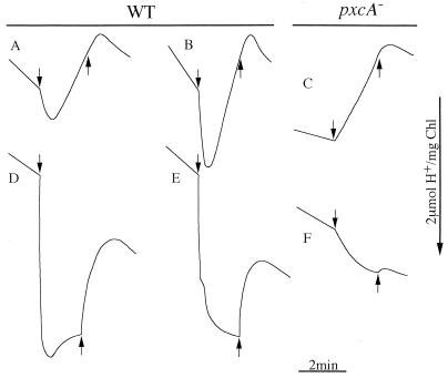
The profiles of net proton exchange measured with the WT and pxcA cells are shown in Fig. 1. For these measurements, the cells were suspended in 0.2 mM TES-KOH buffer (pH 8.0) with (Fig. 1D to F) or without (Fig. 1A to C) 2,5-dimethyl-p-benzoquinone (DMBQ). When WT cells suspended in buffer containing 15 mM KCl (Fig. 1A) or NaCl (Fig. 1B) were illuminated, acidification followed by alkalization of the medium was observed. The acidification was stimulated by 15 mM Na+. In contrast, for the pxcA mutant, only alkalization, not acidification, of the medium was observed upon illumination (Fig. 1C). It has been reported that alkalization of the medium is linked to photosynthetic fixation of CO2 produced by dehydration of HCO3− (10). These results confirm that Na+-stimulated light-induced proton extrusion occurs in the WT strain but not in the mutant (8).
FIG. 1.
Net proton movements in suspensions of WT (A, B, D, and E) and pxcA (C and F) cells upon switching the light on (arrow down) and off (arrow up). The cells were suspended in 0.2 mM TES-KOH buffer (pH 8.0) containing 15 mM KCl (A and D) or NaCl (B, C, E, and F) in the absence (A to C) and presence (D to F) of 1 mM DMBQ. The chlorophyll concentration in the cell suspension was 14 μg/ml.
Acidification of the medium was stimulated when WT cells were illuminated in the presence of DMBQ (Fig. 1D and E). DMBQ can oxidize the plastoquinone pool and may be reduced by PS I; hence, it is an electron acceptor in photosynthetic electron transport. Therefore, proton extrusion is linked to photosynthetic electron transfer. No net alkalization followed the acidification on illumination under these conditions, due to the absence of photosynthetic CO2 fixation. The presence of Na+ showed little effect on the extent of proton extrusion in the presence of DMBQ. Figure 1D and E indicates that the net proton extrusion does not proceed continuously in the light but ceases after 1 min of illumination. After the light was turned off, an influx of protons was observed. This suggests that in the light, both extrusion and influx of protons occur, reaching an equilibrium where there is no net proton exchange, whereas after the light is turned off (causing proton extrusion to cease), proton influx continues for a short time until a new steady-state level is attained. Both light-induced proton extrusion and postillumination proton influx were very low in pxcA cells in the presence of DMBQ (Fig. 1F).
Net proton exchange in mutants defective in PS I, PS II or cytochrome c oxidase.
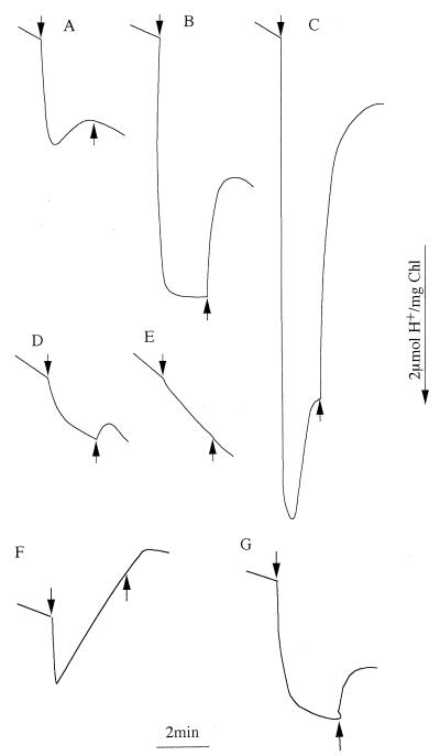
Now that a role of photosynthetic electron transfer in proton extrusion has been established, the next question involves the part(s) of photosynthetic electron transport proton with which extrusion is associated and whether respiratory electron transfer also plays a role. To address this question, mutants lacking either PS I, PS II, or cytochrome c oxidase were investigated. The psaAB (PS I-less) strain showed Na+-stimulated light-induced proton extrusion (Fig. 2A and B). On a per-chlorophyll basis, the amplitude of proton extrusion was two- to threefold larger than that in WT cells (compare with Fig. 1A and B). Since about 85% of the chlorophyll in WT Synechocystis sp. strain PCC6803 is associated with PS I (20), this indicates that PS II-mediated electron transfer can drive a significant amount of proton extrusion. No proton uptake was observed in the PS I-less mutant in the light, consistent with the lack of CO2 fixation in this strain. In the presence of DMBQ, a more extensive acidification followed by proton uptake was observed (Fig. 2C), similar to what was seen in WT cells but again with a two- to threefold-higher amplitude on a per-chlorophyll basis. Thus, PS II-driven electron transport from water to DMBQ or, to a lesser extent, to oxygen (the latter involving oxidase[s]) can lead to proton extrusion.
FIG. 2.
Net proton movement in the suspensions of psaAB (A to C), psbDIC/psbDII (D and E), and coxAB (F and G) cells upon switching the light on (arrow down) and off (arrow up). The cells were suspended in 0.2 mM TES-KOH buffer containing 15 mM KCl (A) and NaCl (B to G). DMBQ was added prior to illumination in panels C, E, and F. The chlorophyll concentration in the cell suspension was 1.4 μg/ml for the psaAB mutant and 14 μg/ml for the psbDIC/psbDII and coxAB mutants.
A small amount of light-induced proton extrusion was observed when a cell suspension of the psbDIC/psbDII strain was illuminated in the absence of DMBQ (Fig. 2D) but not in its presence (Fig. 2E). The initial rate of light-induced proton extrusion in the psbDIC/psbDII strain was about 5% of that in the psaAB stain on a per-chlorophyll basis (the rates were 200 and 4,020 μmol/mg of chlorophyll/h in psbDIC/psbDII and psaAB strains, respectively, in the presence of 15 mM NaCl but in the absence of DMBQ).
The proton exchange profiles obtained for the coxAB mutant in the presence and absence of DMBQ were the same as those obtained for WT cells (Fig. 2F and G). Thus, cytochrome c oxidase is not essential to proton extrusion under these conditions.
Effect of electron transfer inhibitors and acceptors on proton exchange.
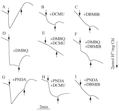
The results presented thus far imply that electron transfer involving PS II is a major factor in light-driven proton extrusion. To further test this, proton extrusion was measured in WT cells after addition of 3-(3-4-dichlorophenyl)-1,1-dimethylurea (DCMU), a PS II electron transport inhibitor. Indeed, DCMU strongly inhibited the proton extrusion and created a pattern similar to that observed in the PS-II less mutant (compare Fig. 3B with Fig. 2D). The proton extrusion was more strongly inhibited by 2,5-dibromo-3-methyl-6-isopropyl-p-benzoquinone (DBMIB), an inhibitor of electron transport at the cytochrome b6/f complex (Fig. 3C). The light-induced proton extrusion of WT cells in the presence of DMBQ was completely inhibited by DCMU (Fig. 3D and E); addition of DBMIB resulted in partial inhibition (Fig. 3F). Addition of DCMU during illumination in the presence of DMBQ caused influx of protons into the cells, and no postillumination proton influx was observed on subsequent removal of the light source (data not shown).
FIG. 3.
Effect of DMBQ, PNDA, DCMU, and DBMIB on net proton movements in WT Synechocystis cells. The light was switched on (arrow down) and off (arrow up) as indicated. The cells were suspended in 0.2 mM TES-KOH buffer (pH 8.0) containing 15 mM NaCl. DMBQ (final concentration, 1 mM) (D to E), PNDA (3 mM) (G to I), DCMU (20 μM) (B, E, and H), and DBMIB (10 μM) (C, F, and I) were added as indicated. All additions were done prior to illumination.
Addition of p-nitrosodimethylaniline (PNDA), a PS I electron acceptor (1), had little effect. However, if both PNDA and DCMU were added, the amount of proton extrusion was somewhat greater than when DCMU alone was added (Fig. 3B and H). DBMIB strongly inhibited the proton extrusion in the presence of PNDA (Fig. 3I). These results indicate that the extrusion of protons was abolished when both water splitting and the cytochrome b6/f complex were inhibited. However, electron transport from water to DMBQ, and, to a lesser extent, from the intracellular reductants to electron acceptors via PS I and/or PS I-dependent cyclic electron flow energizes proton extrusion.
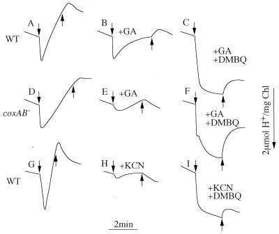
To test the hypothesis that alkalization is driven by HCO3− utilization, photosynthetic CO2 fixation was inhibited by glyceraldehyde (GA) treatment. This treatment reduced the rate of alkalization in both the WT and coxAB cells (Fig. 4A, B, D, and E), indicating that OH− produced as a result of bicarbonate utilization is extruded in the light. Interestingly, GA did not affect the light-induced proton extrusion in the WT strain (Fig. 4A and B) but had a strong inhibitory effect on proton extrusion in the coxAB strain (Fig. 4D and E). The GA inhibition was relieved by addition of DMBQ (Fig. 4F). A similar result was obtained with the WT strain when 5 mM KCN was added (Fig. 4G to I). At this concentration, KCN inhibits both photosynthetic CO2 fixation and oxidase activity. Therefore, in the absence of photosynthetic CO2 fixation, electron flow to oxygen via cytochrome c oxidase is essential for proton extrusion. If this electron flow cannot occur, the quinone pool may be overreduced and continuous electron transfer cannot occur. However, if DMBQ is added, PS II-mediated electron transfer can resume and proton extrusion is observed.
FIG. 4.
Effect of GA, KCN, and DMBQ on net proton movements involving WT (A to C and G to I) and coxAB (D to F) cells of Synechocystis. The light was switched on (arrow down) and off (arrow up). The cells were suspended in 0.2 mM TES-KOH buffer (pH 8.0) containing 15 mM NaCl at a chlorophyll concentration of 14 μg/ml. GA (final concentration, 20 mM) (B, C, E, and F) and KCN (5 mM) (H and I) were added prior to illumination and DMBQ (1 mM) (C, F, and H) was added in the dark to the cell suspensions after the profiles in the presence of the inhibitors were obtained.
Effect of Na+ and pH on the uptake of CO2, HCO3−, and NO3− in WT and pxcA strains.
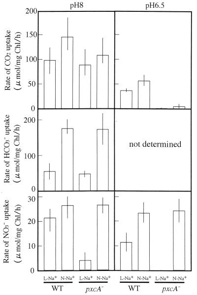
Protons are produced during the transport of CO2 and are consumed when NO3− is reduced to NH4 via NO2− or when HCO3− is converted to CO2. Cells have a mechanism to maintain homeostasis with respect to the intracellular pH and electroneutrality during these processes. To test whether the PxcA-dependent proton exchange is involved in maintaining this homeostasis, the uptake of CO2, HCO3−, and NO3− was monitored as a function of the activity of proton exchange. For this purpose, the uptake of CO2, HCO3−, and NO3− in the WT and the pxcA strains was measured at pH 8.0 and 6.5 in the presence of a normal concentration of NaCl (15 mM, close to the concentration in BG-11 medium) or KCl (15 mM) with a low contaminating concentration of Na+ (∼100 μM Na+). As reported previously (5), HCO3− uptake was high at the normal Na+ concentration and low at the low Na+ concentration in the WT and the pxcA strains (Fig. 5, middle row). Thus, pxcA inactivation did not affect the HCO3− uptake. At the low Na+ concentration, the NO3− uptake was very low in the pxcA strain at pH 8.0 and was zero at pH 6.5 (bottom rows). At the normal Na+ concentration, no significant effect of pxcA inactivation was observed on CO2 and NO3− uptake at pH 8.0 but CO2 uptake activity was reduced significantly at pH 6.5 (top and bottom rows). No CO2 uptake was observed in the mutant at pH 6.5 in the presence of a low Na+ concentration. It is evident that the inactivation of pxcA strongly affected the CO2 uptake under acidic conditions and the NO3− uptake at low Na+ concentrations.
FIG. 5.
Rates of CO2, HCO3−, and NO3− uptake in WT and pxcA cells of Synechocystis at pH 8.0 and pH 6.5 in the presence of 15 mM NaCl (N-Na+) or KCl (L-Na+).
DISCUSSION
The results presented here demonstrate that proton extrusion is driven by PS II coupled to the cytochrome b6/f complex (Fig. 1 to 3). Some proton extrusion can also be driven by PS I. PxcA is an important factor in mediating this proton extrusion. The question now is how this proton extrusion occurs. First, it is unlikely that protons produced by PS II and the cytochrome b6/f complex are directly extruded into the medium, since the lumen and the periplasmic space are presumed to be two different compartments. In addition, protons pumped by these complexes should lead to ATP synthesis and should not be “wasted” by extrusion. Therefore, an energy carrier would be required. ATP seems to be the only candidate for such a carrier that energizes the proton extrusion system; NADPH is not a candidate, because the PS II electron transfer is effective in causing proton extrusion.
The activity of proton extrusion appears to be correlated with the activity of photosynthetic water splitting and electron transport through the cytochrome b6/f complex; both of these processes produce a proton gradient across the thylakoid membrane and thereby can lead to the generation of ATP. This supports the view that the PxcA-dependent proton extrusion is energized by ATP. PxcA does not have an ATP-binding motif and therefore probably is unable to hydrolyze ATP by itself. PxcA may be a regulator of an ATP-dependent proton extrusion pump, and the pump activity is very low in the absence of PxcA.
Besides this PxcA-dependent proton exchange system, cyanobacterial cells possess a Na+/H+ antiport system (14). In fact, the genome of Synechocystis sp. strain PCC6803 contains five genes resembling those coding for Na+/H+ antiporters (4). Two of these gene products contain an ATP-binding motif. It is possible that these gene products are involved in PxcA-dependent proton exchange.
The results presented in Fig. 5 indicate that inactivation of pxcA affects the uptake of CO2 and NO3−. Recently, Rolland et al. reported that inactivation of cemA affects the uptake of inorganic carbon in the chloroplast of Chlamydomonas (15). These results obtained with Synechocystis and Chlamydomonas strongly suggest that cemA and pxcA have the same function in chloroplasts and cyanobacterial cells, respectively.
Based on the results obtained, we propose a working hypothesis involving two complementary proton exchange systems, one of which depends on PxcA, to explain the growth characteristics and inorganic carbon and nitrate uptake of the WT and pxcA strains. This hypothesis has the following features. (i) PxcA-dependent and PxcA-independent proton exchange systems play essential roles in maintaining homeostasis with respect to the intracellular pH and electroneutrality. The proton exchange catalyzed by both systems is stimulated by Na+. (ii) Both systems are essential to growth and CO2 transport at pH 6.5, but the PxcA-independent system alone is sufficient at pH 8 when the activity is high at the normal Na+ concentration. However, both systems are required even at this alkaline pH when the activity of each system is low at the low Na+ concentration. (iii) At the low Na+ concentration, NO3− uptake requires the PxcA-dependent system. However, when PxcA-independent proton exchange is active at the normal Na+ concentration, the PxcA-dependent system is not required for NO3− uptake. (iv) Uptake of HCO3− requires a high activity of PxcA-independent proton exchange at the normal Na+ concentration in both WT and pxcA cells.
Proton exchange catalyzed by the PxcA-independent system should be observed as the pH of the suspension medium of pxcA cells changes. The slow alkalization observed with pxcA cells in the light may be due to proton influx by the PxcA-independent system; it is also possible that rapid influx and efflux of protons via the PxcA-independent system occur with a small net proton movement that cannot be measured by the pH electrode used in this study.
ACKNOWLEDGMENTS
This study was supported by a Grant-in-Aid for Scientific Research (grant 09640767) from the Ministry of Education, Science, Sports and Culture of Japan and by grants from the New Energy and Industrial Technology Development Organization (NEDO) of Japan and from the Human Frontier Science Program.
REFERENCES
- 1.Elstner E F, Zeller H. Bleaching of p-nitrosodimethylaniline by photosystem 1 of spinach lamellae. Plant Sci Lett. 1978;13:15–20. [Google Scholar]
- 2.Hawkesford M J, Rowell P, Stewart W D P. Energy transduction in cyanobacteria. In: Papageorgiou G C, Packer L, editors. Photosynthetic prokaryotes: cell differentiation and function. Amsterdam, The Netherlands: Elsevier; 1963. pp. 199–218. [Google Scholar]
- 3.Hinrichs I, Scherer S, Boger P. Two different mechanisms of light induced proton efflux in the blue-green alga Anabaena variabilis. Physiol Veg. 1985;23:717–724. [Google Scholar]
- 4.Kaneko T, Sato S, Kotani H, Tanaka A, Asamizu E, Nakamura Y, Miyajima N, Hirosawa M, Sugiura M, Sasamoto S, Kimura T, Hosouchi T, Matsuno A, Muraki A, Nakazaki N, Naruo K, Okumura S, Shimpo S, Takeuchi C, Wada T, Watanabe A, Yamada M, Yasuda M, Tabata S. Sequence analysis of the genome of the unicellular cyanobacterium Synechocystis sp. strain PCC6803. II. Sequence determination of the entire genome and assignment of potential protein-coding regions. DNA Res. 1996;3:109–136. doi: 10.1093/dnares/3.3.109. [DOI] [PubMed] [Google Scholar]
- 5.Kaplan A, Schwarz R, Lieman-Hurwitz J, Ronen-Tarazi M, Reinhold L. Physiological and molecular studies on the response of cyanobacteria to changes in the ambient inorganic carbon concentration. In: Bryant D A, editor. The molecular biology of cyanobacteria. Dordrecht, The Netherlands: Kluwer Academic Publishers; 1994. pp. 469–485. [Google Scholar]
- 6.Kaplan A, Scherer S, Lerner M. Nature of the light-induced H+ efflux and Na+ uptake in cyanobacteria. Plant Physiol. 1989;89:1220–1225. doi: 10.1104/pp.89.4.1220. [DOI] [PMC free article] [PubMed] [Google Scholar]
- 7.Katoh A, Lee K S, Fukuzawa H, Ohyama K, Ogawa T. A cemA homologue essential to CO2 transport in the cyanobacterium, Synechocystis PCC6803. Proc Natl Acad Sci USA. 1996;93:4006–4010. doi: 10.1073/pnas.93.9.4006. [DOI] [PMC free article] [PubMed] [Google Scholar]
- 8.Katoh A, Sonoda M, Katoh H, Ogawa T. Absence of light-induced proton extrusion in cotA-less mutant of Synechocystis sp. strain PCC6803. J Bacteriol. 1996;178:5452–5455. doi: 10.1128/jb.178.18.5452-5455.1996. [DOI] [PMC free article] [PubMed] [Google Scholar]
- 9.Lockau W, Pfeffer S. A cyanobacterial ATPase distinct from the coupling factor of photophosphorylation. Z Naturforsch Sect C. 1982;37:658–664. [Google Scholar]
- 10.Miller A G, Colman B. Active transport and accumulation of bicarbonate by a unicellular cyanobacterium. J Bacteriol. 1980;143:1253–1259. doi: 10.1128/jb.143.3.1253-1259.1980. [DOI] [PMC free article] [PubMed] [Google Scholar]
- 11.Ogawa T. A gene homologous to the subunit-2 gene of NADH dehydrogenase is essential to inorganic carbon transport of Synechocystis PCC6803. Proc Natl Acad Sci USA. 1991;88:4275–4279. doi: 10.1073/pnas.88.10.4275. [DOI] [PMC free article] [PubMed] [Google Scholar]
- 12.Ogawa T, Kaplan A. The stoichiometry between CO2 and H+ fluxes involved in the transport of inorganic carbon in cyanobacteria. Plant Physiol. 1987;83:888–891. doi: 10.1104/pp.83.4.888. [DOI] [PMC free article] [PubMed] [Google Scholar]
- 13.Omata T, Ohmori M, Arai N, Ogawa T. Genetically engineered mutant of the cyanobacterium Synechococcus PCC7942 defective in nitrate transport. Proc Natl Acad Sci USA. 1989;86:6612–6616. doi: 10.1073/pnas.86.17.6612. [DOI] [PMC free article] [PubMed] [Google Scholar]
- 14.Ritchie R J. Sodium transport and the origin of the membrane potential in the cyanobacterium Synechococcus R-2 (Anacystis nidulans) PCC7942. J Plant Physiol. 1992;139:320–330. [Google Scholar]
- 15.Rolland N, Dorne A-J, Amoroso G, Sultemeyer D F, Joyard J, Rochaix J-D. Disruption of the plastid ycf10 open reading frame affects uptake of inorganic carbon in the chloroplast of Chlamydomonas. EMBO J. 1997;16:6713–6726. doi: 10.1093/emboj/16.22.6713. [DOI] [PMC free article] [PubMed] [Google Scholar]
- 16.Sasaki Y, Sekiguchi K, Nagano Y, Matsuno R. Chloroplast envelope protein encoded by chloroplast genome. FEBS Lett. 1993;316:93–98. doi: 10.1016/0014-5793(93)81743-j. [DOI] [PubMed] [Google Scholar]
- 17.Scherer S, Hinrichs I, Boger P. Effect of monochromatic light on proton efflux of the blue-green alga Anabaena variabilis. Plant Physiol. 1986;81:939–941. doi: 10.1104/pp.81.3.939. [DOI] [PMC free article] [PubMed] [Google Scholar]
- 18.Scherer S, Riege H, Boger P. Light-induced proton release by the cyanobacterium Anabaena variabilis: dependence on CO2 and Na+ Plant Physiol. 1988;86:769–772. doi: 10.1104/pp.86.3.769. [DOI] [PMC free article] [PubMed] [Google Scholar]
- 19.Schmetterer G, Alge D, Gregor W. Deletion of cytochrome c oxidase genes from the cyanobacterium Synechocystis sp. PCC6803: evidence for alternative respiratory pathways. Photosynth Res. 1994;42:43–50. doi: 10.1007/BF00019057. [DOI] [PubMed] [Google Scholar]
- 20.Shen G, Boussiba S, Vermaas W F J. Synechocystis sp. PCC6803 strains lacking photosystem I and phycobilisome function. Plant Cell. 1993;5:1853–1863. doi: 10.1105/tpc.5.12.1853. [DOI] [PMC free article] [PubMed] [Google Scholar]
- 21.Sonoda M, Kitano K, Katoh A, Katoh H, Ohkawa H, Ogawa T. Size of cotA and identification of the gene Product in Synechocystis sp. strain PCC6803. J Bacteriol. 1997;179:3845–3850. doi: 10.1128/jb.179.12.3845-3850.1997. [DOI] [PMC free article] [PubMed] [Google Scholar]
- 22.Sonoda M, Katoh H, Ohkawa H, Ogawa T. Cloning of cotA gene of Synechococcus PCC7942 and complementation of a cotA-less mutant of Synechocystis PCC6803 with chimeric genes of the two strains. Photosynth Res. 1997b;54:99–105. [Google Scholar]
- 23.Stanier R Y, Kunisawa R, Mandel M, Cohen-Bazire G. Purification and properties of unicellular blue-green algae (order Chroococcales) Bacteriol Rev. 1971;35:171–205. doi: 10.1128/br.35.2.171-205.1971. [DOI] [PMC free article] [PubMed] [Google Scholar]
- 24.Vermaas W F J, Charit J, Eggers B. System for site-directed mutagenesis in the psbD1/C operon of Synechocystis sp. PCC6803. In: Baltscheffsky M, editor. Current research in photosynthesis. Vol. 1. Dordrecht, The Netherlands: Kluwer Academic Publishers; 1990. pp. 231–238. [Google Scholar]
- 25.Volokita M, Zenvirth D, Kaplan A, Reinhold L. Nature of the inorganic carbon species actively taken up by the cyanobacterium Anabaena variabilis. Plant Physiol. 1984;76:599–602. doi: 10.1104/pp.76.3.599. [DOI] [PMC free article] [PubMed] [Google Scholar]


