Abstract
Simple Summary
Necrotic enteritis is an enterotoxemic disease caused by Clostridium perfringens, leading to diarrhea or necrotizing lesions in the intestines of animals, with severe cases leading to death. The butyrate attenuated the inflammatory response and improved intestinal health in piglets challenged with pathogenic bacteria, but it was absorbed by the anterior segment of the gastrointestinal tract. Microencapsulation is a simple and effective method to prevent butyrate from absorption. Thus, we evaluate the effectiveness to two butyrate alleviates clostridium perfringens infections. Our results indicate that dietary supplementation with sodium butyrate or microencapsulated sodium butyrate improves the immune status and morphology of intestinal villi, increases the production of VFAs, and modulates cecal microbiota in chickens challenged with Clostridium perfringens. Moreover, microencapsulated sodium butyrate contains less butyrate than sodium butyrate. These findings indicate that microencapsulated sodium butyrate was more effective than sodium butyrate with the same butyrate supplemental amount.
Abstract
Microencapsulated sodium butyrate (MS-SB) is an effective sodium butyrate additive which can reduce the release of sodium butyrate (SB) in the fore gastrointestinal tract. In this study, we assess the protective effects and mechanisms of MS-SB in Clostridium perfringens (C. perfringens)-challenged broilers. Broiler chickens were pre-treated with SB or MS-SB for 56 days and then challenged with C. perfringens three times. Our results indicate that the addition of MS-SB or SB before C. perfringens infection significantly decreased the thymus index (p < 0.05). Serum IgA, IgY, and IgM concentrations were significantly increased (p < 0.05), while pro-inflammatory IL-1β, IL-6, and TNF-α were significantly decreased (p < 0.05) under MS-SB or SB supplementation. Compared with SB, MS-SB presented a stronger performance, with higher IgA content, as well as a lower IL-1β level when normal or C. perfringens-challenged. While C. perfringens challenge significantly decreased the villus height (p < 0.05), MS-SB or SB administration significantly increased the villus height and villus height/crypt depth (V/C ratio) (p < 0.05). Varying degrees of SB or MS-SB increased the concentrations of volatile fatty acids (VFAs) during C. perfringens challenge, where MS-SB presented a stronger performance, as evidenced by the higher content of isovaleric acid and valeric acid. Microbial analysis demonstrated that both SB or MS-SB addition and C. perfringens infection increase variation in the microbiota community. The results also indicate that the proportions of Bacteroides, Faecalibacterium, Clostridia, Ruminococcaceae, Alistipes, and Clostridia were significantly higher in the MS-SB addition group while, at same time, C. perfringens infection increased the abundance of Bacteroides and Alistipes. In summary, dietary supplementation with SB or MS-SB improves the immune status and morphology of intestinal villi, increases the production of VFAs, and modulates cecal microbiota in chickens challenged with C. perfringens. Moreover, MS-SB was more effective than SB with the same supplemental amount.
Keywords: microencapsulated sodium butyrate, broiler chicken, immunity, cecum microflora, Clostridium perfringens
1. Introduction
Antibiotic growth promoters have been used to improve animal growth and to control or prevent animal disease during rearing; however, many countries have forbidden the dietary use of antimicrobial agents to avoid the emergence of antibiotic-resistant bacteria [1]. Thus, animal nutritionists have been attempting to discover antibiotic alternatives that are low-cost, widely used, and which have an obvious effect. Butyric acid has a positive effect on the normal intestinal mucosa, which can be attributed to a major energy source for intestinal epithelial cells [2,3]. In the intestine, an appropriate concentration of butyric acid is beneficial in protecting against the invasion of micro-organisms, which may be attributed to the effects of VFAs in terms of maintaining the pH of the intestinal lumen [4]. In a previous study, butyrate significantly ameliorated the mice intestine and intestinal epithelial cell inflammatory response and intestinal epithelium barrier dysfunction caused by 2,4,6-trinitrobenzene sulphonic acid (TNBS) [5]. Butyrate also has an immunomodulatory effect; for example, butyrate attenuated steatohepatitis through restoring the dysbiosis of gut microbiota. As such, butyrate has been considered as a potential gut microbiota modulator and therapeutic substance for non-alcoholic fatty liver disease (NAFLD) [6].
In the context of animal rearing, the harsh environment, stress, harmful bacteria, and other factors may result in intestinal health damage, expressed as intestinal inflammatory responses, intestinal barrier injury, and intestinal flora imbalances. Necrotic enteritis is an enterotoxemic disease caused by C. perfringens, leading to diarrhea or necrotizing lesions in the intestines of livestock and humans and, in severe cases, leading to death, with mortality rates of up to 100% in affected piglets [7]. As one of the most economically important diseases affecting poultry worldwide, necrotic enteritis causes $6 billion in annual losses globally [8]. In animals, associated lesions caused by C. perfringens have been found in the whole intestine, and were significantly more severe in the jejunum compared to the duodenum and ileum [9]. In healthy chickens, it is hard to find C. perfringens spores in the gastrointestinal tract, but the total number of vegetative C. perfringens cells increased when necrotic enteritis occurred, presenting a positive correlation between presence in the duodenum, jejunum, and ileum and disease severity [9,10]. In a retrospective study, C. perfringens was also found to cause liver morphological lesions, necrotizing hepatitis, congested lungs, and neurological diseases, manifesting as tremors, stargazing, and incoordination [11].
In a previous study, dietary supplementation with butyrate attenuated the inflammatory response and improved intestinal health in piglets challenged with enterotoxigenic Escherichia coli (ETEC) through inhibiting the activation of NF-κB/MAPK and modulating the hindgut microbiota [12]. Another study used an adherent-invasive Escherichia coli (AIEC) challenge model and showed that butyrate augmented AIEC invasiveness, while concurrently bolstering the intestinal epithelial barrier and reducing intestinal inflammation [13]. However, to the best of our knowledge, there have been few studies on the effect of butyrate in mitigating C. perfringens infection in broilers. In addition, butyrate given orally is quickly absorbed and used as energy by mucosal cells, absorbed and metabolized by the bird ingluvies and throughout the whole gastrointestinal tract, limiting the amount of butyrate that reaches the hindgut and restricting its practical use in the animal production context [14]. In order to prevent butyrate from absorption and metabolization in the anterior segment of the gastrointestinal tract, encapsulation is a simple and effective method. Previous studies have shown that microencapsulation reduces the release of contents into gastrointestinal fluid [15].
Here, we propose the hypothesis that microencapsulated sodium butyrate has a better protective effect than sodium butyrate in broilers challenged with C. perfringens. Consequently, we systematically assess the protective and underlying mechanisms of sodium butyrate (SB) and microencapsulated sodium butyrate (MS-SB) against C. perfringens infection in broilers. This study possesses considerable theoretical significance in relation to protection against the occurrence of necrotic enteritis and providing a strategy for effectively utilizing gastrointestinal-sensitive biological agents.
2. Materials and Methods
2.1. Animals and Diets
A total of 360 1-day-old male yellow feather broiler chickens were obtained from a commercial hatchery. The chickens were completely randomly allocated into 6 groups, with 6 replicates, and each replicate containing 10 chickens. The study adopted a completely randomized design with a 3 × 2 factorial pattern (3 kinds of diet and C. perfringens-challenged or not). The chickens were reared in a temperature-controlled room and maintained on a 24 h constant light schedule, and allowed ad libitum access to feed and water. The experiment last 57 days. The basal diet was formulated based on the NRC (1994) [16] and nutrient requirements for yellow chickens [17]; see Table 1. During the experimental period, chickens were fed a basal diet (basic), a basal diet with 1000 mg/kg sodium butyrate (SB), or 1000 mg/kg microencapsulated sodium butyrate (MS-SB), respectively. At 53 days old, the challenged group of chickens were challenged with 1 mL C. perfringens suspension (109 CFU/mL) via intragastric administration every other day, and the non-challenged group of chickens were given 1 mL media in the same way. The C. perfringens used in this study were kept in our laboratory. The SB contained 98% sodium butyrate, and was purchase from Wuhan Jiyesheng Chemical Co., Ltd. (Wuhan, China). The MS-SB was coated with a polymer enteral material and contained 40% sodium butyrate, and was provided by Zhejiang Vegamax Biotechnology Co., Ltd. (Huzhou, China).
Table 1.
Ingredients and nutrient composition of base diets, as feed basis.
| Ingredients (%) | Starter (Days 1–28) |
Grower (Days 29–56) |
Nutritional Level | Starter (Days 1–28) |
Grower (Days 29–56) |
|---|---|---|---|---|---|
| Corn | 54.4 | 53 | Me (kcal/kg) | 2983 | 3090 |
| Soybean meal | 23.6 | 16 | CP (%) | 20.4 | 17.2 |
| Expanded soybean | 5 | 3 | Lysine (%) | 1.18 | 0.96 |
| Rice DDGS | 5 | 8 | Methionine (%) | 0.55 | 0.44 |
| Rice bran | / | 8 | Met + Cys (%) | 0.90 | 0.74 |
| Corn bran | / | 2 | Tryptophan (%) | 0.22 | 0.20 |
| Soybean oil | 2.2 | 4.5 | Threonine (%) | 0.88 | 0.78 |
| Limestone | 1.5 | 1.9 | Calcium (%) | 0.86 | 0.73 |
| Fermented soybean meal | 2.5 | / | Total P (%) | 0.70 | 0.71 |
| Corn gluten meal | 2.0 | / | Available P (%) | 0.43 | 0.44 |
| CaHPO4 (2H2O) | 2.0 | 1.8 | |||
| NaCl | 0.3 | 0.3 | |||
| Premix a | 1.5 | 1.5 | |||
| Total | 100.00 | 100.00 |
a The following substances were supplied per kilogram of diet: vitamin A, 10,000 IU; vitamin D3, 2500 IU; vitamin E, 20 mg; vitamin B1, 1.5 mg; vitamin B2, 3.5 mg; pantothenic acid, 10 mg; vitamin B12, 0.01 mg; folic acid, 1 mg; niacin 30 mg; Choline chloride, 1000 mg; Cu (CuSO4·5H2O), 8 mg; Fe (FeSO4·7H2O), 80 mg; Zn (MnSO4·7H2O), 60 mg; Se (NaSeO3), 0.15 mg; I (KI), 0.2 mg.
2.2. Sample Collection
At 58 days old, blood was collected from the jugular vein after the chickens were starved overnight and weighted. It was centrifuged at 3500× g for 15 min at 4 °C, then stored at −20 °C for future analyses. The chickens were slaughtered by cervical dislocation. The middle segments of jejunum were collected and fixed with 4% paraformaldehyde. The cecal contents were aseptically collected and assessed for volatile fatty acids (VFAs) and microflora composition.
2.3. Organ Index
The liver, spleen, thymus, and bursa of Fabricius of each sampling broiler were removed, the blood stains were wiped off the surface, then they were weighed. The relative organ weights were calculated as per the following equation:
| Organ weight indexes = Organ weight (g)/Body weight (g) × 100 |
2.4. Serum Immune Indicators
The concentrations of serum immunoglobulin A (IgA) (CAS:ANG-E32004C; 10–600 ng/mL), immunoglobulin M (IgM) (CAS:ANG-E32005C; 0.1405–11.25 μg/mL), immunoglobulin Y (IgY) (CAS:ANG-E32209C; 0.062.5–3.75 ng/mL), interleukin-1β (IL-1β) (CAS:ANG-E32031C; 1.875–112.5 ng/L), interleukin-6 (IL-6) (CAS:ANG-E32013C; 1–60 ng/L), and tumor necrosis factor-α (TNF-α) (CAS:ANG-E32030C; 1.25–75 ng/L) were measured using an enzymatic chromatometric method using the ELISA Kits that were purchased from Angle Gene Biotechnology Co., Ltd. (Nanjing, Jiangsu, China), according to the manufacturer’s instructions.
2.5. Jejunum Morphology Analysis
The fixed and pruned jejunum was dehydrated with gradient ethanol, followed by cleaning with xylene, waxing, embedding, slicing, and staining with hematoxylin–eosin (HE). Finally, we took pictures using a microscope (Nikon, Tokyo, Japan). Ten intact villi for each sample were randomly selected to measure the villus height (V) and crypt depth (C) using the Image Pro Plus 6.0 software (Rockville, MD, USA), and the ratio of villus height to crypt depth (V/C) was calculated.
2.6. Volatile Fatty Acid (VFA) Analysis
The VFAs in cecum content were determined via gas chromatography according to the method of Yu et al. (2023) [18]. In brief, a sample containing about 0.5 g of cecum content was mixed with pre-cooled water at a mass volume ratio of 1:3 and centrifuged at 12,000× g for 10 min at 4 °C. The supernatant was mixed with 25% metaphosphoric acid in a 5:1 ratio and rested for 30 min, followed by centrifugation at 10,000× g for 10 min at 4 °C. Then, the supernatant was transferred into a sample bottle for testing using an Agilent Technologies 7890B GC System (column parameter: 30 m × 0.25 mm × 0.25 μm; Agilent Technologies, Santa Clara, CA, USA). Pure acetic acid, propionic acid, isobutyric acid, butyric acid, isovaleric acid, and valeric acid solutions were used as standards to calculate relevant concentrations in the sample.
2.7. Cecum Microflora
Total bacterial DNA of cecum microbiota was extracted using a DNA extraction kit, and the DNA concentration was determined using agarose gel electrophoresis. The V3–V4 regions of 16S rRNA were amplified with universal primers 515F/806R using the Applied Biosystems GENEAMP 9700 (Thermo Fischer Scientific, Waltham, MA, USA). The amplification products were recovered after purification using an AXYGEN DNA Gel Extraction Kit (Union City, CA, USA). The amplification products were sequenced on an Illumina miseq (PE300) platform provided by Majorbio Co., Ltd. (Shanghai, China), after quantitative analysis conducted using a quantifluortm blue fluorescence quantitative system (Promega, WI, USA). In order to obtain the Amplicon Sequence Variant (ASV) variants and feature lists, the DADA2 variants in QIIME2 were used to optimize the data obtained through sequencing. The alpha diversity component, Shannon, Chao, and Simpson indices were adopted to indicate the diversity of microbiota. Principal component analysis (PCA) and principal co-ordinates analysis (PCoA) were conducted to analyze the species indices, including the β-diversity component between different groups. Finally, linear discriminant analysis coupled with effect size (LEfSe) was used to identify microbial differences among all treatment groups.
2.8. Statistical Analysis
The data were tested for normality and homogeneity of variance through the Levene test. Then, two-factor analysis of variance and Tukey’s HSD were carried out using the JMP Pro software 13.0 (SAS, Carrey, MS, USA). The model equation included the main effects (sodium butyrate addition and C. perfringens challenge) and their interactions. The differences among treatments were tested using Tukey’s test when the main effects or interactions were significant. The statistical significance was set to p < 0.05. All values are shown as mean ± SEM. The resulting data were plotted using the GraphPad Prism 8.0 software (GraphPad Software, San Diego, CA, USA).
3. Results
3.1. Microencapsulated Sodium Butyrate Alleviated C. perfringens Infection
At 53–57 days old, the broilers were challenged with C. perfringens three times through gavage administration every other day. As shown in Table 2, the C. perfringens challenge significantly decreased the thymus index and significantly increased the spleen index (p < 0.05). The addition of SB significantly decreased the thymus index (p < 0.05), and MS-SB had the same effect as sodium butyrate. It is worth noting that the addition of SB and MS-SB decreased the thymus index under C. perfringens infection.
Table 2.
Effect of microencapsulated sodium butyrate on the organ index of broiler chickens challenged with clostridium perfringens.
| Diet | Challenge | Liver Index | Spleen Index | Bursa of Fabricius Index | Thymus Index |
|---|---|---|---|---|---|
| Control | CON | 16.99 | 1.37 | 1.14 | 2.34 a |
| SB | 18.04 | 1.49 | 1.23 | 1.84 ab | |
| MS-SB | 17.67 | 1.77 | 0.76 | 2.43 a | |
| Control | CP | 17.77 | 1.84 | 0.53 | 1.46 bc |
| SB | 19.74 | 1.89 | 0.75 | 0.98 c | |
| MS-SB | 17.25 | 1.62 | 0.92 | 0.97 c | |
| SEM | 0.65 | 0.11 | 0.19 | 0.14 | |
| Main effect | |||||
| Diet | Control | 17.38 | 1.60 | 0.83 | 1.90 a |
| SB | 18.86 | 1.70 | 0.99 | 1.41 b | |
| MS-SB | 17.46 | 1.69 | 0.84 | 1.63 b | |
| Challenge | CON | 17.56 | 1.54 b | 0.73 | 2.24 a |
| CP | 18.25 | 1.78 a | 1.04 | 1.37 b | |
| p-value | |||||
| Diet | 0.057 | 0.743 | 0.694 | 0.005 | |
| Challenge | 0.221 | 0.034 | 0.063 | <0.001 | |
| Diet × Challenge | 0.304 | 0.051 | 0.128 | 0.023 |
Note. Number with different letters in the same column are statistically significant.
3.2. Microencapsulated Sodium Butyrate Alleviated Reduced Systemic Inflammation Caused by C. perfringens
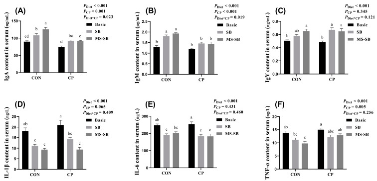
The levels of inflammatory factors and immunoglobulin in sera are shown in Figure 1. The C. perfringens challenge had no influence on inflammatory factor and immunoglobulin content in sera. However, SB addition significantly increased the immunoglobulin content in sera, including IgA, IgM, and IgY (p < 0.05). In addition, SB significantly decreased the content of IL-1β, IL-6, and TNF-α (p < 0.05). Compared with SB, MS-SB presented a stronger performance, with higher IgA content, as well as a lower IL-1β level when normal or C. perfringens-challenged.
Figure 1.
MS-SB modulates immunity through globulins and immune factor content in serum during C. perfringens infection. (A) IgA content; (B) IgM content; (C) IgY content; (D) IL-1β content; (E) IL-6 content; (F) TNF-α content. Bars with different letters are statistically significant (p ˂ 0.05) in different groups, n = 6.
3.3. Microencapsulated Sodium Butyrate Repaired Intestinal Morphology Damaged by C. perfringens
We also measured the parameters of jejunum villi through HE stains, and the results are shown in Figure 2. The results revealed that SB administration significantly increased the villus height and V/C ratio (p < 0.05), especially the MS-SB addition. However, C. perfringens challenge significantly decreased the villus height (p < 0.05) without affecting the V/C ratio.
Figure 2.
MS-SB repaired intestinal morphology damaged by C. perfringens. (A) Villus height of jejunum; (B) crypt depth of jejunum; (C) ratio of the villus height and crypt depth of jejunum. Bars with different letters are statistically significant (p ˂ 0.05) in different groups, n = 6.
3.4. Microencapsulated Sodium Butyrate Ameliorated VFAs under C. perfringens Challenge
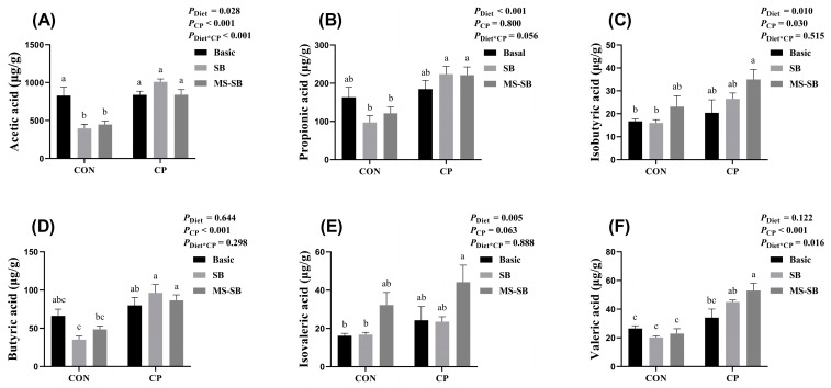
For this study, we also detected the VFA content in the broiler cecum, and the results are shown in Figure 3. The results indicated that the C. perfringens challenge had no effect on the VFA content. However, the addition of SB significantly decreased the content of acetic acid (p < 0.05) under C. perfringens non-challenge; at same time, SB or MS-SB presented varying degrees of increased fatty acid concentrations during C. perfringens challenge. It should be noted that MS-SB had a stronger performance, with a higher content of isobutyric acid, isovaleric acid, and valeric acid.
Figure 3.
Microencapsulated sodium butyrate modulates the VFA content when challenged with C. perfringens. (A) Acetic acid; (B) propionic acid; (C) isobutyric acid; (D) butyric acid; (E) isovaleric acid; (F) valeric acid content in cecum of broiler chickens. Bars with different letters are statistically significant (p ˂ 0.05) in different groups, n = 6.
3.5. Microencapsulated Sodium Butyrate Modulated Gut Microbiota Community Variation Caused by C. perfringens
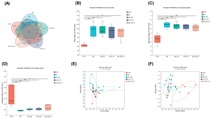
The changes in the diversity of cecum microbiota are summarized in Figure 4. A total of 921 ASVs were shared among the five treatment groups based on the Venn diagram, with non-overlapping regions indicating unique OTUs in the CON group (n = 569), SB group (n = 658), MS-SB group (n = 743), CON-CP group (n = 592), and MS-SB-CP group (n = 611); see Figure 4A. We adopted the Chao index, Shannon index, and Simpson index to assess the microbiota community diversity. The results indicated that C. perfringens infection and SB addition individually presented more significant diversity in cecum microbiota when compared to the CON group, but there was no difference between them (Figure 4B–D). Principal component analysis (PCA) and principal coordinate analysis (PCoA) were performed to investigate the differences in species complexity and structural alterations in microbial communities. PCoA and PCA plots revealed the degree of diversity discrepancy in cecum microbiota between different treatments; in particular, the PCoA plot indicated that the CON group was separated from the other groups (Figure 4E,F).
Figure 4.
Analysis of the diversity of gut microbiota. (A) The Venn diagram summarizing the numbers of common and unique OTUs in cecum microflora community. (B–D) The Chao index, Shannon index, and Simpson index reflecting species alpha diversity between groups. (E,F) The principal component analysis (PCA) and principal co-ordinates analysis (PCoA) reflecting beta diversity within and between groups at the species level. The column with * are statistically significant, ** means p ˂ 0.01, *** means p ˂ 0.001, n = 6.
3.6. Microencapsulated Sodium Butyrate Modulated Gut Microbiota Community Composition Caused by C. perfringens
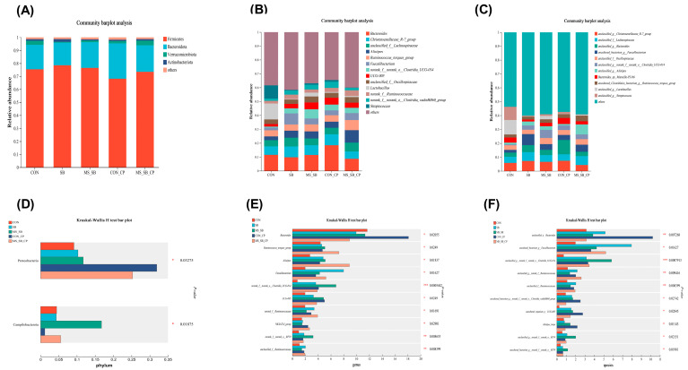
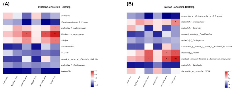
In order to explore the change in cecum microbiota structure, we analyzed the relative abundance of cecum microbiota at phylum to species levels. At the phylum level, about 4 major phyla were detected, including Firmicutes, Bacteroidota, Verrucomicrobiota, and Actinobacteriota (Figure 5A), and the relative abundance of Proteobacteria was higher in the C. perfringens infection groups than non-infection groups. The addition of MS-SB significantly increased the relative abundance of Campilobacteria (Figure 5D). At the genus level, about 13 major phyla were detected as dominant genera (Figure 5B). The relative abundances of Bacteroides in the C. perfringens infection groups were higher than in the non-infection groups, and the relative abundances of Ruminococcus_torques and Alistipes in the MS-SB-CP groups were higher than in other groups (Figure 5E). At the species level, about 11 major phyla were detected as dominant flora (Figure 5C). In addition, the relative abundances of 6 bacteria were higher with MS-SB addition than in the CON group, including Bacteroides, Faecalibacterium, Clostridia_UCG-014, Ruminococcaceae, Alistipes_inops, and Clostridia_vadinBB60. The C. perfringens infection increased the abundance of Bacteroides and Alistipes_inops (Figure 5F). Finally, we analyzed the association between VFAs and microbiota composition. The results demonstrated that Ruminococcaceae_torques and Alistipes were positively correlated with isobutyric acid concentration, while Lachnospiraceae, Ruminococcaceae_torques, and Alistipes were positively correlated with valeric acid (Figure 6A). At the species level, Ruminococcaceae_torques and Alistipes were positively correlated with isobutyric acid, and Lachnospiraceae, Ruminococcaceae_torques, and Alistipes were positively correlated with valeric acid (Figure 6B).
Figure 5.
The abundance of the microbial community in cecum content. (A–C) The top relative abundance of the microflora community between groups (phylum level, genus level, and species level). (D–F) The bacteria with significant differences between groups (phylum level, genus level, and species level). The column with * are statistically significant, * means p ˂ 0.05, ** means p ˂ 0.01, *** means p ˂ 0.001, n = 6.
Figure 6.
Correlation analysis between gut microbiota and VFAs. (A) Genus level and VFAs. (B) Species level and VFAs. The column with * are statistically significant, * means p ˂ 0.05, ** means p ˂ 0.01, *** means p ˂ 0.001, n = 6.
4. Discussion
In intensive animal production, the animals are faced with a variety of external factors at any time, such as production environment stress and physiological stress. In healthy animals, bacteria and health stand at either end of a pair of scales; once this balance is broken, harmful bacteria in the environment and body can cause damage to animal health, including intestinal inflammation, immune stress, and so on [19]. C. perfringens is consumed by chickens from environmental sources during rearing, including contaminated feed, water, and the farm environment [20]. As previously reported, the immune organ changes in response to infection with C. perfringens, shown in terms of the morphology and weight of the bursa of Fabricius, spleen, and thymus [21]. In this study, the results indicated that the spleen index increased, which is consistent with previous findings, as well as showing expected changes in the thymus index. This may be attributed to the age and species, as broilers near the end of growth were used in this study. Thus, these results indicate the validity of the C. perfringens infection model.
Moreover, we demonstrated that the addition of SB or MS-SB had no promotive or protective effects on the immune organs in this study. The results are not consistent with the results of a previous study, in which the thymus, spleen, and bursa weighed more in the SB addition group compared with control group [22]. Moreover, in a study in quails, dietary supplementation with 1000 mg/kg SB significantly increased the thymus and bursa of Fabricius [23]. As a matter of fact, we have also confirmed that SB and MS-SB could enhance immune organ development throughout the growth period in a previous study (unpublished). Thus, according to the results of this study, SB or MS-SB have no protective effect against the immune organ changes caused by C. perfringens infection, evidenced by the fact that SB or MS-SB were unable to reverse the organ changes induced by C. perfringens.
The immune organs, immune cells, and immune molecules make up the immune system of animals, and can be categorized into two parts—the innate immune system and the adaptive immune system—according to the manner in which they act against invading pathogens [24]. As our results indicated that SB and MS-SB could enhance immune organ development during the growth period (unpublished), we further determined the levels of immunoglobulins and immunomodulatory cytokines in sera. Immunoglobulins are synthesized and secreted by B-cells after immune cells are activated by antigens, which can bind to specific antigens in order to defend against invading pathogens [25]. In this study, the results showed that C. perfringens infection decreased the immunoglobulin levels in sera, while SB and MS-SB improved the secretion of immunoglobulins, consistent with the results of previous studies [26]. However, it should be noted that the enhancement brought by SB and MS-SB was diminished under C. perfringens infection, when compared to normal conditions. Immunomodulatory cytokines are produced by immune cells and act on other immune cells, which can be classified as pro-inflammatory or anti-inflammatory according to their function [27]. Pro-inflammatory cytokines (e.g., IL-1α/β, TNF-α/β, and IL-6) up-regulate inflammatory reactions, while anti-inflammatory cytokines (e.g., IL-10) down-regulate inflammatory responses and promote tissue healing [28]. In rat models, necrotic enteritis causes enteric inflammation accompanied by serum proinflammatory cytokine production [29]. Thus, we proceeded to determine the content of inflammatory factors in sera. The results demonstrated that SB or MS-SB exhibited a potent anti-inflammatory effect, evidenced by reductions in IL-1β, IL-6, and TNF-α, especially in the case of the slight increase in inflammatory factors caused by C. perfringens. Similar to the results of our study, the study published by Sun et al. (2021) reported that sodium butyrate inhibited intestinal inflammation through the HMGB1-TLR4/NF-κB pathway [3]. Therefore, we speculate that SB or MS-SB may enhance immune function by regulating serum inflammatory cytokines in broiler chickens. Moreover, compared with SB, MS-SB presented a better anti-inflammatory effect.
As an organ for nutrient digestion and absorption, the intestinal tract is also an important barrier to maintain the homeostasis of the internal environment, and is the first barrier to deal with foreign harmful bacteria [30]. Normal intestinal permeability prevents water and electrolyte loss, promotes the absorption of dietary nutrients, and prevents the entry of antigens and micro-organisms into the body, which are dependent on the integrity of the intestinal villi [31]. In a previous study, necrotic enteritis caused by C. perfringens was mostly observed in the jejunum, manifesting as intestinal morphological damage and inflammation [9]. In addition, SCFA can improve the proliferation of gut epithelial cells and increase their villi height, which subsequently helps to improve the capacity of the intestine for nutrient absorption [32]. Thus, we detected the morphology of the jejunum using HE staining, and the results indicated that C. perfringens negatively affected the intestinal villi morphology, decreasing both the villus height and crypt depth. These results are similar to those previously reported in broilers [33,34] and mice [35]. We cannot ignore that the addition of SB or MS-SB alleviated the morphological damage to the intestinal villi, and the performance of MS-SB was particularly prominent compared with that of SB. Similar results have also been confirmed in a study using a mouse model [5].
In previous research, VFAs have been shown to inhibit pathogenic micro-organisms and increase the absorption of nutrients, which may contribute to a reduction in the luminal pH [36]. Acetic acid is the shortest fatty acid with a carbon chain, which has been shown to be an intermediary involved in bifidobacteria inhibiting the proliferation of intestinal pathogens [37]. Propionate and butyrate have been reported to assist in controlling intestinal inflammation by inducing the differentiation of T-regulatory cells, and the inhibition of histone deacetylation may be involved in the relevant regulatory mechanism [2,38]. In addition, recent studies have shown that valerate inhibits the proliferation of Clostridium difficile in the intestinal tract, thereby protecting or treating intestinal diseases [39]. Moreover, butyric acid is usually produced in the large intestine by intestinal bacteria and plays important roles, such as fueling intestinal epithelial cells and increasing mucin production, which may result in changes in bacterial adhesion and improved tight-junction integrity [40,41]. Thus, VFAs seem to play an important role in the maintenance of gut barrier function [42]. We also measured the concentrations of VFAs in the cecum, such as acetic acid, butyric acid (isobutyric acid), and valeric acid (isovaleric acid). The results indicated that C. perfringens infection has no effect on the VFA content in the cecum. However, SB or MS-SB increased the concentrations of fatty acids to varying degrees under the C. perfringens challenge. Our results are partially consistent with previous findings in broilers [43], and the dietary composition and butyric acid originating from foregut may be the cause of this result, as a result of butyric acid and other VFAs being produced through the bacterial fermentation of unabsorbed carbohydrates or food scraps [44]. Meanwhile, MS-SB was used in our study, which can significantly delay the enteric release of butyric acid, thus reducing small intestinal absorption and enhancing colonic delivery [44,45,46]. This may explain the difference between our results and those reported in previous studies.
Numerous studies have revealed that the gut microbiota that improve growth and metabolism promote host nutrient absorption and modulate the immune system, which are all behaviors providing irreplaceable functionality [47,48]. Furthermore, the diversity of the microbial community helps to maintain the homeostasis of the intestinal microbiome and improve resistance to pathogens in the host [49]. Our results on alpha diversity and beta diversity in the cecum content revealed a degree of diversity discrepancy in the cecum microbiota. Both the C. perfringens challenge and addition of SB improved the diversity of bacterial flora. This result is not consistent with previous studies, such as that of Pammi et al. (2017), in which the intestinal flora diversity in necrotizing enterocolitis patients was lower than that in unaffected patients, such as the lower relative abundance of Firmicutes and Bacteroides and higher relative abundance of Proteobacteria [50]. Meanwhile, our results were consistent with those of Zhang et al. (2018), who revealed the α-diversity index of broiler gut microbial community after C. perfringens infection [51]. This may be explained by the fact that C. perfringens infection can destroy the ecological balance of intestinal flora, resulting in intestinal ecological imbalance [52]. In addition, C. perfringens strains and dietary components, as well as the timing and duration of the C. perfringens challenge, may have contributed to the observed discrepancy [53]. In this study, the results indicated that the proportions of Bacteroides, Faecalibacterium, Clostridia, Ruminococcaceae, Alistipes, and Clostridia were significantly higher in the MS-SB addition group; at the same time, C. perfringens infection increased the abundance of Bacteroides and Alistipes. In previous studies, the genera Alistipes and Bacteroides have been identified as butyrate producers in the gut and have demonstrated good anti-inflammatory effects through butyrate [54]. The results of the correlation analysis considering VFAs and microbiota indicated that the isobutyric acid content increased and was positively correlated with the abundance of Alistipes and Clostridia. Thus, our findings demonstrate that SB and MS-SB exhibit a protective role to suppress C. perfringens-induced intestinal damage and microbiota disturbances.
5. Conclusions
In summary, sodium butyrate ameliorated C. perfringens infection by reducing inflammation, repairing intestinal damage, and modulating the cecum microbiota. Compared to sodium butyrate, microencapsulated sodium butyrate presented a better effect. This study highlights the effectiveness of microencapsulated sodium butyrate and provides a novel strategy for protection against C. perfringens infection through the effective utilization of gastrointestinal-sensitive biological agents.
Author Contributions
B.D. designed the experiments. S.X., Y.L. and X.W. performed the experiments. C.Y., J.L., S.X. and Z.D. analyzed the data. T.Y. and Y.S. wrote the main manuscript. Z.D., S.Y. and R.Z. revised the main manuscript. All authors have read and agreed to the published version of the manuscript.
Institutional Review Board Statement
The animal study protocol was approved by the Animal Health and Care Committee of Zhejiang Agricultural and Forestry University (No. ZAFUAC2022005, 15 October 2022).
Informed Consent Statement
Not applicable.
Data Availability Statement
The data that support the findings of this study were not deposited in an official repository, but they are available from the authors upon request.
Conflicts of Interest
Jinsong Liu, Shiping Xiao, Yulan Liu, and Caimei Yang are employees of Zhejiang Vegamax Biotechnology. This paper reflects the views of the scientists, and not the company. We certify that there is no conflict of interest with any financial organization regarding the material discussed in the manuscript, and the authors declare no conflict of interest.
Funding Statement
This work was supported by the Leading Innovation and Entrepreneurship Team Project of Zhejiang Province (2020R01015), Zhejiang Provincial Key R&D Program of China (2021C02008), the Natural Science Foundation of China (32260847, 32202687), Jiangxi Modern Agricultural Research Collaborative Innovation Project (JXXTCXBSJJ202208), and Hangzhou Key Projects for Agricultural and Social Development (202203A08).
Footnotes
Disclaimer/Publisher’s Note: The statements, opinions and data contained in all publications are solely those of the individual author(s) and contributor(s) and not of MDPI and/or the editor(s). MDPI and/or the editor(s) disclaim responsibility for any injury to people or property resulting from any ideas, methods, instructions or products referred to in the content.
References
- 1.Li Z., Wang W., Liu D., Guo Y. Effects of Lactobacillus acidophilus on the growth performance and intestinal health of broilers challenged with Clostridium perfringens. J. Anim. Sci. Biotechnol. 2018;9:25. doi: 10.1186/s40104-018-0243-3. [DOI] [PMC free article] [PubMed] [Google Scholar]
- 2.Salvi P.S., Cowles R.A. Butyrate and the Intestinal Epithelium: Modulation of Proliferation and Inflammation in Homeostasis and Disease. Cells. 2021;10:1775. doi: 10.3390/cells10071775. [DOI] [PMC free article] [PubMed] [Google Scholar]
- 3.Sun Q., Ji Y.-C., Wang Z.-L., She X., He Y., Ai Q., Li L.-Q. Sodium Butyrate Alleviates Intestinal Inflammation in Mice with Necrotizing Enterocolitis. Mediat. Inflamm. 2021;2021:6259381. doi: 10.1155/2021/6259381. [DOI] [PMC free article] [PubMed] [Google Scholar]
- 4.Manrique Vergara D., González Sánchez M.E. Short chain fatty acids (butyric acid) and intestinal diseases. Nutr. Hosp. 2017;34:58–61. doi: 10.20960/nh.1573. [DOI] [PubMed] [Google Scholar]
- 5.Chen G., Ran X., Li B., Li Y., He D., Huang B., Fu S., Liu J., Wang W. Sodium Butyrate Inhibits Inflammation and Maintains Epithelium Barrier Integrity in a TNBS-induced Inflammatory Bowel Disease Mice Model. EBioMedicine. 2018;30:317–325. doi: 10.1016/j.ebiom.2018.03.030. [DOI] [PMC free article] [PubMed] [Google Scholar]
- 6.Zhou D., Pan Q., Xin F.Z., Zhang R.N., He C.X., Chen G.Y., Liu C., Chen Y.W., Fan J.G. Sodium butyrate attenuates high-fat diet-induced steatohepatitis in mice by improving gut microbiota and gastrointestinal barrier. World J. Gastroenterol. 2017;23:60–75. doi: 10.3748/wjg.v23.i1.60. [DOI] [PMC free article] [PubMed] [Google Scholar]
- 7.Posthaus H., Kittl S., Tarek B., Bruggisser J. Clostridium perfringens type C necrotic enteritis in pigs: Diagnosis, pathogenesis, and prevention. J. Vet. Diagn. Investig. Off. Publ. Am. Assoc. Vet. Lab. Diagn. Inc. 2020;32:203–212. doi: 10.1177/1040638719900180. [DOI] [PMC free article] [PubMed] [Google Scholar]
- 8.Wade B., Keyburn A. The true cost of necrotic enteritis. World Poult. 2015;31:16–17. [Google Scholar]
- 9.Hustá M., Tretiak S., Ducatelle R., Van Immerseel F., Goossens E. Clostridium perfringens strains proliferate to high counts in the broiler small intestinal tract, in accordance with necrotic lesion severity, and sporulate in the distal intestine. Vet. Microbiol. 2023;280:109705. doi: 10.1016/j.vetmic.2023.109705. [DOI] [PubMed] [Google Scholar]
- 10.Daneshmand A., Kermanshahi H., Mohammed J., Sekhavati M.H., Javadmanesh A., Ahmadian M., Alizadeh M., Razmyar J., Kulkarni R.R. Intestinal changes and immune responses during Clostridium perfringens-induced necrotic enteritis in broiler chickens. Poult. Sci. 2022;101:101652. doi: 10.1016/j.psj.2021.101652. [DOI] [PMC free article] [PubMed] [Google Scholar]
- 11.Marcano V., Gamble T., Maschek K., Stabler L., Fletcher O., Davis J., Troan B.V., Villegas A.M., Tsai Y.Y., Barbieri N.L., et al. Necrotizing Hepatitis Associated with Clostridium perfringens in Broiler Chicks. Avian Dis. 2022;66:337–344. doi: 10.1637/aviandiseases-D-22-00033. [DOI] [PubMed] [Google Scholar]
- 12.Tian M., Li L., Tian Z., Zhao H., Chen F., Guan W., Zhang S. Glyceryl butyrate attenuates enterotoxigenic Escherichia coli-induced intestinal inflammation in piglets by inhibiting the NF-κB/MAPK pathways and modulating the gut microbiota. Food Funct. 2022;13:6282–6292. doi: 10.1039/D2FO01056A. [DOI] [PubMed] [Google Scholar]
- 13.Pace F., Rudolph S.E., Chen Y., Bao B., Kaplan D.L., Watnick P.I. The Short-Chain Fatty Acids Propionate and Butyrate Augment Adherent-Invasive Escherichia coli Virulence but Repress Inflammation in a Human Intestinal Enteroid Model of Infection. Microbiol. Spectr. 2021;9:e0136921. doi: 10.1128/Spectrum.01369-21. [DOI] [PMC free article] [PubMed] [Google Scholar]
- 14.Kaczmarek S.A., Barri A., Hejdysz M., Rutkowski A. Effect of different doses of coated butyric acid on growth performance and energy utilization in broilers. Poult. Sci. 2016;95:851–859. doi: 10.3382/ps/pev382. [DOI] [PubMed] [Google Scholar]
- 15.Pourjafar H., Noori N., Gandomi H., Basti A.A., Ansari F. Viability of microencapsulated and non-microencapsulated Lactobacilli in a commercial beverage. Biotechnol. Rep. 2020;25:e00432. doi: 10.1016/j.btre.2020.e00432. [DOI] [PMC free article] [PubMed] [Google Scholar]
- 16.Pesti G.M. Nutrient Requirements of Poultry. National Academy Press; Washington, DC, USA: 1994. [Google Scholar]
- 17.Sciences G.A.o.A. Nutrient Requirements of Yellow Chickens. Ministry of Agriculture and Rural Affairs, PRC; Beijing, China: 2020. [Google Scholar]
- 18.Yu X., Dai Z., Cao G., Cui Z., Zhang R., Xu Y., Wu Y., Yang C. Protective effects of Bacillus licheniformis on growth performance, gut barrier functions, immunity and serum metabolome in lipopolysaccharide-challenged weaned piglets. Front. Immunol. 2023;14:1140564. doi: 10.3389/fimmu.2023.1140564. [DOI] [PMC free article] [PubMed] [Google Scholar]
- 19.Cong J., Zhou P., Zhang R. Intestinal Microbiota-Derived Short Chain Fatty Acids in Host Health and Disease. Nutrients. 2022;14:1977. doi: 10.3390/nu14091977. [DOI] [PMC free article] [PubMed] [Google Scholar]
- 20.He W., Goes E.C., Wakaruk J., Barreda D.R., Korver D.R. A Poultry Subclinical Necrotic Enteritis Disease Model Based on Natural Clostridium perfringens Uptake. Front. Physiol. 2022;13:788592. doi: 10.3389/fphys.2022.788592. [DOI] [PMC free article] [PubMed] [Google Scholar]
- 21.Villagrán-de la Mora Z., Vázquez-Paulino O., Avalos H., Ascencio F., Nuño K., Villarruel-López A. Effect of a Synbiotic Mix on Lymphoid Organs of Broilers Infected with Salmonella typhimurium and Clostridium perfringens. Animals. 2020;10:886. doi: 10.3390/ani10050886. [DOI] [PMC free article] [PubMed] [Google Scholar]
- 22.Sikandar A., Zaneb H., Younus M., Masood S., Aslam A., Khattak F., Ashraf S., Yousaf M.S., Rehman H. Effect of sodium butyrate on performance, immune status, microarchitecture of small intestinal mucosa and lymphoid organs in broiler chickens. Asian-Australas. J. Anim. Sci. 2017;30:690–699. doi: 10.5713/ajas.16.0824. [DOI] [PMC free article] [PubMed] [Google Scholar]
- 23.Elnesr S., Ropy A., Abdel-Razik A. Effect of dietary sodium butyrate supplementation on growth, blood biochemistry, haematology and histomorphometry of intestine and immune organs of Japanese quail. Anim. Int. J. Anim. Biosci. 2019;13:1234–1244. doi: 10.1017/S1751731118002732. [DOI] [PubMed] [Google Scholar]
- 24.Ferreira N.S., Tostes R.C., Paradis P., Schiffrin E.L. Aldosterone, inflammation, immune system, and hypertension. Am. J. Hypertens. 2021;34:15–27. doi: 10.1093/ajh/hpaa137. [DOI] [PMC free article] [PubMed] [Google Scholar]
- 25.Hand T.W., Reboldi A. Production and function of immunoglobulin A. Annu. Rev. Immunol. 2021;39:695–718. doi: 10.1146/annurev-immunol-102119-074236. [DOI] [PubMed] [Google Scholar]
- 26.Zhang R., Qin S., Yang C., Niu Y., Feng J. The protective effects of Bacillus licheniformis against inflammatory responses and intestinal barrier damage in broilers with necrotic enteritis induced by Clostridium perfringens. J. Sci. Food Agric. 2023;13:6958–6965. doi: 10.1002/jsfa.12781. [DOI] [PubMed] [Google Scholar]
- 27.Azad M.A.K., Sarker M., Wan D. Immunomodulatory effects of probiotics on cytokine profiles. BioMed Res. Int. 2018;2018:8063647. doi: 10.1155/2018/8063647. [DOI] [PMC free article] [PubMed] [Google Scholar]
- 28.Crawford C.K., Lopez Cervantes V., Quilici M.L., Armién A.G., Questa M., Matloob M.S., Huynh L.D., Beltran A., Karchemskiy S.J., Crakes K.R. Inflammatory cytokines directly disrupt the bovine intestinal epithelial barrier. Sci. Rep. 2022;12:14578. doi: 10.1038/s41598-022-18771-y. [DOI] [PMC free article] [PubMed] [Google Scholar]
- 29.Yu R., Jiang S., Tao Y., Li P., Yin J., Zhou Q. Inhibition of HMGB1 improves necrotizing enterocolitis by inhibiting NLRP3 via TLR4 and NF-κB signaling pathways. J. Cell. Physiol. 2019;234:13431–13438. doi: 10.1002/jcp.28022. [DOI] [PubMed] [Google Scholar]
- 30.Di Tommaso N., Gasbarrini A., Ponziani F.R. Intestinal barrier in human health and disease. Int. J. Environ. Res. Public Health. 2021;18:12836. doi: 10.3390/ijerph182312836. [DOI] [PMC free article] [PubMed] [Google Scholar]
- 31.Bischoff S.C., Barbara G., Buurman W., Ockhuizen T., Schulzke J.-D., Serino M., Tilg H., Watson A., Wells J.M. Intestinal permeability–a new target for disease prevention and therapy. BMC Gastroenterol. 2014;14:189. doi: 10.1186/s12876-014-0189-7. [DOI] [PMC free article] [PubMed] [Google Scholar]
- 32.Pérez-Reytor D., Puebla C., Karahanian E., García K. Use of short-chain fatty acids for the recovery of the intestinal epithelial barrier affected by bacterial toxins. Front. Physiol. 2021;12:650313. doi: 10.3389/fphys.2021.650313. [DOI] [PMC free article] [PubMed] [Google Scholar]
- 33.Fasina Y.O., Lillehoj H.S. Characterization of intestinal immune response to Clostridium perfringens infection in broiler chickens. Poult. Sci. 2019;98:188–198. doi: 10.3382/ps/pey390. [DOI] [PMC free article] [PubMed] [Google Scholar]
- 34.Ibrahim D., Ismail T.A., Khalifa E., Abd El-Kader S.A., Mohamed D.I., Mohamed D.T., Shahin S.E., Abd El-Hamid M.I. Supplementing Garlic Nanohydrogel Optimized Growth, Gastrointestinal Integrity and Economics and Ameliorated Necrotic Enteritis in Broiler Chickens Using a Clostridium perfringens Challenge Model. Animal. 2021;11:2027. doi: 10.3390/ani11072027. [DOI] [PMC free article] [PubMed] [Google Scholar]
- 35.Navarro M.A., Li J., McClane B.A., Morrell E., Beingesser J., Uzal F.A. NanI sialidase is an important contributor to Clostridium perfringens type F strain F4969 intestinal colonization in mice. Infect. Immun. 2018;86:10–1128. doi: 10.1128/IAI.00462-18. [DOI] [PMC free article] [PubMed] [Google Scholar]
- 36.Dąbek-Drobny A., Kaczmarczyk O., Woźniakiewicz M., Paśko P., Dobrowolska-Iwanek J., Woźniakiewicz A., Piątek-Guziewicz A., Zagrodzki P., Zwolińska-Wcisło M. Association between Fecal Short-Chain Fatty Acid Levels, Diet, and Body Mass Index in Patients with Inflammatory Bowel Disease. Biology. 2022;11:108. doi: 10.3390/biology11010108. [DOI] [PMC free article] [PubMed] [Google Scholar]
- 37.Fukuda S., Toh H., Hase K., Oshima K., Nakanishi Y., Yoshimura K., Tobe T., Clarke J.M., Topping D.L., Suzuki T., et al. Bifidobacteria can protect from enteropathogenic infection through production of acetate. Nature. 2011;469:543–547. doi: 10.1038/nature09646. [DOI] [PubMed] [Google Scholar]
- 38.Yang J., Wei H., Zhou Y., Szeto C.H., Li C., Lin Y., Coker O.O., Lau H.C.H., Chan A.W.H., Sung J.J.Y., et al. High-Fat Diet Promotes Colorectal Tumorigenesis Through Modulating Gut Microbiota and Metabolites. Gastroenterology. 2022;162:135–149.e132. doi: 10.1053/j.gastro.2021.08.041. [DOI] [PubMed] [Google Scholar]
- 39.McDonald J.A., Mullish B.H., Pechlivanis A., Liu Z., Brignardello J., Kao D., Holmes E., Li J.V., Clarke T.B., Thursz M.R. Inhibiting growth of Clostridioides difficile by restoring valerate, produced by the intestinal microbiota. Gastroenterology. 2018;155:1495–1507.e1415. doi: 10.1053/j.gastro.2018.07.014. [DOI] [PMC free article] [PubMed] [Google Scholar]
- 40.Pietrzak A., Banasiuk M., Szczepanik M., Borys-Iwanicka A., Pytrus T., Walkowiak J., Banaszkiewicz A. Sodium Butyrate Effectiveness in Children and Adolescents with Newly Diagnosed Inflammatory Bowel Diseases-Randomized Placebo-Controlled Multicenter Trial. Nutrients. 2022;14:3283. doi: 10.3390/nu14163283. [DOI] [PMC free article] [PubMed] [Google Scholar]
- 41.Jung T.-H., Park J.H., Jeon W.-M., Han K.-S. Butyrate modulates bacterial adherence on LS174T human colorectal cells by stimulating mucin secretion and MAPK signaling pathway. Nutr. Res. Pract. 2015;9:343–349. doi: 10.4162/nrp.2015.9.4.343. [DOI] [PMC free article] [PubMed] [Google Scholar]
- 42.Liu P., Wang Y., Yang G., Zhang Q., Meng L., Xin Y., Jiang X. The role of short-chain fatty acids in intestinal barrier function, inflammation, oxidative stress, and colonic carcinogenesis. Pharmacol. Res. 2021;165:105420. doi: 10.1016/j.phrs.2021.105420. [DOI] [PubMed] [Google Scholar]
- 43.Mallo J.J., Sol C., Puyalto M., Bortoluzzi C., Applegate T.J., Villamide M.J. Evaluation of sodium butyrate and nutrient concentration for broiler chickens. Poult. Sci. 2021;100:101456. doi: 10.1016/j.psj.2021.101456. [DOI] [PMC free article] [PubMed] [Google Scholar]
- 44.Krix-Jachym K., Onyszkiewicz M., Swiatkiewicz M., Fiedorowicz M., Sapierzynski R., Grieb P., Rekas M. Evaluation of butyric acid as a potential supportive treatment in anterior uveitis. Ophthalmol. J. 2022;7:117–126. doi: 10.5603/OJ.2022.0016. [DOI] [Google Scholar]
- 45.Pham V.T., Fehlbaum S., Seifert N., Richard N., Bruins M.J., Sybesma W., Rehman A., Steinert R.E. Effects of colon-targeted vitamins on the composition and metabolic activity of the human gut microbiome—A pilot study. Gut Microbes. 2021;13:1875774. doi: 10.1080/19490976.2021.1875774. [DOI] [PMC free article] [PubMed] [Google Scholar]
- 46.Makowski Z., Lipiński K., Mazur-Kuśnirek M. The Effects of sodium butyrate, coated sodium butyrate, and butyric acid glycerides on nutrient digestibility, gastrointestinal function, and fecal microbiota in turkeys. Animals. 2022;12:1836. doi: 10.3390/ani12141836. [DOI] [PMC free article] [PubMed] [Google Scholar]
- 47.Xu Y., Yu Y., Shen Y., Li Q., Lan J., Wu Y., Zhang R., Cao G., Yang C. Effects of Bacillus subtilis and Bacillus licheniformis on growth performance, immunity, short chain fatty acid production, antioxidant capacity, and cecal microflora in broilers. Poult. Sci. 2021;100:101358. doi: 10.1016/j.psj.2021.101358. [DOI] [PMC free article] [PubMed] [Google Scholar]
- 48.Yao H., Zhang D., Yu H., Yuan H., Shen H., Lan X., Liu H., Chen X., Meng F., Wu X. Gut microbiota regulates chronic ethanol exposure-induced depressive-like behavior through hippocampal NLRP3-mediated neuroinflammation. Mol. Psychiatry. 2023;28:919–930. doi: 10.1038/s41380-022-01841-y. [DOI] [PMC free article] [PubMed] [Google Scholar]
- 49.Li C., Wang S., Chen S., Wang X., Deng X., Liu G., Chang W., Beckers Y., Cai H. Screening and characterization of Pediococcus acidilactici LC-9-1 toward selection as a potential probiotic for poultry with antibacterial and antioxidative properties. Antioxidants. 2023;12:215. doi: 10.3390/antiox12020215. [DOI] [PMC free article] [PubMed] [Google Scholar]
- 50.Pammi M., Cope J., Tarr P.I., Warner B.B., Morrow A.L., Mai V., Gregory K.E., Kroll J.S., McMurtry V., Ferris M.J. Intestinal dysbiosis in preterm infants preceding necrotizing enterocolitis: A systematic review and meta-analysis. Microbiome. 2017;5:31. doi: 10.1186/s40168-017-0248-8. [DOI] [PMC free article] [PubMed] [Google Scholar]
- 51.Zhang B., Lv Z., Li Z., Wang W., Li G., Guo Y. Dietary l-arginine Supplementation Alleviates the Intestinal Injury and Modulates the Gut Microbiota in Broiler Chickens Challenged by Clostridium perfringens. Front. Microbiol. 2018;9:1716. doi: 10.3389/fmicb.2018.01716. [DOI] [PMC free article] [PubMed] [Google Scholar]
- 52.Gong L., Wang B., Zhou Y., Tang L., Zeng Z., Zhang H., Li W. Protective effects of Lactobacillus plantarum 16 and Paenibacillus polymyxa 10 against Clostridium perfringens infection in broilers. Front. Immunol. 2021;11:628374. doi: 10.3389/fimmu.2020.628374. [DOI] [PMC free article] [PubMed] [Google Scholar]
- 53.Liu Y., Zhang S., Luo Z., Liu D. Supplemental Bacillus subtilis PB6 improves growth performance and gut health in broilers challenged with Clostridium perfringens. J. Immunol. Res. 2021;2021:2549541. doi: 10.1155/2021/2549541. [DOI] [PMC free article] [PubMed] [Google Scholar]
- 54.Wu M., Cao W., Chen X., She J. Intelligent Optimization and Control of Complex Metallurgical Processes. Springer; Singapore: 2020. Intelligent Optimization and Control for Reheating Furnaces; pp. 223–271. [DOI] [Google Scholar]
Associated Data
This section collects any data citations, data availability statements, or supplementary materials included in this article.
Data Availability Statement
The data that support the findings of this study were not deposited in an official repository, but they are available from the authors upon request.


