Abstract
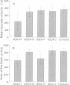
Slow saccades are often found in degenerative ataxia. Experimental studies have shown that horizontal saccades are generated in the paramedian pontine reticular formation and that lesions in this area produce slow saccades. Based on these findings, saccade slowing should be a frequent feature of olivopontocerebellar atrophy, a type of cerebellar degeneration with prominent involvement of the pons. To test this hypothesis, saccade velocity was measured in 31 patients with autosomal dominant cerebellar ataxia (ADCA) and 17 patients with idiopathic cerebellar ataxia (IDCA). Saccade velocity was reduced in most patients with ADCA whereas it was normal in IDCA although olivopontocerebellar atrophy occurred in both groups. Saccade velocities correlated with pontine size in ADCA but not in IDCA. The data disprove the hypothesis that saccadic slowing is a clinical hallmark of olivopontocerebellar atrophy. Instead, only patients with ADCA and morphological features of olivopontocerebellar atrophy have slow saccades.
Full text
PDF


Images in this article
Selected References
These references are in PubMed. This may not be the complete list of references from this article.
- Bürk K., Abele M., Fetter M., Dichgans J., Skalej M., Laccone F., Didierjean O., Brice A., Klockgether T. Autosomal dominant cerebellar ataxia type I clinical features and MRI in families with SCA1, SCA2 and SCA3. Brain. 1996 Oct;119(Pt 5):1497–1505. doi: 10.1093/brain/119.5.1497. [DOI] [PubMed] [Google Scholar]
- Harding A. E. The clinical features and classification of the late onset autosomal dominant cerebellar ataxias. A study of 11 families, including descendants of the 'the Drew family of Walworth'. Brain. 1982 Mar;105(Pt 1):1–28. doi: 10.1093/brain/105.1.1. [DOI] [PubMed] [Google Scholar]
- Imbert G., Saudou F., Yvert G., Devys D., Trottier Y., Garnier J. M., Weber C., Mandel J. L., Cancel G., Abbas N. Cloning of the gene for spinocerebellar ataxia 2 reveals a locus with high sensitivity to expanded CAG/glutamine repeats. Nat Genet. 1996 Nov;14(3):285–291. doi: 10.1038/ng1196-285. [DOI] [PubMed] [Google Scholar]
- Kawaguchi Y., Okamoto T., Taniwaki M., Aizawa M., Inoue M., Katayama S., Kawakami H., Nakamura S., Nishimura M., Akiguchi I. CAG expansions in a novel gene for Machado-Joseph disease at chromosome 14q32.1. Nat Genet. 1994 Nov;8(3):221–228. doi: 10.1038/ng1194-221. [DOI] [PubMed] [Google Scholar]
- Keller E. L. Participation of medial pontine reticular formation in eye movement generation in monkey. J Neurophysiol. 1974 Mar;37(2):316–332. doi: 10.1152/jn.1974.37.2.316. [DOI] [PubMed] [Google Scholar]
- Klockgether T., Schroth G., Diener H. C., Dichgans J. Idiopathic cerebellar ataxia of late onset: natural history and MRI morphology. J Neurol Neurosurg Psychiatry. 1990 Apr;53(4):297–305. doi: 10.1136/jnnp.53.4.297. [DOI] [PMC free article] [PubMed] [Google Scholar]
- Luschei E. S., Fuchs A. F. Activity of brain stem neurons during eye movements of alert monkeys. J Neurophysiol. 1972 Jul;35(4):445–461. doi: 10.1152/jn.1972.35.4.445. [DOI] [PubMed] [Google Scholar]
- Murphy M. J., Goldblatt D. Slow eye movements, with absent saccades, in a patient with hereditary ataxia. Arch Neurol. 1977 Mar;34(3):191–195. doi: 10.1001/archneur.1977.00500150077016. [DOI] [PubMed] [Google Scholar]
- Newman N., Gay A. J., Stroud M. H., Brooks J. Defective rapid eye movements in progressive supranuclear palsy. An ocular electromyographic study. Brain. 1970;93(4):775–784. doi: 10.1093/brain/93.4.775. [DOI] [PubMed] [Google Scholar]
- Niakan E., Bertorini T. E., Lemmi H., Medeiros M., Drewry R., Kish E. Spinocerebellar degeneration and slow saccades in three generations of a kinship: clinical and electrophysiologic findings. Arq Neuropsiquiatr. 1984 Sep;42(3):232–241. doi: 10.1590/s0004-282x1984000300007. [DOI] [PubMed] [Google Scholar]
- Orr H. T., Chung M. Y., Banfi S., Kwiatkowski T. J., Jr, Servadio A., Beaudet A. L., McCall A. E., Duvick L. A., Ranum L. P., Zoghbi H. Y. Expansion of an unstable trinucleotide CAG repeat in spinocerebellar ataxia type 1. Nat Genet. 1993 Jul;4(3):221–226. doi: 10.1038/ng0793-221. [DOI] [PubMed] [Google Scholar]
- Papp M. I., Kahn J. E., Lantos P. L. Glial cytoplasmic inclusions in the CNS of patients with multiple system atrophy (striatonigral degeneration, olivopontocerebellar atrophy and Shy-Drager syndrome). J Neurol Sci. 1989 Dec;94(1-3):79–100. doi: 10.1016/0022-510x(89)90219-0. [DOI] [PubMed] [Google Scholar]
- Perenin M. T., Prablanc C. Anomalies des saccades oculaires dans un cas d'hérédodégénérescence spino-cérébelleuse. Rev Electroencephalogr Neurophysiol Clin. 1974 Jul-Sep;4(3):489–494. doi: 10.1016/s0370-4475(74)80064-x. [DOI] [PubMed] [Google Scholar]
- Pulst S. M., Nechiporuk A., Nechiporuk T., Gispert S., Chen X. N., Lopes-Cendes I., Pearlman S., Starkman S., Orozco-Diaz G., Lunkes A. Moderate expansion of a normally biallelic trinucleotide repeat in spinocerebellar ataxia type 2. Nat Genet. 1996 Nov;14(3):269–276. doi: 10.1038/ng1196-269. [DOI] [PubMed] [Google Scholar]
- Sanpei K., Takano H., Igarashi S., Sato T., Oyake M., Sasaki H., Wakisaka A., Tashiro K., Ishida Y., Ikeuchi T. Identification of the spinocerebellar ataxia type 2 gene using a direct identification of repeat expansion and cloning technique, DIRECT. Nat Genet. 1996 Nov;14(3):277–284. doi: 10.1038/ng1196-277. [DOI] [PubMed] [Google Scholar]
- Schroth G., Naegele T., Klose U., Mann K., Petersen D. Reversible brain shrinkage in abstinent alcoholics, measured by MRI. Neuroradiology. 1988;30(5):385–389. doi: 10.1007/BF00404102. [DOI] [PubMed] [Google Scholar]
- Starr A. A disorder of rapid eye movements in Huntington's chorea. Brain. 1967 Sep;90(3):545–564. doi: 10.1093/brain/90.3.545. [DOI] [PubMed] [Google Scholar]
- Wadia N. H., Swami R. K. A new form of heredo-familial spinocerebellar degeneration with slow eye movements (nine families). Brain. 1971;94(2):359–374. doi: 10.1093/brain/94.2.359. [DOI] [PubMed] [Google Scholar]
- Wadia R. S., Amin R. B., Divate U. P., Divate P. G., Sainani G. S., Sardesai H. V. Autosomal dominant spinocerebellar ataxia with slow eye movements-a common hereditary ataxia in Western India. J Assoc Physicians India. 1976 Jun;24(6):367–371. [PubMed] [Google Scholar]
- Wüllner U., Klockgether T., Petersen D., Naegele T., Dichgans J. Magnetic resonance imaging in hereditary and idiopathic ataxia. Neurology. 1993 Feb;43(2):318–325. doi: 10.1212/wnl.43.2.318. [DOI] [PubMed] [Google Scholar]


