Abstract
Using the MAP1-B enzyme-linked immunosorbent assay, we tested 1,318 serum samples collected from sheep and goats at 28 sites in the five divisions of The Gambia to determine the Ehrlichia ruminantium seroprevalence rates and to assess the risk for heartwater. About half (51.6%) of 639 sheep were positive, with seroprevalence rates per site varying between 6.9% and 100%. The highest seroprevalence was detected in the western part of the country (88.1% in the Western Division and 62.1% in the Lower River Division). Sheep in the two easterly divisions (Central River and Upper River divisions) showed the lowest seroprevalence of 29.3% and 32.4%, respectively, while those in the North Bank Division showed an intermediate prevalence of 40.6%. In goats, less than one-third (30.3%) of 679 animals tested were positive. The highest seroprevalence was detected in goats in the North Bank Division (59%) and Western Division (44.1%). Goats in the Lower River Division showed an intermediate level of 21.9%, whereas the lowest rates were found in the eastern part of the country (4.8% in the Central River Division and 2.3% in the Upper River Division). At nearly all sites, seroprevalence rates were higher in sheep than in goats. The results show a gradient of increasing heartwater risk for susceptible small ruminants from the east to the west of The Gambia. These findings need to be taken into consideration when future livestock-upgrading programs are implemented.
Heartwater (cowdriosis) is a major tick-borne disease of ruminants caused by the rickettsia Ehrlichia ruminantium and is transmitted by ticks of the genus Amblyomma. Amblyomma variegatum is the major vector in West Africa (22), is distributed throughout most of sub-Saharan Africa, and occurs on some islands in the Caribbean (23). Heartwater represents a major constraint to improvement of the livestock industry in sub-Saharan Africa. In The Gambia and neighboring Senegal, serological evidence of a high prevalence of E. ruminantium infection has been reported in cattle (8, 10, 17), which indicates a potential risk of heartwater disease for susceptible livestock. In contrast to what occurs in indigenous cattle, which appear more resistant to heartwater (6, 16), the disease has been known in small ruminants in The Gambia as “fayo,” referring to cases of sudden death, characteristic of acute forms of the disease (23). Mortality occurs in indigenous local dwarf sheep and goats and is estimated at 10% in areas of the country where the disease is endemic (R. Mattioli and J. Jaitner, unpublished data). In addition, frequent occurrences of sudden death due to heartwater have been observed in indigenous sheep and goats following translocation from the east to the west of the country (B. Faburay, unpublished observation). These observations suggest the existence of a gradient of heartwater disease risk for susceptible livestock species and the possibility that a significant proportion of the small-ruminant population in the east of the country has not been exposed to E. ruminantium infection. We carried out a countrywide serological survey to determine the distribution of E. ruminantium infection in small ruminants and to assess the heartwater risk for susceptible livestock.
MATERIALS AND METHODS
Survey.
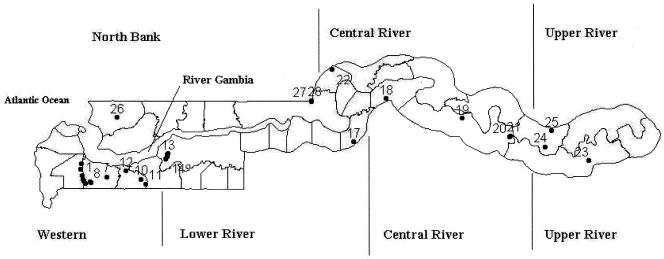
Serum samples were collected from Djallonké sheep and West African dwarf goats of both sexes in a cross-sectional study at 28 sites in all five divisions of The Gambia: the Western, Lower River, North Bank, Central River, and Upper River divisions (Fig. 1). The sites were located in three main agro-ecological zones: Sudanian, Sudano-Sahelian, and Sahelian. The sites were chosen in consultation with livestock assistants based on owner cooperation and accessibility to the animals. Adult animals between 1 and 3 years of age were sampled in April 2004. All animals were maintained under a traditional husbandry system without acaricide treatment. Blood samples were kept on ice, and serum was separated after 2 to 4 h by centrifugation and stored at −20°C until use. A total of 1,318 indigenous small ruminants comprising 639 sheep and 679 goats were sampled. The sites were selected in a transect layout to make the results representative of the country. The numbers sampled at different sites for each species are shown in Table 1. The largest number of sampling sites was in the Western Division, as this area is experiencing an expansion of a livestock-upgrading program carried out by the Gambian government in collaboration with the International Trypanotolerance Centre (ITC), involving cattle breeds (Holstein and Jersey) highly susceptible to heartwater disease.
FIG. 1.
Map of The Gambia showing the five divisions and the distribution of sampling sites.
TABLE 1.
Ehrlichia ruminantium seroprevalence in sheep and goats at 28 different sites in The Gambia
| Division | Village | Site | Seroprevalence (%)
|
||
|---|---|---|---|---|---|
| Sheep | Goats | Total | |||
| Western Division | Basori | 1 | 75 (8) | 51.6 (31) | 56.4 (39) |
| Berefet | 12 | 90.3 (31) | 31.7 (60) | 51.6 (91) | |
| Bitta | 11 | 94.7 (38) | 0 (12) | 72 (50) | |
| Duwasu | 8 | 88.9 (9) | 33.3 (9) | 61.1 (18) | |
| Giboro Kuta | 3 | 81.8 (33) | 47.8 (46) | 62 (79) | |
| Gida | 2 | 100 (2)c | 42.1 (19) | 47.6 (21) | |
| Jenunkunda | 9 | 100 (2)c | 45.5 (11) | 53.8 (13) | |
| Mandinaba | 5 | 85.7 (7) | 70 (10) | 76.5 (17) | |
| Somita | 10 | 75 (16) | 15.2 (33) | 34.7 (49) | |
| Talokotob | 4 | 80 (5) | 80 (5) | ||
| Toubakutab | 6 | 77.8 (9) | 77.8 (9) | ||
| Toumani Tenda | 7 | 100 (14) | 68 (50) | 75 (64) | |
| Lower River Division | Bodeyel | 17 | 30.9 (55) | 20 (5) | 30 (60) |
| Burong | 16 | 100 (9) | 42.9 (7) | 75 (16) | |
| Julakunda | 13 | 100 (14) | 0 (12) | 53.8 (26) | |
| Missira | 15 | 100 (13) | 40 (5) | 83.3 (18) | |
| Taborongkoto | 14 | 85 (20) | 33.3 (3)c | 78.3 (23) | |
| Central River Division | Jimballa Kerr Chendu | 22 | 41 (61) | 7.7 (52) | 25.7 (113) |
| Mamutfana | 18 | 6.9 (72) | 0 (20) | 54.3 (92) | |
| Sare Sofie | 21 | 31.3 (16) | 0 (8) | 20.8 (24) | |
| Sinchan Faranba | 20 | 29.6 (27) | 1.5 (69) | 9.4 (96) | |
| Yorro Beri Kunda | 19 | 100 (12) | 17.7 (17) | 51.7 (29) | |
| Upper River Division | Kulkullay | 23 | 22.2 (54) | 4.2 (48) | 13.7 (102) |
| Missira Sandou | 24 | 47.6 (42) | 0 (20) | 32.3 (62) | |
| Sare Demba Torro | 25 | 26.7 (15) | 0 (18) | 12.1 (33) | |
| North Bank Division | Kolli Kunda | 26 | 68.8 (16) | 48.7 (37) | 54.7 (53) |
| Mbappa Ba | 27 | 50 (16) | 50 (22) | 50 (38) | |
| Mbappa Marigaa | 28 | 24.3 (37) | 73.2 (41) | 50 (80) | |
Village with a high introgression of Sahelian sheep genes into the local population.
No sheep were sampled in the Talokoto and Toubakuta villages.
Prevalence was based on a small sample size, as there were very few animals presented for sampling.
MAP1-B ELISA.
The indirect MAP1-B enzyme-linked immunosorbent assay (ELISA), based on a recombinant truncated form of major antigenic protein 1 of E. ruminantium, was carried out as described previously (18, 21). Although the assay does not detect antibodies to ehrlichial agents infecting domestic ruminants, such as Ehrlichia bovis and Ehrlichia ovina, antibodies are detected against Ehrlichia canis (which infects dogs) and Ehrlichia chaffeensis (a human pathogen) (21). The MAP1-B ELISA has been shown to perform satisfactorily for small ruminants (5, 15), with specificities of 98.9% and 99.4% for caprine and ovine sera, respectively (19, 21). Each serum sample was tested in duplicate. For sheep, each test included duplicate negative-control sera obtained from a heartwater-naïve sheep of the Tesselaar breed. Duplicate positive-control sera were obtained from Tesselaar sheep 229 infected with the Gambian isolate Kerr Seringe 1 of E. ruminantium at the Faculty of Veterinary Medicine, Utrecht, The Netherlands. This stock of E. ruminantium was isolated from a naturally infected goat in The Gambia. In goats, each MAP1-B ELISA test also included duplicate positive-control sera, which were obtained from a Saanen goat infected with the Senegal isolate of E. ruminantium at Utrecht University. The negative-control serum was obtained from the same animal prior to infection. Species-specific second-step immunoglobulin G antibodies conjugated with horseradish peroxidase were used.
The cutoff point for the ELISA was determined by the addition of 2 standard deviations (SD) to the mean optical density (OD) values of reference local noninfected sheep (n = 24) and goat (n = 18) populations (18). The sheep and goat populations were considered negative based on the following: (i) they originated from northern Senegal, which is known to be free from Amblyomma ticks, and (ii) they were highly susceptible to E. ruminantium infection, as demonstrated by a high rate of mortality due to confirmed cases of heartwater upon first exposure to A. variegatum-infected ticks under natural conditions in an area of enzooticity (7). The cutoff point for sheep was determined at 0.53 (SD = 0.10) and for goats as 0.58 (SD = 0.11). OD values of samples that were equal to or greater than the cutoff value were considered positive for E. ruminantium infection. Variations between OD values of duplicate negative-control sera or between duplicate positive-control sera on each plate were acceptable only if such variations were lower than 10%.
Statistical analysis.
Plate-to-plate variation was considered by a statistical test for significance in difference, using the general linear model (SAS), among OD values of the positive controls included in each plate. Variation among the positive controls for the accepted plates was not significant for sheep (P = 0.4457; coefficient of variation, 14.7%) or for goats (P = 0.4514; coefficient of variation, 11.8%). The mean OD values of the positive controls included in the accepted plates was 1.5 ± 0.22 for sheep and 1.49 ± 0.18 for goats. Comparison for statistical significance of differences in the proportions of E. ruminantium-seropositive samples was carried out at two levels: (i) between species within a division using the Wilcoxon two-sample test and (ii) between divisions cumulatively (sheep and goats combined) and within species using the general linear model procedure (SAS statistical program) and Kruskal-Wallis one-way analysis of variance, respectively.
RESULTS
Of the 639 sheep samples tested, 51.6% were positive for E. ruminantium infection, with seroprevalence at individual sites ranging from 6.9% to 100% (Table 1). The highest seroprevalence was seen in the two westerly divisions, Western (88.1%) and Lower River (63.1%) (Table 2). Sheep populations in the two easterly divisions, Central River (29.3%) and Upper River (32.4%), showed the lowest levels of E. ruminantium seroprevalence, whereas animals sampled in the North Bank Division (40.6%) showed an intermediate level of seroprevalence (Table 2).
TABLE 2.
Proportions of total small-ruminant populations in the five divisions of The Gambia and overall heartwater seroprevalence rates
| Division | No. of sites | % of total livestocka
|
% of seropositive animals (total no. sampled)
|
Probabilityb | |||
|---|---|---|---|---|---|---|---|
| Sheep | Goats | Total | Sheep | Goats | |||
| Western Division | 12 | 10.9 | 11.9 | 11.5 | 88.1 (160) | 44.1 (295) | <0.001 |
| Lower River Division | 5 | 7.9 | 11.8 | 10.2 | 63.1 (111) | 21.9 (32) | <0.001 |
| North Bank Division | 3 | 12.7 | 23.5 | 19.0 | 40.6 (69) | 59.0 (100) | 0.019 |
| Central River Division | 5 | 43.5 | 34.8 | 38.4 | 29.3 (188) | 4.8 (166) | <0.001 |
| Upper River Division | 3 | 25.0 | 18.0 | 20.9 | 32.4 (111) | 2.3 (86) | <0.001 |
Deduced from reference 1.
Probability determined by comparing differences between the proportions of seropositive sheep and seropositive goats in a division (a P value of 0.05 or less is significant).
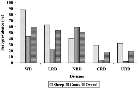
In contrast to the results for sheep, of the 679 goat samples collected, only about one-third (30.5%) were positive for E. ruminantium infection (Table 1). Overall, the highest seroprevalences were detected in goat populations in the North Bank (59%) and Western divisions (44.1%), with more than half of the animals sampled in the North Bank Division testing positive for E. ruminantium (Table 2). Seroprevalence in goats in the Lower River Division (21.9%) showed an intermediate level, with the two easterly divisions, Central River (4.8%; range, 0% to 17.7%) and Upper River (2.3%; range, 0% to 4.2%) showing the lowest level of seroprevalence (Table 2; Fig. 2). In all divisions, except for the North Bank Division, overall seroprevalence was significantly higher (P < 0.001) in sheep than in goats (Table 2). Moreover, at all sample sites (except Mbappa Mariga in the North Bank Division), the proportion of seropositive samples was consistently higher in sheep than in goats. Differences observed in the proportion of E. ruminantium-positive samples among sheep populations in the different divisions of The Gambia were statistically significant (P < 0.001). The same conclusion applied to goats. Similarly, differences in the overall seroprevalences between divisions were statistically significant (P < 0.001) (Table 2).
FIG. 2.
Distribution of E. ruminantium infection in sheep and goats as determined by serology in the five divisions of The Gambia. WD, Western Division; LRD, Lower River Division; NBD, North Bank Division; CRD, Central River Division; URD, Upper River Division.
DISCUSSION
Ehrlichia ruminantium seroprevalence in small ruminants was found to be highest in the western part of The Gambia, with the Western Division showing the highest prevalence of nearly 60% and the Lower River and North Bank divisions showing seroprevalences of more than 50%. Although the two easterly divisions, Central River and Upper River, account for the highest proportions of the small-ruminant population in The Gambia of 38.4% and 20.9%, respectively (Table 2), the overall seroprevalence of E. ruminantium in small-ruminant populations in these regions was significantly lower than in the westerly divisions (Western, North Bank, and Lower River) (Fig. 2). At most sites in the Central River and Upper River divisions, the serological prevalence was generally lower than 50%, resulting in a substantial population of sheep and goats being susceptible to heartwater.
In a recent study (E. Hoeven et al., unpublished data) a relatively high introgression of Sahelian genes was found in indigenous goats in the Central River Division as opposed to those in the Western Division. Interestingly, the easterly region accounts for over 60% of the small-ruminant population in The Gambia (Table 2), of which Sahelian sheep and goats and their crosses with local dwarf sheep and dwarf goats constitute a significant proportion. Generally, Sahelian sheep and goats found in the easterly part of The Gambia originate from Amblyomma/heartwater-free areas in northern Senegal and are therefore susceptible to heartwater disease. However, due to their larger size, farmers in this region show preference for them and use them for crossbreeding with local sheep and goats. The above factors therefore suggest the existence of a lower risk of E. ruminantium infection in most of the eastern part of the country. Thus, small ruminants, including those of susceptible Sahelian genotypes, may have a greater chance of survival in these areas. This, among other factors, possibly allowed the proliferation of large populations consisting of local dwarf sheep and goats and crossbred (Djallonké sheep/West African dwarf goat × Sahelian sheep/goats) as well as Sahelian small ruminants.
Heartwater, described as a case of sudden death in small ruminants, is perceived as a major problem by farmers in The Gambia. Our unpublished observations confirmed that small ruminants have suffered mortality due to confirmed cowdriosis after translocation from the Central River Division to the coast in the Western Division. Although possible antigenic variation between stocks of E. ruminantium (11, 12) in the different regions of The Gambia may be a possible cause of the mortalities (since there was no record on the immune status of the translocated animals), it is postulated that small ruminants that died from heartwater, after translocation from the east to the west of the country, constituted a naïve group that had no previous exposure to E. ruminantium infection. Furthermore, in a 3-year period (1997 to 1999) of monitoring by postmortem analysis the major causes of mortality in local dwarf sheep and goats at an ITC field station in Keneba in the Lower River Division, heartwater, confirmed in brain-crush smears, accounted for 36% of deaths in sheep and 25% in goats (R. Mattioli and J. Jaitner, unpublished data). Analysis of similar data collected from October 1996 to January 1999 from local dwarf sheep and goats at the ITC Kerr Seringe station in the Western Division showed that deaths due to heartwater were 17.9% in sheep and 12.5% in goats. The higher seroprevalence in sheep observed in this study, combined with lower incidences of clinical disease in goats in The Gambia as indicated above, appears to agree with the observations made elsewhere in West Africa by Koney et al. (14) that local dwarf goats in Ghana are more resistant to heartwater than dwarf sheep. The higher seroprevalence in sheep could also be an indication that local dwarf sheep are more tolerant of E. ruminantium infection than dwarf goats or merely that sheep are more frequently infested with A. variegatum ticks than goats. A longitudinal study in Ghana of E. ruminantium seropositivity using a competitive ELISA (20) with sensitivity comparable to that of the MAP1-B ELISA (4) revealed high antibody levels detectable for longer periods in sheep than in goats (3). Therefore, it is also possible that the higher seroprevalence in sheep could be due to longer persistence of antibodies. Interestingly, although both sheep and goats are vulnerable to the disease, peracute cases of heartwater are reported to be more common in the latter (23). This requires further investigation.
Surveys of E. ruminantium seroprevalence in small ruminants have been carried out in other parts of Africa. In Ghana, Koney et al. (14) reported a seroprevalence of 51% for sheep and 28% for goats, figures that are comparable with those of 51.6% for sheep and 30.3% for goats in the present study. In north Cameroon, using a modified polyclonal ELISA, a mean E. ruminantium prevalence of 58 to 66% was reported for sheep and 65 to 66% for goats (1a). In a comparable study in southern Africa, using the MAP1-B ELISA, a lower E. ruminantium seroprevalence was detected in indigenous goats in the northern part of Mozambique (8.1%) than in the southern part (65.6%), resulting in mortalities due to heartwater after translocation of animals from the north to the south (2). These findings suggest that a substantial proportion of small-ruminant populations in parts of heartwater-endemic areas in Africa are at risk.
However, serological cross-reactions between Ehrlichia species have also been reported. As far as the MAP1-B ELISA is concerned, the assay does not detect antibodies to known ehrlichial agents infecting domestic ruminants, such as E. bovis and E. ovina, but antibodies are detected against E. canis (which infects dogs) and E. chaffeensis (a human pathogen) (21). Although the MAP1-B ELISA has been shown to perform satisfactorily for small ruminants, cross-reactions with unknown Ehrlichia species have been detected based on levels of seropositivity among sheep and goats in Amblyomma-free areas in South Africa and Zimbabwe (5, 13, 15). Similar Ehrlichia species of low pathogenicity may occur in The Gambia, since they have been found in neighboring Senegal (9). Such cross-reactions may falsely indicate previous exposure to heartwater, whereas in fact such animals are highly susceptible, and thereby the risk of small ruminants contracting the disease is underestimated.
In conclusion, our serological results in conjunction with recorded cases of heartwater-related mortalities indicate that E. ruminantium is widespread in The Gambia, and this poses a threat to susceptible livestock species. An estimated 50% of the sheep and 70% of the goats have not been exposed to E. ruminantium infection and constitute a group at risk from the disease. There appears to be a gradient of risk for livestock, increasing from the eastern part of the country towards the western coastal region. This gradient may be positively correlated with the distribution of A. variegatum ticks on sheep and goats in the country; ticks were significantly lower in abundance in the eastern part of the country (0.01 tick per animal) than in the western part (0.76 tick per animal) (B. Faburay et al., unpublished data). It is recommended that, in future livestock-upgrading programs in The Gambia, susceptible small ruminants should be protected by prophylactic treatment using oxytetracyclines or vaccination using attenuated or inactivated rickettsiae, possibly in conjunction with tick control, prior to their translocation from the eastern to the western region of the country.
Acknowledgments
This work was supported by The European Development Fund, contract no. REG/6061/006, the project Concerté de Recherche-Développement sur l'Elevage en Afrique de l'Ouest (PROCORDEL), and the International Foundation for Science (IFS), Stockholm, Sweden. We appreciate the support of the Utrecht Scholarship Programme and the ICTTD-2 concerted action project through the INCO-DEV program of the European Commission under contract no. ICA4T-CT-2000-30006.
We thank Kwaku Agyemang, Director General of ITC, for his support. We are grateful for the support of Cornelis Bekker, Amar Taoufik, Saja Kora, Nerry Corr, and M. Mbake. Gerrit Uilenberg is thanked for his critical comments on the manuscript.
REFERENCES
- 1.Anonymous. 2000. Statistical yearbook of Gambian agriculture, p. 47. 2000/2001 National Agricultural Sample Survey, Department of Planning, Department of State for Agriculture, The Republic of The Gambia.
- 1a.Awa, D. N. 1997. Serological survey of heartwater relative to the distribution of the vector Amblyomma variegatum and other tick species in north Cameroon. Vet. Parasitol. 68:165-173. [DOI] [PubMed] [Google Scholar]
- 2.Bekker, C. P. J., D. Vink, C. M. Lopes Pereira, W. Wapenaar, A. Langa, and F. Jongejan. 2001. Heartwater (Cowdria ruminantium infection) as a cause of postrestocking mortality of goats in Mozambique. Clin. Diagn. Lab. Immunol. 8:843-846. [DOI] [PMC free article] [PubMed] [Google Scholar]
- 3.Bell-Sakyi, E. B. M. Koney, O. Dogbey, and A. R. Walker. 2004. Ehrlichia ruminantium seroprevalence in domestic ruminants in Ghana. I. Longitudinal survey in the Greater Accra Region. Vet. Microbiol. 100:175-188. [DOI] [PubMed] [Google Scholar]
- 4.Bell-Sakyi, L., E. B. M. Koney, O. Dogbey, K. J. Sumption, A. R. Walker, A. Bath, and F. Jongejan. 2003. Detection by two enzyme-linked immunosorbent assays of antibodies to Ehrlichia ruminantium in field sera collected from sheep and cattle in Ghana. Clin. Diagn. Lab. Immunol. 10:917-925. [DOI] [PMC free article] [PubMed] [Google Scholar]
- 5.De Waal, D. T., O. Matthee, and F. Jongejan. 2000. Evaluation of the MAP1-B ELISA for the diagnosis of heartwater in South Africa. Ann. N. Y. Acad. Sci. 916:622-627. [DOI] [PubMed] [Google Scholar]
- 6.Gueye, A., M. Mbengue, B. Kebbe, and A. Diouf. 1982. Note epizootologique sur la cowdriose bovine dans les Niayes au Sénégal. Rev. Elev. Med. Vet. Pays Trop. 35:217-219. [PubMed] [Google Scholar]
- 7.Gueye, A., M. Mbengue, A. Diouf, and F. Vassiliades. 1989. Prophylaxie de la cowdriose et observation sur la pathologie ovine dans la région des Niayes au Sénégal. Rev. Elev. Med. Vet. Pays Trop. 42:497-503. [PubMed] [Google Scholar]
- 8.Gueye, A., M. Mbengue, T. Dieye, A. Diouf, M. Seye, and M. H. Seye. 1993. La cowdriose au Sénégal: quelques aspects epidemiologiques. Rev. Elev. Med. Vet. Pays Trop. 46:217-221. [PubMed] [Google Scholar]
- 9.Gueye, A., M. Mbengue, and A. Diouf. 1994. Ticks and haemoparasites of livestock in Sénégal. VI. The Soudano-Sahelian zone. Rev. Elev. Med. Vet. Pays Trop. 47:39-46. [PubMed] [Google Scholar]
- 10.Gueye, A., D. Martinez, M. Mbengue, T. Dieye and A. Diouf. 1993. Epidemiologie de la cowdriose au Sénégal. II. Resultats de suivis sero-epidemiologiques. Rev. Elev. Med. Vet. Pays Trop. 46:449-454. [PubMed] [Google Scholar]
- 11.Jongejan, F., G. Uilenberg, F. F. Franssen, A. Gueye, and J. Nieuwenhuijs. 1988. Antigenic differences between stocks of Cowdria ruminantium. Res. Vet. Sci. 44:186-189. [PubMed] [Google Scholar]
- 12.Jongejan, F., M. J. C. Thielemans, C. Briere, and G. Uilenberg. 1991. Antigenic diversity of Cowdria ruminantium isolates determined by cross-immunity. Res. Vet. Sci. 51:24-28. [DOI] [PubMed] [Google Scholar]
- 13.Kakono, O., T. Hove, D. Geysen, and S. Mahan. 2003. Detection of antibodies to the Ehrlichia ruminantium MAP1-B antigen in goat sera from communal land areas of Zimbabwe by an indirect enzyme-linked immunosorbent assay. Onderstepoort J. Vet. Res. 70:243-249. [PubMed] [Google Scholar]
- 14.Koney, E. B. M., O. Dogbey, A. R. Walker, and L. Bell-Sakyi. 2004. Ehrlichia ruminantium seroprevalence in domestic ruminants in Ghana. II. Point prevalence survey. Vet. Microbiol. 103:183-193. [DOI] [PubMed] [Google Scholar]
- 15.Mahan, S. M., S. Semu, T. Peter, and F. Jongejan. 1998. Evaluation of MAP1-B ELISA for cowdriosis with field sera from livestock in Zimbabwe. Ann. N. Y. Acad. Sci. 849:259-261. [DOI] [PubMed] [Google Scholar]
- 16.Mattioli, R. C., M. Bah, J. Faye, S. Kora, and M. Cassama. 1993. A comparison of field tick infestation on N′Dama, Gobra zebu and N′Dama × Gobra zebu cattle. Parasitology 47:139-148. [DOI] [PubMed] [Google Scholar]
- 17.Mattioli, R. C., M. Bah, R. Reibel, and F. Jongejan. 2000. Cowdria ruminantium antibodies in acaricide-treated and untreated cattle exposed to Amblyomma variegatum ticks in The Gambia. Exp. Appl. Acarol. 24:957-969. [DOI] [PubMed] [Google Scholar]
- 18.Mboloi, M. M., C. P. J. Bekker, C. Kruitwagen, M. Greiner, and F. Jongejan. 1999. Validation of the indirect MAP1-B enzyme-linked immunosorbent assay for diagnosis of experimental Cowdria ruminantium infection in small ruminants. Clin. Diagn. Lab. Immunol. 6:66-72. [DOI] [PMC free article] [PubMed] [Google Scholar]
- 19.Mondry, R., D. Martinez, E. Camus, A. Liebisch, J. B. Katz, R. Dewald, A. H. M. van Vliet, and F. Jongejan. 1998. Validation and comparison of three enzyme-linked immunosorbent assays for the detection of antibodies to Cowdria ruminantium infection. Ann. N. Y. Acad. Sci. 849:262-272. [DOI] [PubMed] [Google Scholar]
- 20.Sumption, K. J., E. A. Paxton, and L. Bell-Sakyi. 2003. Development of a polyclonal competitive enzyme-linked immunosorbent assay for detection of antibodies to Ehrlichia ruminantium. Clin. Diagn. Lab. Immunol. 10:910-916. [DOI] [PMC free article] [PubMed] [Google Scholar]
- 21.Van Vliet, A. H. M., B. A. M. Van Der Zeijst, E. Camus, S. M. Mahan, D. Martinez, and F. Jongejan. 1995. Use of a specific immunogenic region on the Cowdria ruminantium MAP1 protein in a serological assay. J. Clin. Microbiol. 33:2405-2410. [DOI] [PMC free article] [PubMed] [Google Scholar]
- 22.Walker, J. B., and A. Olwage. 1987. The tick vectors of Cowdria ruminantium (Ixodoidae, Ixodidae, genus Amblyomma) and their distribution. Onderstepoort J. Vet. Res. 54:353-379. [PubMed] [Google Scholar]
- 23.Yunker, C. E. 1996. Heartwater in sheep and goats: a review. Onderstepoort J. Vet. Res. 63:159-170. [PubMed] [Google Scholar]


