Abstract
Mice deficient for Id2, a negative regulator of basic helix–loop–helix (bHLH) transcription factors, exhibit a defect in lactation due to impaired lobuloalveolar development during pregnancy, similar to the mice lacking the CCAAT enhancer binding protein (C/EBP) β. Here, we show that Id2 is a direct target of C/EBPβ. Translocation of C/EBPβ into the nucleus, which was achieved by using a system utilizing the fusion protein between C/EBPβ and the ligand-binding domain of the human estrogen receptor (C/EBPβ-ERT), demonstrated the rapid induction of endogenous Id2 expression. In reporter assays, transactivation of the Id2 promoter by C/EBPβ was observed and, among three potential C/EBPβ binding sites found in the 2.3 kb Id2 promoter region, the most proximal element was responsible for the transactivation. Electrophoretic mobility shift assay (EMSA) identified this element as a core sequence to which C/EBPβ binds. Chromatin immunoprecipitation (ChIP) furthermore confirmed the presence of C/EBPβ in the Id2 promoter region. Northern blotting showed that Id2 expression in C/EBPβ-deficient mammary glands was reduced at 10 days post coitus (d.p.c.), compared with that in wild-type mammary glands. Thus, our data demonstrate that Id2 is a direct target of C/EBPβ and provide insight into molecular mechanisms underlying mammary gland development during pregnancy.
INTRODUCTION
Transcription factors play a pivotal role in gene networks that control various events related to differentiation, proliferation and responses to extra- and intracellular stimuli. To understand the molecular bases underlying complex biological processes, it is essential to elucidate the functional relationships of transcription factors and their regulators that are involved in a process of interest.
The mammary gland provides a good experimental model system to investigate mechanisms underlying cell proliferation, differentiation, apoptosis and tissue remodeling (1). In pregnancy, under the influence of various hormones and growth factors, mammary epithelial cells undergo rapid proliferation and subsequent differentiation into an expanded glandular structure via the process of lobuloalveolar development (1). Loss-of-function experiments in mice have revealed several genes that are involved in the distinct steps of mammary gland development (2). These genes include not only nuclear hormone receptors, i.e. the estrogen receptor and progesterone receptor, but also other transcription factors and cell cycle regulators (2).
Id proteins are negative regulators of basic helix–loop–helix (bHLH) transcription factors that are exemplified by MyoD and Neurogenin (3). They have a helix–loop–helix (HLH) motif but lack a DNA-binding domain. Due to these structural features, Id proteins dimerize with bHLH factors (mainly with ubiquitously expressed bHLH factors known as E proteins) in a dominant-negative manner, and prevent bHLH factors from forming hetero- and homodimers (3). Four members of the Id protein family, Id1 through Id4, have been identified in mammals (3). Since bHLH factors are involved in the regulation of multiple processes of cell differentiation, proliferation and cellular functions, Id proteins also participate in these biological situations. We previously reported that mice deficient for Id2 show a lactation defect due to impaired lobuloalveolar development of the mammary gland during pregnancy (4). The observed defect is due mostly to the impaired proliferation and enhanced apoptosis of mammary epithelial cells in the early and late phases of pregnancy, respectively (4).
The CCAAT enhancer binding proteins (C/EBPs) are transcription factors bearing a basic leucine zipper motif. Six members of the family, C/EBPα, C/EBPβ, C/EBPγ, C/EBPδ, C/EBPε and C/EBPζ, have been identified, and homo- and heterodimers formed by the members and their isoforms generated by differential usage of the start codon (5–7). C/EBPs act as important regulators of cellular differentiation, proliferation and function in a variety of tissues (5–7). Mice deficient for C/EBPβ, also known as NF-IL6 (8), display impaired lobuloalveolar development, which is associated with perturbed proliferation of mammary epithelial cells in the early phase of pregnancy (9,10), as in the case of Id2-null mice (4). Elevated expression of the progesterone receptor in mammary epithelial cells is found in both these types of null mutant mice (4,11).
To understand the molecular mechanisms underlying pregnancy-induced mammary gland development, we investigated the relationship between Id2 and C/EBPβ, and found that Id2 is under the control of C/EBPβ. Our present work revealed a gene network that regulates normal mammary gland development in response to gestation.
MATERIALS AND METHODS
Mice and animal experiments
Both Cebpb mutant and Rag2 mutant mice were maintained under specific pathogen-free conditions. Transplantation of mammary glands to Rag2-null mice and whole mount staining of mammary glands with carmine were done as described previously (4). All experimental procedures followed the guidelines of University of Fukui for animal experiments.
Plasmid construction
pC/EBPβ-ERT and pC/EBPβ-6Myc were constructed with fragments corresponding to nucleotides 897–994 and 1–994 of the mouse Cebpb cDNA, respectively, that were amplified by PCR with pMSV-C/EBPβ (12) as a template. For pC/EBPβ-ERT, the PCR fragment was used to substitute the corresponding region of pMSV-C/EBPβ and the XhoI–EcoRI fragment (the region encoding the human estrogen receptor ligand-binding domain, ERT) of pBS-ERT2 (gift from T. Nakano) was inserted into the XhoI and EcoRI sites of the resultant plasmid. For pC/EBPβ-6Myc, the endogenous NheI site present in the end of the coding region of the Cebpb cDNA was changed to BamHI or XhoI sites by using appropriate reverse primers. pC/EBPβ-6Myc was generated by inserting the amplified fragment into the SmaI and BamHI sites of pCMV-6Myc (13).
Construction of a series of reporter plasmids bearing various lengths of the Id2 promoter was done using pGL2-basic (Promega) and the plasmid pBS-Eco10K, which harbors a mouse genomic fragment (∼10 kb) containing the whole Id2 gene locus (14). Endogenous restriction sites for BamHI, HindIII, SmaI and SacI in the Id2 promoter region were utilized to generate pGL2-Id2/A, pGL2-Id2/C, pGL2-Id2/D and pGL2-Id2/E, respectively. Plasmids pGL2-Id2/F and pGL2-Id2/G were generated by subcloning the corresponding PCR fragments prepared with appropriate primer sets into pGL2-basic. Reporter plasmids bearing proximal deletion mutants, pGL2-Id2/E Δ1 through Δ3, were generated by substituting the EcoRI–ApaLI fragment of pGL2-Id2/E with mutated fragments prepared by annealing synthesized sense and antisense oligonucleotides. All the reporter constructs contained the sequence 5′ to the translation start codon of the Id2 gene, including the endogenous TATA box. Authenticity of the plasmids was verified by sequencing.
Cell cultures and DNA transfection
NIH3T3 cells were maintained in DMEM supplemented with 10% fetal calf serum plus 100 U/ml penicillin and 100 μg/ml streptomycin. For transient transfection, NIH3T3 cells were plated at densities of 4 × 10⁴ cells/1.6-cm well or 1 × 10⁶ cells/10-cm dish 24 h before transfection. Transfections were performed by the lipofection method using TransIT-LT1™ (Pan Vera Corp.) according to the manufacturer's instructions.
Establishment of a cell line constitutively expressing a C/EBPβ-ERT chimeric protein
NIH3T3 cells were co-transfected with pPGKneo (15) and pC/EBPβ-ERT using the calcium-phosphate DNA coprecipitation method in a molar ratio of 1:10. One day after transfection, the cells were subjected to neomycin selection for 10 days. Neomycin-resistant clones were transferred to 96-well plates and C/EBPβ protein expression were evaluated by immunostaining with polyclonal anti-C/EBPβ Ab (C-19) (Santa Cruz Biotechnology). Nuclear translocation of the C/EBPβ-ERT chimeric protein was induced by treating the cells with 1 μM 4-hydroxytamoxifen (4HT) (Sigma).
Northern blot analysis
Total RNA was extracted from cultured cells and mouse organs using an RNeasy kit (QIAGEN) according to the manufacturer's instructions. Ten to twenty micrograms of total RNA were separated by electrophoresis on a 1.0% agarose–formaldehyde gel, transferred onto nylon filters, and cross-linked in a UV chamber. Radioactive DNA probes for Id2, β-actin (Actb) and cytokeratin 18 (Cyk18) were prepared by random-primed labeling of the mouse Id2, Actb and Cyk18 cDNAs with α-[32P]dCTP, respectively. Hybridization and washing were performed under high-stringency condition. The filters were exposed to X-ray films with an intensifying screen or an imaging plate for BAS2000 (Fuji Film) to calculate the radioactivity.
Luciferase assay
Using the Dual-Luciferase™ reporter assay system (Promega), total cell lysates were prepared 48 h after transfection and luciferase assays were performed as described (13). As an internal control to normalize firefly luciferase activity, the PGK promoter-driven sea-pansy luciferase plasmid, pRL-PGK, which was constructed by inserting the PGK promoter into pRL-null (Promega), was used. Total light production was measured with a Luminescencer-JHR (ATTO) or Luminescencer-PSN (ATTO).
Nuclear extracts preparation, EMSA and western blot analysis
Nuclear extracts of NIH3T3 cells were prepared, as described previously (16). The protein content was determined using the BCA Protein Assay Reagent (Pierce). For EMSA, oligonucleotides labeled with α-[32P]dCTP using the Klenow fragment were reacted with 5 μg of nuclear extract and separated on a 6.0% polyacrylamide gel, as described previously (17). The sequence of the HpxA oligonucleotide was 5′-GATCCTATTTGCAGTGATGTAATCAGCG-3′ (18). For the inhibition assay, a 200-fold excess of unlabeled competitor oligonucleotide was added in the reaction. Competitor oligonucleotides were synthesized according to the sequences reported previously: Oct, YY and NF-Y (19), Myb (20), Sox (21) and NF-κB (22). For the supershift assay, 2 μg of rabbit polyclonal IgG Ab (Ab) against c-Myc (A-14) (Santa Cruz Biotechnology) was preincubated with the nuclear extracts for 30 min at room temperature and the mixture was subjected to EMSA. Western blot analysis was done essentially as described previously (13). Antibodies used were anti-c-Myc (A-14) (Santa Cruz Biotechnology) and anti-C/EBPβ (C-19) (Santa Cruz Biotechnology).
ChIP assay
The ChIP assay was done as described previously (23), with the following modification. NIH3T3 cells (∼2 × 10⁶ cells) were transfected with 12 μg of pC/EBPβ-6Myc using the FuGENE 6 reagent (Roche Molecular Biochemicals), according to the manufacturer's instructions, and incubated for 48 h. After cross-linkage with 1% formaldehyde in the medium for 2 min at 37°C, cells were rinsed with ice-cold PBS twice, resuspended in 1 ml of SDS lysis buffer (23), and incubated on ice for 10 min. Cell lysates were then sonicated with a Bioruptor (Cosmo Bio) for 30 s at the maximum setting eight times with 1-min intervals, yielding chromatin fragments between 100 and 500 bp in size. The sonicated lysates were centrifuged for 10 min and the resulting supernatants were diluted in 1.8 ml of ChIP dilution buffer (23) and precleared by adding 30 μl of protein A-Sepharose supplemented with 100 μg/ml salmon sperm DNA at 4°C for 1 h. Immunoprecipitation was performed with 5 μg of polyclonal anti-Myc Ab (A-14) or anti-C/EBPβ Ab (C-19) (Santa Cruz Biotechnology) at 4°C for 2 h. As controls, we omitted Ab or included normal rabbit IgG (Sigma). Immune complexes were then mixed with 30 μl of protein A-Sepharose at 4°C for 2 h and washed sequentially with low salt buffer, high salt buffer and LiCl wash buffer (23). After precipitates were washed with TE and extracted with elution buffer, reversal of cross-linking and purification of DNA were done as described (23). Precipitated DNA was resuspended in 100 μl of TE for input DNA and 20 μl of TE for ChIP DNA. Obtained DNA fragments were analyzed by PCR using the following primer pairs: Id2, forward, 5′-GAATTCGCCTGGTATGATGGACGG-3′ and reverse, 5′-ATGTTGTAGAGCAGACTCATCGGG-3′; IGF-1, forward, 5′-GTCTGCTAACCCTGTCAGAGACAC-3′ and reverse, 5′-GGCTCTATCTGCTCTGAATTTAGC-3′.
RESULTS
Induction of endogenous Id2 gene expression by C/EBPβ
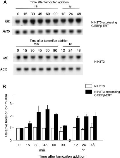
To determine if exogenous C/EBPβ expression can transactivate the endogenous Id2 gene expression, we established a stable line of NIH3T3 cells expressing the fusion protein of C/EBPβ with the ligand-binding domain of human estrogen receptor (C/EBPβ-ERT). The C/EBPβ-ERT protein is sequestered in the cytoplasm by heat shock proteins and the application of 4-hydroxytamoxifen (4HT) to the cells induces rapid translocation of C/EBPβ-ERT from the cytoplasm to the nucleus (24), which enables us to follow the effect of C/EBPβ on gene expression. As shown in Figure 1, the level of Id2 mRNA was increased by >2-fold in a biphasic manner, showing a sharp peak at 30 min and a broad rise in the later phase, after the addition of 4HT. In contrast, Id2 mRNA expression in the parental NIH3T3 cells was not changed significantly by the treatment (Figure 1). This indicates that the endogenous Id2 gene expression is induced by C/EBPβ. The observed rapid and biphasic changes in the Id2 mRNA level after the 4HT treatment suggested the short half-life of Id2 mRNA and a complex regulation of the endogenous Id2 gene expression by C/EBPβ.
Figure 1.
Induction of endogenous Id2 expression by the C/EBPβ-ERT fusion protein. A permanent cell line expressing C/EBPβ-ERT was established with NIH3T3 cells and treated with 1 μM 4HT to induce the translocation of the C/EBPβ-ERT fusion protein from the cytosol. RNA was extracted from the cells at various times and subjected to northern blot analysis. Representative results are shown in (A). The probes used are indicated on the left of each panel. Relative Id2 expression is shown in (B). Actb expression was used as an internal control. Each Id2 mRNA level is expressed as fold increase, compared to that in cells incubated without 4HT. White and black bars indicate relative Id2 mRNA levels in the parental NIH3T3 cells and NIH3T3 cells stably expressing C/EBPβ-ERT, respectively. Results are shown as the mean and standard error values of the mean (n = 3).
C/EBPβ activates Id2 gene expression through the proximal promoter region
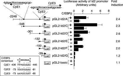
We next examined whether the promoter region of the Id2 gene contains the C/EBPβ consensus binding site, TT/GNNGNAAT/G (8), and found three potential sites at positions −445 to −436, −81 to −73 and −73 to −65 (designated C/EBPβ related element CβE1, CβE2 and CβE3, respectively), as shown in Figure 2. In addition, a luciferase reporter construct containing the upstream region of the Id2 gene, spanning from −2248 to +84 relative to the transcription initiation site, showed a 2.5-fold increase in activity in NIH3T3 cells co-transfected with a C/EBPβ expression vector, indicating that the Id2 promoter is responsive to the transcription factor (Figure 2). We then constructed a series of deletion mutants of the Id2 promoter and analyzed their activities in response to C/EBPβ using the same system. Deletion of the promoter region from −2248 to −101 bp resulted in retention of the responsiveness of the promoter to C/EBPβ, while further deletion from positions −100 to −40 abolished the response to C/EBPβ. These results demonstrated that the C/EBPβ-responsive element resides in the region containing CβE2 and CβE3 that spans −100 to −40 bp in the Id2 promoter.
Figure 2.
Transactivation of the Id2 promoter by C/EBPβ. In the Id2 promoter region spanning positions −2248 to +84 from the transcription initiation site, there are three potential C/EBPβ binding sites, named CβE1 (−445 to −436), CβE2 (−81 to −73) and CβE3 (−73 to −65), as indicated by arrows in the schematic representation of pGL2-Id2/A. The numbers shown on the left of the respective reporter constructs indicate the positions from the transcription initiation site. The sequence comparison of these sites with the consensus binding site (8), TT/GNNGNAAT/G, is shown in the inset. In the reporter assay, each of the reporter plasmids, which contained various lengths of the Id2 promoter, was co-transfected into NIH3T3 cells with pPGK-renilla, together with pMSV-C/EBPβ or pMSV-mock, as indicated by + and −, respectively, in the middle, and luciferase activities were determined after 48 h. Luciferase activities of the respective reporter constructs were determined by normalizing by the respective renilla luciferase activity. Fold induction of luciferase activity by C/EBPβ is indicated on the right. The mean and standard error of the mean values of three independent experiments are shown.
C/EBPβ binds directly to the Id2 promoter sequence containing its consensus binding sites
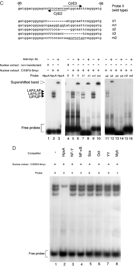
To examine whether the transactivation of the Id2 promoter by C/EBPβ is direct or indirect, we next performed an EMSA using oligonucleotide probes derived from the Id2 promoter region. As the reporter assay demonstrated that the region spanning positions −100 to −40 from the transcription initiation site is responsible for the transactivation of the Id2 promoter by C/EBPβ, we designed four oligonucleotides to cover this region in an overlapping manner (Figure 3A). Nuclear extracts were prepared from NIH3T3 cells transfected with an expression vector of C/EBPβ tagged with 6xMyc or its parental vector. As shown in Figure 3A (lanes 1–3), three retarded bands were detected with an oligonucleotide derived from the promoter of the HpxA gene, which is one of the direct targets of C/EBPβ (18). It is known that a single Cebpb mRNA can generate three isoforms of the C/EBPβ protein, liver-enriched activating proteins (LAP1 and LAP2) and liver-enriched inhibitory protein (LIP), through leaky ribosomal scanning (7,18,25). Western blot analysis confirmed that transient transfection with the vectors of untagged C/EBPβ or 6xMyc-tagged C/EBPβ leads to the expression of the three isoforms in NIH3T3 cells, while endogenous C/EBPβ proteins were barely detectable in the parental NIH3T3 cells (Figure 3B). Therefore, according to the previous report (25), the detected bands seemed to correspond to dimers of LAP/LAP, LAP/LIP and LIP/LIP, as indicated in Figure 3A. Similarly, probe II containing the region covering positions −95 and −56 demonstrated the binding of C/EBPβ (lanes 7–9 in Figure 3A). In both cases, inclusion of Ab against the Myc-tag or C/EBPβ (data not shown) caused supershift of the retarded bands, demonstrating the specificity of the reaction. Furthermore, the formation of protein–DNA complexes with probe II was blocked by competition with a 200-fold molar excess of the cold HpxA oligonucleotide (lanes 16 and 17 in Figure 3A), although, as detected in lanes 7–9, nonspecific binding at the position of LAP/LAP was observed with nuclear extracts of both non-transfectants and transfectants of pC/EBPβ-6Myc (lanes 16 and 17). In contrast to the binding to probe II, no specific binding was detected in EMSA with probes I, III and IV (lanes 4–6 and 10–15 in Figure 3A). These results suggested that C/EBPβ binds directly to the Id2 promoter via CβE2 and CβE3.
Figure 3.
EMSA for C/EBPβ with the Id2 promoter. EMSA was performed with 32P-labeled probes and nuclear extracts obtained from NIH3T3 cells transiently transfected with pC/EBPβ-6Myc or parental NIH3T3 cells. Oligonucleotide HpxA was used as a positive control for C/EBPβ binding. DNA–protein complexes were separated on a 6% polyacrylamide gel, and the gel was dried and exposed to X-ray film. Three retarded bands derived from dimers of C/EBPβ isoforms are indicated by filled arrowheads (LAP/LAP, LAP/LIP and LIP/LIP). The supershifted band formed with anti-Myc Ab is indicated by an open arrowhead. (A) On the top, probes used in EMSA are aligned with their positions in the Id2 promoter sequence. Arrows indicate the positions of CβE2 and CβE3. The probes used were HpxA (lanes 1–3), probe I (lanes 4–6), probe II (lanes 7–9), probe III (lanes 10–12) and probe IV (lanes 13–15). Nuclear extracts were prepared from NIH3T3 cells transfected with pC/EBPβ-6Myc (lanes 2, 3, 5, 6, 8, 9, 11, 12, 14, 15) or the parental NIH3T3 cells (lanes 1, 4, 7, 10, 13). For the detection of a supershifted band, anti-Myc Ab was included in the incubation mixture (lanes 3, 6, 9, 12, 15). In lanes 16 and 17, a 200-fold molar excess of the HpxA oligonucleotide was added as cold competitor. (B) Expression of C/EBPβ isoforms in NIH3T3 cells. Left and right panels show the results with anti-C/EBPβ and anti-Myc Abs, respectively. Origins of nuclear extracts are shown on the top. Positions of the respective C/EBPβ isoforms are indicated by arrowheads (untagged-C/EBPβ) and arrows (6Myc-tagged-C/EBPβ) on the right of each panel. Positions of size markers are indicated on the left of each panel. (C) Mutant oligonucleotides designed based on probe II were used in EMSA. The positions of CβE2 and CβE3 are indicated by arrows. Probes Δ1 and Δ3 are CβE2 and CβE3 deletion mutants of probe II, respectively. Deleted regions are indicated by dashes. Base-substituted regions are underlined. The probes used were HpxA (lanes 1–3), probe II (lanes 4–6), Δ1 (lanes 7 and 8), m1 (lanes 9 and 10), Δ2 (lanes 11 and 12), Δ3 (lanes 13 and 14) and m2 (lanes 15 and 16). Nuclear extracts were prepared from NIH3T3 cells transfected with pC/EBPβ-6Myc (lanes 2, 3, 5–16) or the parental NIH3T3 cells (lanes 1 and 4). For the detection of a supershifted band, anti-Myc Ab was included in the incubation mixture (lanes 3, 6, 8, 12, 14 and 16). (D) 32P-labeled probe II was incubated with nuclear extracts containing C/EBPβ-6Myc, together with 200-fold molar excess of the cold competitors that contain the binding sites of the transcription factors as indicated on the top (see Materials and Methods).
To delineate the binding site of C/EBPβ more precisely, we synthesized five oligonucleotides bearing deletions or mutations in the domain related to CβE2 and/or CβE3, and performed EMSA. As shown in Figure 3C, C/EBPβ bound normally to oligonucleotides Δ1 and m1 (lanes 7–10), which had a deletion and base substitutions in CβE2, respectively. In addition, C/EBPβ formed a complex with oligonucleotide Δ2 (lanes 11 and 12 in Figure 3C), in which the spacer between CβE2 and CβE3 was deleted. On the other hand, deletion and base substitutions in CβE2 (oligonucleotides Δ3 and m2, respectively) failed to form a retarded complex with C/EBPβ (lanes 13–16 in Figure 3C). These results strongly suggest that C/EBPβ binds to CβE3, although CβE3 displays relatively low sequence conservation with the C/EBPβ consensus binding site (inset in Figure 2).
CβE3 contains sequences that are similar to the consensus binding sites of other transcription factors including NF-Y, NF-κB, Sox, Oct, YY and Myb. To exclude the possibility that the retarded bands shown in Figure 3A and C contain these transcription factors, we performed competitive EMSA using the oligonucleotides that contain the consensus binding sites of these transcription factors. As shown in Figure 3D, none of the oligonucleotides competed for nor altered the retarded bands generated by probe II and the nuclear extract of transfectants of pC/EBPβ-6Myc (lanes 1 and 3–8), while the HpxA oligonucleotide did (lane 2). Therefore, it is unlikely that transcription factors other than C/EBPβ bind CβE3 directly or indirectly.
Transactivation by C/EBPβ depends on CβE3
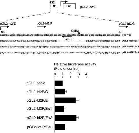
To confirm that CβE3 is responsible for the transactivation of the Id2 promoter by C/EBPβ, we constructed three reporter plasmids, pGL2-Id2/EΔ1, pGL2-Id2/EΔ2 and pGL2-Id2/EΔ3, with deletion of both CβE2 and CβE3, CβE2 or CβE3, respectively, and transfected them with or without a C/EBPβ expression vector into NIH3T3 cells. As shown in Figure 4, pGL2-Id2/EΔ2 expressed luciferase activity comparable to that of the wild-type pGL2-Id2/E plasmid, whereas no appreciable response to C/EBPβ was observed with pGL2-Id2/EΔ1 or pGL2-Id2/EΔ3. These results, together with the data shown in Figure 3B, indicate that the regulation of Id2 expression by C/EBPβ occurs via the most proximal CβE in the Id2 promoter region.
Figure 4.
Transactivation of the Id2 promoter by C/EBPβ is via CβE3. Mutant reporter plasmids were constructed based on pGL2-Id2/E. pGL2-Id2/EΔ1 lacks the region containing both CβE2 and CβE3. Core sequences of CβE2 and CβE3 are deleted in pGL2-Id2/EΔ2 and pGL2-Id2/EΔ3, respectively. Deleted regions are indicated by dashes. NIH3T3 cells were co-transfected with reporter plasmids and an internal control vector, pGL-renilla, together with pMSV-C/EBPβ or pMSV-mock. Luciferase activity was determined as in the experiments shown in Figure 2. Transactivation of the respective reporter plasmids by C/EBPβ is presented as fold induction of transactivation of pGL2-basic by C/EBPβ. The mean and standard errors of the mean values from three separate assays are shown in the histograms.
Direct binding of C/EBPβ to the endogenous Id2 promoter
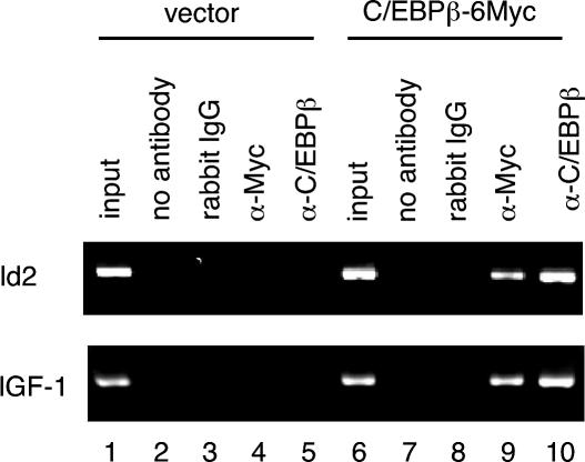
We next performed the chromatin immunoprecipitation (ChIP) assay to demonstrate that C/EBPβ is present on the Id2 promoter region containing CβE2 and CβE3. To do this, NIH3T3 cells were transfected with the C/EBPβ-6xMyc expression vector and the cell lysate prepared after fixation was precipitated with an Ab against Myc or C/EBPβ, followed by PCR with primers for the Id2 promoter. As shown in Figure 5 (lanes 9 and 10), both antibodies enriched the chromatin-containing DNA of the Id2 promoter region compared to incubation with normal rabbit IgG or without Ab (lanes 7 and 8), similar to the case of the insulin-like growth factor-1 (Igf1) gene, which is known to be a direct downstream target of C/EBPβ (26). On the other hand, no such enrichment was detected in cells transfected with the parental expression vector (Figure 5, lanes 2–5). These data indicate that C/EBPβ is bound to the endogenous Id2 promoter.
Figure 5.
Binding of C/EBPβ to the Id2 promoter in vivo. The ChIP assay was performed using NIH3T3 cells transiently transfected with empty vector (lanes 1–5) or pC/EBPβ-6Myc (lanes 6–10). The chromatin-associated DNA was incubated without Ab (lanes 2 and 7) or with normal rabbit IgG (lanes 3 and 8), anti-Myc Ab (lanes 4 and 9), or anti-C/EBPβ Ab (lanes 5 and 10). An aliquot (2.5%) of the total chromatin DNA was used for input (lanes 1 and 6). Immunoprecipitates were subjected to PCR with a primer-pair specific to the Id2 promoter that amplified a 311 bp fragment (upper panel). As a positive control, PCR was carried out with a primer-pair specific to the Igf-1 promoter, which amplified a 287 bp DNA fragment (lower panel). After 32 cycles of amplification, PCR products were electrophoresed through a 3% agarose gel and visualized by ethidium bromide staining.
Reduced expression of Id2 in C/EBPβ-deficient mammary glands during pregnancy
We finally examined Id2 gene expression in pregnant mammary glands of C/EBPβ-null mice during pregnancy. Since C/EBPβ-null mice are infertile, we transplanted mammary epithelial cells into the inguinal fat pads of Rag2-null mice in which mammary glands had been precleared by removing mammary glands of the host at the age of 3 weeks (4). After 7 weeks of transplantation, mice were mated with male mice and RNA was purified from the mammary glands at 10 days post coitus (d.p.c.). In morphological examination, mammary glands were successfully reconstituted in all recipients and the impaired proliferation of mammary epithelial cells was observed in the gland reconstituted with C/EBPβ-null mammary epithelial cells, as expected (Figure 6A). Northern blot analysis demonstrated that, in the non-pregnant state, there was no significant difference in the basal expression level of Id2 between mammary glands reconstituted with mammary epithelial cells of C/EBPβ-null mice and those reconstituted with cells of wild-type mice. In contrast, mammary glands reconstituted with C/EBPβ-null mammary epithelial cells were devoid of the pregnancy-induced elevation of Id2 mRNA expression. Thus, it was suggested that Id2 expression is under the influence of C/EBPβ in mammary glands during pregnancy, but not in the non-pregnant state.
Figure 6.
Id2 expression in mammary glands deficient for C/EBPβ. Epithelial cells of wild-type or C/EBPβ-null mammary glands were transplanted into precleared mammary glands of female Rag2-null mice. After 7 weeks, reconstituted mammary glands of non-pregnant and pregnant (10 d.p.c.) mice were subjected to whole mount analysis and RNA purification. (A) Morphology of reconstituted mammary glands in the non-pregnant and pregnant mice. Reconstituted mammary glands were dissected out, mounted on a slide glass and stained with carmine. The genotype of a donor and the pregnant state of a recipient are indicated on the left and the top, respectively. Both wild-type and C/EBPβ-null mammary epithelial cells reconstituted the mammary gland of a recipient successfully and no significant difference was observed in the non-pregnant state. In the mammary gland at 10 d.p.c., disturbed epithelial proliferation is evident in the gland reconstituted with C/EBPβ-null mammary epithelial cells. (B) Northern blot analysis of Id2 mRNA expression. RNA was purified from reconstituted mammary glands. Genotypes of transplanted mammary epithelial cells are indicated on the top. The lower panel shows the representative data. The probes used are indicated on the left of the lower panels. The upper panel shows the relative Id2 expression normalized by Cyk18 expression. The mean and standard error of the mean values obtained from three independent samples are shown in the histograms.
DISCUSSION
Mammary gland development is divided into several stages that are related to sexual development and reproduction: the embryonic, prepubertal, pubertal, pregnancy, lactation and involution stages (2). Each stage shows distinct requirements for specific genes for completion of the stage and/or progression to the next stage, as revealed or confirmed by gene inactivation studies in mice (2). Among these stages, the pregnancy stage, where robust proliferation and differentiation of mammary epithelial cells lead to lobuloalveolar development, is controlled by various molecules including the prolactin receptor, Stat5, receptor activator of NF-κB (RANK), receptor activator of NF-κB ligand (RANKL), cyclin D1, C/EBPβ and Id2 (2). The present study provides evidence linking C/EBPβ and Id2 in a gene network that is involved in lobuloalveolar development at the pregnancy stage of the mammary gland. Furthermore, RANKL, also known as osteoprotegerin ligand (OPGL), has been shown to induce rapid nuclear translocation of C/EBPβ from the cytosol in mammary epithelial cells and to regulate gene expression of β-casein (Csnb) gene, one of the most abundant milk proteins (27). Therefore, it is conceivable that an intracellular signaling cascade provoked by RANKL/OPGL induces Id2 gene expression via C/EBPβ during lobuloalveolar development of the mammary gland during pregnancy.
Besides a lactation defect, mice deficient for Id2 display multiple defects, mainly in the immune system, including agenesis of lymph nodes and Peyer's patches, impaired natural killer cell development (14), selective loss of CD8α+ dendritic cells (28,29), Th2 dominance with an increased serum IgE level (28,30) and reduction of intestinal lymphocytes (31). These defects are not observed in C/EBPβ-null mice. On the contrary, C/EBPβ-null mice exhibit impaired macrophage function against bacteria and tumors (32,33), and are sterile as a result of defective differentiation of ovarian granulosa cells (34). These phenotypes are not shared with Id2-null mice, although a fraction of Id2-null female mice show sterility for unknown reasons (Y. Yokota, unpublished data). Currently, therefore, the regulation of Id2 expression by C/EBPβ seems to be specific to mammary epithelial cells during lobuloalveolar development. However, we cannot exclude the possibility that redundancy within the respective gene family compensates for the function of the mutant genes and prevents the mice from developing a defect in other cell types.
It has been shown that Id1, a founding member of the Id gene family, is regulated by C/EBPβ in pro-B cells (35,36). Interestingly, IL-3-dependent expression of Id1 relies on C/EBPβ and Stat5 via the pro-B-cell enhancer element, and deacetylation of C/EBPβ by a histone deacetylase recruited by Stat5 is required for transcription of Id1 (36). Since Stat5 is an important transcription factor that is activated by prolactin signaling, it will be interesting to know whether a similar mechanism operates in the regulation of Id2 expression.
Among the molecules that are required for lobuloalveolar development, some have been found to be involved in mammary carcinogenesis, as might be predicted based on the fact that robust cell proliferation occurs during lobuloalveolar development. Cyclin D1 is a typical example: overexpression of cyclin D1 protein is observed in more than half of human breast cancers (37–39) and amplification of the gene locus is detected in 15–20% of such cancers (40). Through computational analysis of the expression patterns of genes across tumor specimens, a recent study demonstrated that C/EBPβ plays a role in the consequences of cyclin D1 overexpression (41). It is therefore tempting to speculate that the cyclin D1-C/EBPβ-Id2 axis is involved in the development of mammary tumors.
Acknowledgments
We are grateful to T. Kaisho, M. Takiguchi, S.L. Mcknight and T. Nakano for materials, and K. Yamada and members of the Yokota lab for valuable suggestions and discussion. This work was supported by Grants-in-Aid from the Ministry of Education, Culture, Sports, Science and Technology, Japan, by the 21st Century COE program, by Takeda Science Foundation and by The Naito Foundation. Funding to pay the Open Access publication charges for this article was provided by The Naito Foundation.
Conflict of interest statement. None declared.
REFERENCES
- 1.Neville M.C., Daniel C.W. The Mammary gland: Development, Regulation, and Function. New York, NY: Plenum Press; 1987. [Google Scholar]
- 2.Hennighausen L., Robinson G.W. Signaling pathways in mammary gland development. Dev. Cell. 2001;1:467–475. doi: 10.1016/s1534-5807(01)00064-8. [DOI] [PubMed] [Google Scholar]
- 3.Ruzinova M.B., Benezra R. Id proteins in development, cell cycle and cancer. Trends Cell Biol. 2003;13:410–418. doi: 10.1016/s0962-8924(03)00147-8. [DOI] [PubMed] [Google Scholar]
- 4.Mori S., Nishikawa S., Yokota Y. Lactation defect in mice lacking the helix–loop–helix inhibitor Id2. EMBO J. 2000;19:5772–5781. doi: 10.1093/emboj/19.21.5772. [DOI] [PMC free article] [PubMed] [Google Scholar]
- 5.Takiguchi M. The C/EBP family of transcription factors in the liver and other organs. Int. J. Exp. Pathol. 1998;79:369–391. doi: 10.1046/j.1365-2613.1998.00082.x. [DOI] [PMC free article] [PubMed] [Google Scholar]
- 6.Grimm S.L., Rosen J.M. The role of C/EBPbeta in mammary gland development and breast cancer. J. Mammary Gland Biol. Neoplasia. 2003;8:191–204. doi: 10.1023/a:1025900908026. [DOI] [PubMed] [Google Scholar]
- 7.Lekstrom-Himes J., Xanthopoulos K.G. Biological role of the CCAAT/enhancer-binding protein family of transcription factors. J. Biol. Chem. 1998;273:28545–28548. doi: 10.1074/jbc.273.44.28545. [DOI] [PubMed] [Google Scholar]
- 8.Akira S., Isshiki H., Sugita T., Tanabe O., Kinoshita S., Nishio Y., Nakajima T., Hirano T., Kishimoto T. A nuclear factor for IL-6 expression (NF-IL6) is a member of a C/EBP family. EMBO J. 1990;9:1897–1906. doi: 10.1002/j.1460-2075.1990.tb08316.x. [DOI] [PMC free article] [PubMed] [Google Scholar]
- 9.Seagroves T.N., Krnacik S., Raught B., Gay J., Burgess-Beusse B., Darlington G.J., Rosen J.M. C/EBPbeta, but not C/EBPalpha, is essential for ductal morphogenesis, lobuloalveolar proliferation, and functional differentiation in the mouse mammary gland. Genes Dev. 1998;12:1917–1928. doi: 10.1101/gad.12.12.1917. [DOI] [PMC free article] [PubMed] [Google Scholar]
- 10.Robinson G.W., Johnson P.F., Hennighausen L., Sterneck E. The C/EBPbeta transcription factor regulates epithelial cell proliferation and differentiation in the mammary gland. Genes Dev. 1998;12:1907–1916. doi: 10.1101/gad.12.12.1907. [DOI] [PMC free article] [PubMed] [Google Scholar]
- 11.Seagroves T.N., Lydon J.P., Hovey R.C., Vonderhaar B.K., Rosen J.M. C/EBPbeta (CCAAT/enhancer binding protein) controls cell fate determination during mammary gland development. Mol. Endocrinol. 2000;14:359–368. doi: 10.1210/mend.14.3.0434. [DOI] [PubMed] [Google Scholar]
- 12.Cao Z., Umek R.M., McKnight S.L. Regulated expression of three C/EBP isoforms during adipose conversion of 3T3-L1 cells. Genes Dev. 1991;5:1538–1552. doi: 10.1101/gad.5.9.1538. [DOI] [PubMed] [Google Scholar]
- 13.Narumi O., Mori S., Boku S., Tsuji Y., Hashimoto N., Nishikawa S., Yokota Y. OUT, a novel basic helix–loop–helix transcription factor with an Id-like inhibitory activity. J. Biol. Chem. 2000;275:3510–3521. doi: 10.1074/jbc.275.5.3510. [DOI] [PubMed] [Google Scholar]
- 14.Yokota Y., Mansouri A., Mori S., Sugawara S., Adachi S., Nishikawa S., Gruss P. Development of peripheral lymphoid organs and natural killer cells depends on the helix–loop–helix inhibitor Id2. Nature. 1999;397:702–706. doi: 10.1038/17812. [DOI] [PubMed] [Google Scholar]
- 15.Soriano P., Montgomery C., Geske R., Bradley A. Targeted disruption of the c-src proto-oncogene leads to osteopetrosis in mice. Cell. 1991;64:693–702. doi: 10.1016/0092-8674(91)90499-o. [DOI] [PubMed] [Google Scholar]
- 16.Andrews N.C., Faller D.V. A rapid micropreparation technique for extraction of DNA-binding proteins from limiting numbers of mammalian cells. Nucleic Acids Res. 1991;19:2499. doi: 10.1093/nar/19.9.2499. [DOI] [PMC free article] [PubMed] [Google Scholar]
- 17.Dumais N., Bounou S., Olivier M., Tremblay M.J. Prostaglandin E(2)-mediated activation of HIV-1 long terminal repeat transcription in human T cells necessitates CCAAT/enhancer binding protein (C/EBP) binding sites in addition to cooperative interactions between C/EBPbeta and cyclic adenosine 5′-monophosphate response element binding protein. J. Immunol. 2002;168:274–282. doi: 10.4049/jimmunol.168.1.274. [DOI] [PubMed] [Google Scholar]
- 18.Poli V., Mancini F.P., Cortese R. IL-6DBP, a nuclear protein involved in interleukin-6 signal transduction, defines a new family of leucine zipper proteins related to C/EBP. Cell. 1990;63:643–653. doi: 10.1016/0092-8674(90)90459-r. [DOI] [PubMed] [Google Scholar]
- 19.Shou Z., Yamada K., Inazu T., Kawata H., Hirano S., Mizutani T., Yazawa T., Sekiguchi T., Yoshino M., Kajitani T., Okada K., Miyamoto K. Genomic structure and analysis of transcriptional regulation of the mouse zinc-fingers and homeoboxes 1 (ZHX1) gene. Gene. 2003;302:83–94. doi: 10.1016/s0378-1119(02)01093-4. [DOI] [PubMed] [Google Scholar]
- 20.Ramsay G.R., Ishii S., Gondall T.J. Interaction of the Myb protein with specific DNA binding sites. J. Biol. Chem. 1992;267:5656–5662. [PubMed] [Google Scholar]
- 21.Connor F., Wright E., Denny P., Koopmanl P., Ashworth A. The Sry-related HMG box-containing gene Sox6 is expressed in the adult testis and developing nervous system of the mouse. Nucleic Acids Res. 1995;23:3365–3372. doi: 10.1093/nar/23.17.3365. [DOI] [PMC free article] [PubMed] [Google Scholar]
- 22.Kunsch C., Ruben S.M., Rosen C.A. Selection of optimal kappa B/Rel DNA-binding motifs: interaction of both subunits of NF-κB with DNA is required for transcriptional activation. Mol. Cell. Biol. 1992;12:4412–4421. doi: 10.1128/mcb.12.10.4412. [DOI] [PMC free article] [PubMed] [Google Scholar]
- 23.Gonda H., Sugai M., Nambu Y., Katakai T., Agata Y., Mori K.J., Yokota Y., Shimizu A. The balance between Pax5 and Id2 activities is the key to AID gene expression. J. Exp. Med. 2003;198:1427–1437. doi: 10.1084/jem.20030802. [DOI] [PMC free article] [PubMed] [Google Scholar]
- 24.Mattioni T., Louvion J.F., Picard D. Regulation of protein activities by fusion to steroid binding domains. Methods Cell Biol. 1994;43:335–352. doi: 10.1016/s0091-679x(08)60611-1. [DOI] [PubMed] [Google Scholar]
- 25.Descombes P., Schibler U. A liver-enriched transcriptional activator protein, LAP, and a transcriptional inhibitory protein, LIP, are translated from the same mRNA. Cell. 1991;67:569–579. doi: 10.1016/0092-8674(91)90531-3. [DOI] [PubMed] [Google Scholar]
- 26.Nolten L.A., van Schaik F.M., Steenbergh P.H., Sussenbach J.S. Expression of the insulin-like growth factor I gene is stimulated by the liver-enriched transcription factors C/EBP alpha and LAP. Mol. Endocrinol. 1994;8:1636–1645. doi: 10.1210/mend.8.12.7708053. [DOI] [PubMed] [Google Scholar]
- 27.Kim H.J., Yoon M.J., Lee J., Penninger J.M., Kong Y.Y. Osteoprotegerin ligand induces beta-casein gene expression through the transcription factor CCAAT/enhancer-binding protein beta. J. Biol. Chem. 2002;277:5339–5344. doi: 10.1074/jbc.M108342200. [DOI] [PubMed] [Google Scholar]
- 28.Kusunoki T., Sugai M., Katakai T., Omatsu Y., Iyoda T., Inaba K., Nakahata T., Shimizu A., Yokota Y. TH2 dominance and defective development of a CD8+ dendritic cell subset in Id2-deficient mice. J. Allergy Clin. Immunol. 2003;111:136–142. doi: 10.1067/mai.2003.29. [DOI] [PubMed] [Google Scholar]
- 29.Hacker C., Kirsch R.D., Ju X.S., Hieronymus T., Gust T.C., Kuhl C., Jorgas T., Kurz S.M., Rose-John S., Yokota Y., Zenke M. Transcriptional profiling identifies Id2 function in dendritic cell development. Nature Immunol. 2003;4:380–386. doi: 10.1038/ni903. [DOI] [PubMed] [Google Scholar]
- 30.Sugai M., Gonda H., Kusunoki T., Katakai T., Yokota Y., Shimizu A. Essential role of Id2 in negative regulation of IgE class switching. Nature Immunol. 2003;4:25–30. doi: 10.1038/ni874. [DOI] [PubMed] [Google Scholar]
- 31.Kim J.K., Takeuchi M., Yokota Y. Impairment of intestinal intraepithelial lymphocytes in Id2 deficient mice. Gut. 2004;53:480–486. doi: 10.1136/gut.2003.022293. [DOI] [PMC free article] [PubMed] [Google Scholar]
- 32.Tanaka T., Akira S., Yoshida K., Umemoto M., Yoneda Y., Shirafuji N., Fujiwara H., Suematsu S., Yoshida N., Kishimoto T. Targeted disruption of the NF-IL6 gene discloses its essential role in bacteria killing and tumor cytotoxicity by macrophages. Cell. 1995;80:353–361. doi: 10.1016/0092-8674(95)90418-2. [DOI] [PubMed] [Google Scholar]
- 33.Screpanti I., Romani L., Musiani P., Modesti A., Fattori E., Lazzaro D., Sellitto C., Scarpa S., Bellavia D., Lattanzio G., Francesco B., Frati L., Cortese R., Gulino A., Ciliberto G., Costantini F., Poli V. Lymphoproliferative disorder and imbalanced T-helper response in C/EBP beta-deficient mice. EMBO J. 1995;14:1932–1941. doi: 10.1002/j.1460-2075.1995.tb07185.x. [DOI] [PMC free article] [PubMed] [Google Scholar]
- 34.Sterneck E., Tessarollo L., Johnson P.F. An essential role for C/EBPbeta in female reproduction. Genes Dev. 1997;11:2153–2162. doi: 10.1101/gad.11.17.2153. [DOI] [PMC free article] [PubMed] [Google Scholar]
- 35.Saisanit S., Sun X.H. Regulation of the pro-B-cell-specific enhancer of the Id1 gene involves the C/EBP family of proteins. Mol. Cell. Biol. 1997;17:844–850. doi: 10.1128/mcb.17.2.844. [DOI] [PMC free article] [PubMed] [Google Scholar]
- 36.Xu M., Nie L., Kim S.H., Sun X.H. STAT5-induced Id-1 transcription involves recruitment of HDAC1 and deacetylation of C/EBPbeta. EMBO J. 2003;22:893–904. doi: 10.1093/emboj/cdg094. [DOI] [PMC free article] [PubMed] [Google Scholar]
- 37.Bartkova J., Lukas J., Muller H., Lutzhoft D., Strauss M., Bartek J. Cyclin D1 protein expression and function in human breast cancer. Int. J. Cancer. 1994;57:353–361. doi: 10.1002/ijc.2910570311. [DOI] [PubMed] [Google Scholar]
- 38.Gillett C., Fantl V., Smith R., Fisher C., Bartek J., Dickson C., Barnes D., Peters G. Amplification and overexpression of cyclin D1 in breast cancer detected by immunohistochemical staining. Cancer Res. 1994;54:1812–1817. [PubMed] [Google Scholar]
- 39.McIntosh G.G., Anderson J.J., Milton I., Steward M., Parr A.H., Thomas M.D., Henry J.A., Angus B., Lennard T.W., Horne C.H. Determination of the prognostic value of cyclin D1 overexpression in breast cancer. Oncogene. 1995;11:885–891. [PubMed] [Google Scholar]
- 40.Dickson C., Fantl V., Gillett C., Brookes S., Bartek J., Smith R., Fisher C., Barnes D., Peters G. Amplification of chromosome band 11q13 and a role for cyclin D1 in human breast cancer. Cancer Lett. 1995;90:43–50. doi: 10.1016/0304-3835(94)03676-a. [DOI] [PubMed] [Google Scholar]
- 41.Lamb J., Ramaswamy S., Ford H.L., Contreras B., Martinez R.V., Kittrell F.S., Zahnow C.A., Patterson N., Golub T.R., Ewen M.E. A mechanism of cyclin D1 action encoded in the patterns of gene expression in human cancer. Cell. 2003;114:323–334. doi: 10.1016/s0092-8674(03)00570-1. [DOI] [PubMed] [Google Scholar]


