Abstract
Inflammatory bowel disease (IBD), including ulcerative colitis and Crohn’s disease, is a complex gastrointestinal disorder with a multifactorial etiology, including environmental triggers, autoimmune mechanisms, and genetic predisposition. Despite advancements in therapeutic strategies for IBD, its associated mortality rate continues to rise, which is often attributed to unforeseen side effects of conventional treatments. In this context, we explored the potential of Ecklonia cava extract (ECE), derived from an edible marine alga known for its anti-inflammatory and antioxidant properties, in mitigating IBD. This study investigated the effectiveness of ECE as a preventive agent in a murine model of dextran sulfate sodium (DSS)-induced colitis. Our findings revealed that pretreatment with ECE significantly ameliorated colitis severity, as evidenced by increased colon length, reduced spleen weight, and histological improvements demonstrated by immunohistochemical analysis. Furthermore, ECE significantly attenuated the upregulation of inflammatory cytokines and mediators and the infiltration of immune cells known to be prominent features of colitis in mice. Notably, ECE alleviated dysbiosis of intestinal microflora and aided in the recovery of damaged intestinal mucosa. Mechanistically, ECE exhibited protective effects against pathogenic colitis by inhibiting the NLRP3/NF-κB pathways known to be pivotal regulators in the inflammatory signaling cascade. These compelling results suggest that ECE holds promise as a potential candidate for IBD prevention. It might be developed into a functional food for promoting gastrointestinal health. This research sheds light on the preventive potential of natural compounds like ECE in the management of IBD, offering a safer and more effective approach to combating this challenging disease.
Keywords: Ecklonia cava extract, DSS-induced colitis, intestinal barrier, pathogenic inflammation
1. Introduction
Inflammatory bowel disease (IBD), a composite of ulcerative colitis (UC) and Crohn’s disease (CD), comprises a cluster of protracted inflammatory conditions affecting the gastrointestinal tract. These disorders, characterized by chronic and unbridled inflammation alongside epithelial deterioration of the intestinal mucosa, ensue from a complex interplay of genetic predisposition and a milieu of environmental risk factors [1,2,3]. Although the precise mechanisms of IBD’s pathogenesis remain to be discovered, an emerging consensus implicates that a disordered immune modulation within the gastrointestinal microenvironment and a concomitant breakdown in the homeostasis of the epithelial barrier might play important roles in IBD [4]. Untreated IBD carries a marked elevation in the risk of colorectal cancer, underscoring the imperativeness of efficacious IBD management [5].
During the development of IBD, many pathological phenomena are accompanied by defective changes in intestinal environments. Due to excess inflammatory stimuli, immunological dysregulation is initiated with damage to the epithelial barrier, infiltration of immune cells, and dysbiosis of intestinal flora [6,7]. Activated intestinal cells are known to express high levels of inflammatory cytokines to accelerate pathological processes. The intestinal epithelial barrier serves to protect the host by blocking the entry of pathogenic microorganisms and foreign antigens into the body. It is composed of enterocytes that are tightly connected through intercellular junctions [8]. Intestinal barrier dysfunction in IBD refers to uncontrolled inflammation due to increased intestinal permeability, decreased tight junction (TJ) barrier function, and impaired immune regulation [9,10]. Mucin 2 (MUC2) secreted by goblet cells can also prevent colonization by pathogenic microorganisms and the transfer of enterotoxins from bacteria to the internal environment [11]. With a defective mucosal barrier, compositions of the intestinal microbiome can change into those of dysbiotic strains. These pathogenic bacteria can induce more destructive immune responses by interacting with Toll-like receptor to activate inflammatory signaling through nucleotide-binding oligomerization domain-like receptor protein 3 (NLRP3) and NF-κB-mediated signal transduction [12,13].
Traditional IBD therapeutics, from antibiotics to biologics and immunosuppressants, are frequently accompanied by a series of significant side effects [14]. The recent direction of IBD etiological research has ushered in the potential for innovative therapeutic modalities, including naturally occurring compounds. These compounds proffer the prospect of redressing perturbations in the gut microbiome while simultaneously expediting the restorative process of the mucosal layer [15,16]. Among such natural compounds, complex marine polysaccharides have been extensively studied as pharmaceuticals [17,18]. Based on previous reports, algae extracts and polysaccharides are excellent substances for treating and preventing intestinal inflammation such as IBD due to the anti-inflammatory functions they provide as fermentation substrates for beneficial intestinal microbiomes [19,20]. Among this category, Ecklonia cava (E. cava), an edible brown alga distributed along the coasts of Korea, China, and Japan, is composed of various physiologically active substances such as fucoidan, sulfated polysaccharide, and phlorotannin [21,22]. Compared to other brown algae, E. cava is rich in a unique polyphenol with polymerized phloroglucinol units called phlorotannin, among various components. Phlorotannins, including dieckol (DK), eckol, and phlorofucofuroeckol A (PFFA) in E. cava, are known to exhibit many biological potentials against viral infection, diabetic complications, hypertension, and obesity-associated phenomena [23,24,25,26]. These phlorotannins in E. cava suggest that they can be used as a natural good source with potential applications in various diseases [27,28].
Studies on E. cava’s anti-inflammatory properties have been conducted in several fields. In a periodontal disease-related study, E. cava prevented alveolar bone loss by reducing inflammatory cell infiltration and IL-1β production in gingival tissue, and it also attenuated endothelial cell dysfunction by regulating the inflammation of perivascular adipose tissue in cardiovascular disease [29,30]. It has also been reported that it can be used as a preventative and therapeutic compound for diabetes-related diseases by reducing inflammation-associated receptors such as TLR4 and RAGE [31]. Despite extensive investigations into these anti-inflammatory activities, the specific role of E. cava in IBD remains a largely unexamined area.
Thus, we tried to assess the preventive efficacy of E. cava extract (ECE) using a colitis animal model employing various colitis disease activity indices. Ultimately, we aim to illuminate the potential of ECE as a safe and efficient agent for promoting intestinal health, thus paving the way for its development as a functional food without adverse long-term effects.
2. Results
2.1. Ecklonia cava Extract Protects against DSS-Induced Colitis
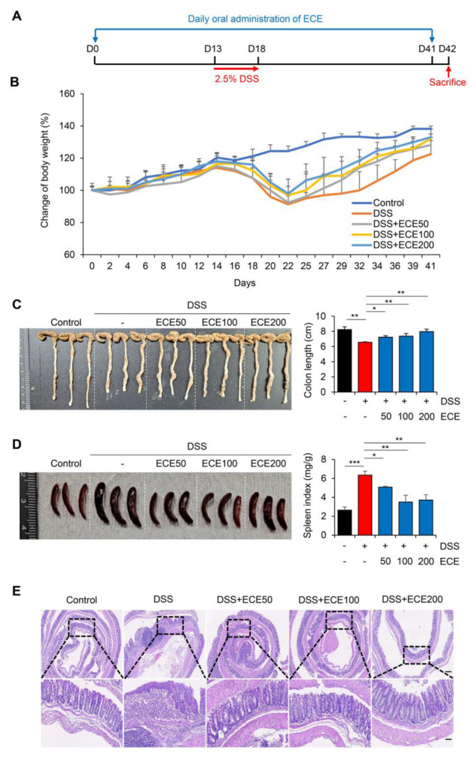
To determine whether ECE could prevent colonic damage and inflammation, we prepared a dextran sulfate sodium (DSS)-induced colitis model (Figure 1A). ECE was orally administered daily from 14 days before DSS treatment until mice were sacrificed. Mice were fed 2.5% DSS in drinking water for 5 days. After that, the mice were fed normal drinking water. Weight loss was confirmed in the DSS group compared to the normal group. There was a little restoration of body weight between the DSS group and the ECE-treated groups in a dose-dependent manner (Figure 1B). Colon length, an indicator of severity of colitis, was found to be longer in the ECE-treated group than in the DSS group (Figure 1C). Moreover, the spleen index (spleen weight/body weight) was markedly increased in the DSS group compared to in the normal group. However, it was significantly restored in the ECE-treated groups (Figure 1D). Histopathological examination of the colon revealed that colonic crypt damage and mucosal infiltration of immune cells in the DSS group were improved in the ECE-treated groups (Figure 1E).
Figure 1.
Ecklonia cava extract protects against dextran sodium sulfate (DSS)-induced colitis: (A) Experimental design for DSS-induced colitis and Ecklonia cava extract (ECE) pretreatment by oral administration. Different doses (50, 100, 200 mg/kg body weight) of ECE were orally administrated to mice every day until the end of experiment. After 14 days, mice were fed with drinking water containing 2.5% DSS for 5 days. Mice were sacrificed at 42 days after treatment to analyze disease activity index. (B) Body weight changes in control or DSS-treated mice and ECE-pretreated colitis mice. (C) Gross morphology of colon (left) and quantification of colon length (right). Colon length was measured, except for the cecum. (D) Representative image of spleen (left) and quantitative analysis of spleen weight-to-body weight ratio. (E) Representative image of H&E-stained colon tissue. Scale bar = 300 μm (top, 4×), 60 μm (bottom, 20×). All p-values were calculated using unpaired two-tailed Student’s t-tests. Results are presented as mean ± SD from at least triplicate samples. *, p < 0.05; **, p < 0.01; ***, p < 0.001.
To determine a preventive effect of ECE against colitis with therapeutic potential, an animal model was prepared by orally administering ECE after DSS treatment (Figure S1A). In contrast with the preventive model, body weight change was not significant between the DSS and ECE groups during therapeutic treatment (Figure S1B). Colon length and spleen index were not dramatically changed by treatment with ECE (Figure S1C,D). Consistent with phenotypical changes, hematoxylin–eosin (H&E) staining showed no difference in submucosal damage or destruction of colonic structure between the DSS and ECE groups (Figure S1E). These data suggest that ECE is more effective against DSS-induced colitis through preventive use rather than as a therapeutic treatment.
2.2. Ecklonia cava Extract Reduces Inflammatory Response in DSS-Induced Colitis
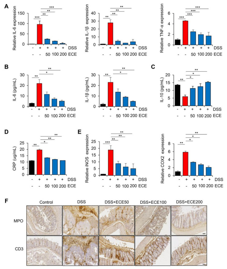
DSS-induced colitis is known to be closely associated with the production of pro-inflammatory cytokines [32]. To determine whether the DSS-induced expression of pro-inflammatory cytokines was affected by ECE, we first measured levels of pro-inflammatory cytokines in colon tissues and sera samples. As shown in Figure 2A, mRNA levels of pro-inflammatory cytokines such as IL-6, IL-1β, and TNF-α in colon tissues were increased in DSS-treated mice but significantly suppressed by ECE in a dose-dependent manner. It was also confirmed that protein levels of IL-6 and IL-1β in sera samples were decreased in the ECE-treated groups (Figure 2B). Notably, pretreatment with ECE enhanced levels of the anti-inflammatory cytokine IL-10 in the sera of DSS-treated mice (Figure 2C).
Figure 2.
Ecklonia cava extract reduces inflammatory response in DSS-induced colitis: (A) IL-6, IL-1β, and TNF-α mRNA expression levels in colon tissues were analyzed using real-time PCR. GAPDH was used as a control for normalization. (B) Measurements of IL-6 and IL-1β protein levels in serum using ELISA kit. (C) Measurement of IL-10 protein levels in serum using ELISA kit. (D) ELISA quantification of CRP concentration in serum. (E) COX2 and iNOS mRNA expression levels in colon tissues were analyzed using real-time PCR. GAPDH was used as a normalization control. (F) Representative images of immunostaining of MPO and CD3 in colon tissues. IHC scores of MPO and CD3 were quantified using Image J software (version 1.37). Scale bar = 60 μm. All p-values were calculated using unpaired two-tailed Student’s t-tests. Results are presented as mean ± SD from at least triplicate samples. *, p < 0.05; **, p < 0.01; ***, p < 0.001.
C-reactive protein (CRP), another inflammatory cytokine, is synthesized in the liver in response to tissue damage, microbial infection, and autoimmune diseases [33]. IL-6 and IL-1β are known to strongly induce CRP expression. They were reported to be increased in a DSS-induced colitis mouse model [34,35]. As expected, the serum CRP concentration increased by DSS was significantly decreased in the ECE-treated groups to normal levels (Figure 2D). Moreover, mRNA levels of iNOS and COX2, two inflammatory enzymes increased by DSS, were decreased in colon tissues of the ECE-treated groups (Figure 2E).
It was found that there is a positive relationship between colon inflammation and increased immune cell infiltration in a DSS-induced colitis mouse model [36,37]. Therefore, we performed immunohistochemical (IHC) staining of colon tissue to determine whether ECE could affect immune cell infiltration. Severe infiltration of T cells (CD3) and neutrophils (MPO) appeared in DSS-treated mice. However, they were significantly reduced by ECE in a dose-dependent manner (Figure 2F and Figure S2). These data suggest that ECE has an anti-inflammatory effect on DSS-induced colitis by suppressing the production of pro-inflammatory mediators and the infiltration of immune cells by changing the intestinal environment.
2.3. Ecklonia cava Extract Ameliorates Gut Microbiome Imbalance with DSS-Induced Colitis
The gut microbiome is involved in immune homeostasis and gut maintenance. Thus, it has been of great interest in IBD research and biologic therapy in recent years [38]. Several studies have shown that compositions of the gut microbiome are different between people with IBD and those without IBD, particularly regarding the abundance and diversity of certain bacteria [39,40]. The destruction of gut microbiome homeostasis in patients with colitis is characterized by dysbiosis, which can decrease beneficial microorganisms such as Firmicutes bacteria and increase harmful microorganisms such as Bacteroidetes bacteria [41,42].
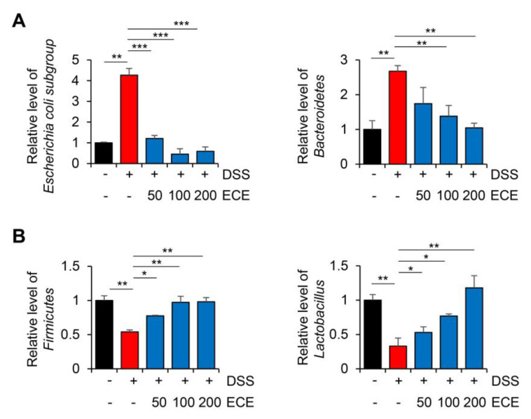
To determine whether ECE could modulate the distribution of the gut microbiome in DSS-induced colitis, the relative level of intestinal microbiota was determined using cecum 16S rRNA-specific PCR. As shown in Figure 3, in mice with DSS-induced colitis, an imbalance of the gut microbiome was observed with an increase in harmful microbiomes and a decrease in beneficial ones. Compared with the DSS-treated group, the ECE-treated groups showed a decreased abundance of the Escherichia coli subgroup and Bacteroidetes. In addition, the abundance of Firmicutes and Lactobacillus, which had been reduced by DSS, was significantly increased in the ECE-treated groups. These results reveal that ECE could ameliorate intestinal dysbiosis in DSS-induced colitis by modulating the balance between beneficial bacteria and harmful ones.
Figure 3.
Ecklonia cava extract ameliorates gut microbiome imbalance in mice with DSS-induced colitis. DNA was extracted from the cecum of each group and used as a template. Real-time PCR was then performed. The relative abundance of bacterial groups (A); beneficial bacterial group (B); harmful bacterial group) is expressed as a percentage of eubacteria. All p-values were calculated using unpaired two-tailed Student’s t-tests. Results are presented as mean ± SD from at least triplicate samples. *, p < 0.05; **, p < 0.01; ***, p < 0.001.
2.4. Ecklonia cava Extract Restores Stability of Intestinal Barrier
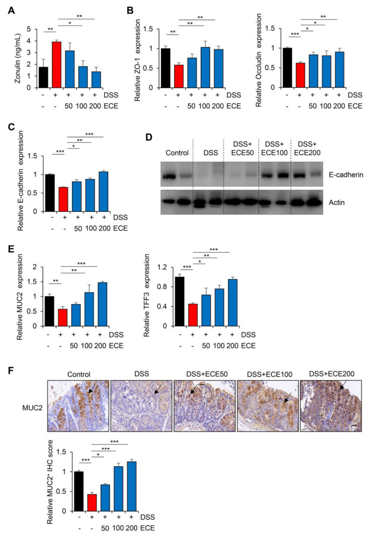
The intestinal barrier functions to maintain mucosal homeostasis by filling in the gap between the intestinal immune system and intestinal microbes [43]. Zonulin, a marker of barrier integrity, can reversibly increase intestinal permeability by modulating tight junctions between cells [44,45]. To investigate the effect of ECE on the intestinal barrier integrity of mice with DSS-induced colitis, the protein concentration of Zonulin was measured in serum with an ELISA kit. As shown in Figure 4A, serum levels of Zonulin, which were increased in the DSS-treated group, were significantly reduced by ECE in dose-dependent manner.
Figure 4.
Ecklonia cava extract restores stability of intestinal barrier: (A) Determination of Zonulin protein level in mouse serum using ELISA kit. (B) mRNA quantification of tight junction proteins (ZO-1 and occludin) in colon tissues was performed using real-time PCR. GAPDH was used as a normalization control. (C,D) Comparison of E-cadherin expression in colon tissue was performed using real-time PCR and Western blot. GAPDH and actin were used as normalization controls, respectively. (E) mRNA quantification of MUC2 and TFF3 in colon tissues was performed using real-time PCR. GAPDH was used as a normalization control. (F) Representative immunohistochemical staining images (top) and IHC quantification with Image J software (version 1.37) (bottom) of MUC2 in colon tissues. Black arrow indicates the stained MUC2 protein. Scale bar = 30 μm. All p-values were calculated using unpaired two-tailed Student’s t-tests. Results are presented as mean ± SD from at least triplicate samples. *, p < 0.05; **, p < 0.01; ***, p < 0.001.
DSS can also elevate intestinal permeability by disrupting epithelial cell tight junctions and adhesive junctions, thereby reducing mucus level [46,47]. Therefore, expression levels of ZO-1 and occludin, which are TJ proteins, and E-cadherin, which is an adhesion molecule, in colon tissues were examined. As expected, decreased mRNA expression levels of TJ proteins and E-cadherin by DSS were restored in the ECE-treated groups (Figure 4B,C). Consistent with restoration of mRNA level, the reduced protein expression of E-cadherin in the DSS-treated group also recovered in the ECE-treated groups (Figure 4D).
Among the mucins constituting the mucus layer that acts as a barrier against harmful substances in the intestine, the recovery of the expression level of MUC2, which forms a gel only in the colon, is an indicator of improvement in colitis [48]. TFF3 expressed in goblet cells of the colon is known to protect the mucous membrane from damage and stabilize the mucosal layer [49]. Thus, the expression levels of MUC2 and TFF3 in colon tissues were determined by real-time PCR and IHC staining, respectively. As shown in Figure 4E, the expression levels of MUC2 and TFF3, which are related to MUC2 secretion, were markedly restored by ECE treatment. Moreover, IHC staining showed that the protein expression of MUC2 was decreased in the DSS-treated group, whereas its level in the ECE-treated group recovered to a level similar to that in the control group (Figure 4F). Taken together, these results suggest that the preventive effect of ECE on the intestinal epithelium might be derived from improvements in barrier function through a restoration of mucosal protection-related genes damaged by DSS-induced colitis.
2.5. Ecklonia cava Extract Suppresses the Pathological Inflammatory Signaling
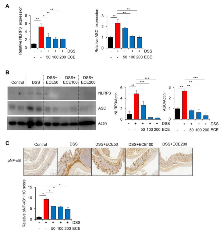
The NLRP3 inflammasome is an intracellular complex that can induce inflammation in IBD. Its expression is increased by DSS [50,51]. In addition, it has been reported that the NLRP3 inflammasome is inhibited by ECE in non-alcoholic fatty liver disease and muscle atrophy [52,53]. To determine whether ECE could affect the NLRP3 inflammasome pathway in DSS-induced colitis, we measured NLRP3 activation in colon tissues using qRT-PCR and Western blot, respectively. The results showed that the expression levels of NLRP3 and ASC mRNAs and proteins were increased in the DSS-treated group, but attenuated in the ECE-treated groups (Figure 5A,B).
Figure 5.
Ecklonia cava extract suppresses NLRP3 inflammasome and NF-κB pathway. (A) mRNA quantification of NLRP3 and ASC in colon tissue was checked using real time PCR. GAPDH was used as a normalization control. (B) Protein expression levels of NLRP3 and ASC in colon tissues were evaluated by Western blot (top). Intensity of Western blot band was quantified using ImageJ (bottom). Actin was used as normalization control. (C) Representative images of pNF-κB immunostaining (top) in colon tissues are presented and relative staining levels (bottom) are quantified using ImageJ software (version 1.37). Scale bar = 100 μm. All p-values were calculated using unpaired two-tailed Student’s t-tests. Results are presented as mean ± SD from at least triplicate samples. *, p < 0.05; **, p < 0.01; ***, p < 0.001.
Next, we confirmed activation of NF-κB, which increased NLRP3 expression, through IHC analysis. As shown in Figure 5C, phosphorylation of NF-κB, which was increased in the DSS-treated group, was dramatically decreased by ECE in a dose-dependent manner. These data suggest that ECE can prevent colitis-induced changes in intestinal permeability, microbiota distribution, and inflammatory markers by modulating the activity of upstream NLRP3 and NF-κB mediators.
3. Discussion
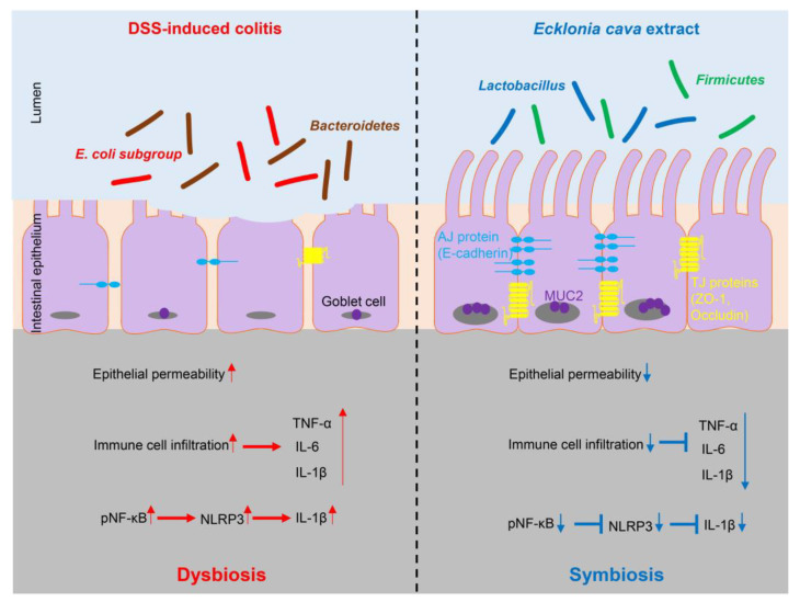
In our study, pretreatment with ECE alleviated the severity of colitis by improving colon shortening, reducing spleen weight gain, and mitigating histological damage in DSS-induced colitis (Figure 1). The balance between pro-inflammatory cytokines, including IL-6, IL-1β, and TNF-α, usually plays a crucial pathological role in colitis by mediating inflammatory responses [32]. We observed a significant reduction in the increase in pro-inflammatory cytokines induced by DSS in ECE-treated mice. Conversely, serum levels of IL-10, an anti-inflammatory cytokine acting as a negative regulator in colitis, were increased by ECE. Additionally, T cell and neutrophil infiltration were notably reduced in ECE-treated mice (Figure 2). The gut microbiome significantly influences gut health, and mucosal-associated bacteria directly impact the integrity of the intestinal epithelial barrier layer by increasing the mucus layer’s thickness and promoting intestinal barrier repair. Several studies report that an imbalance of commensal bacteria is closely associated with the development of various diseases such as IBD [38,39,40]. Our results revealed an increased abundance of colitis-associated Escherichia coli subgroup and Bacteroidetes species, alongside a relative decrease in Firmicutes and Lactobacillus (Figure 3). Tight junctions are pivotal in maintaining the integrity of the intestinal epithelial barrier, crucial for intestinal homeostasis. A disruption of tight junctions and epithelial permeability are linked with IBD progression [44]. Pretreatment with ECE reduced the concentration of Zonulin, a factor increasing barrier permeability, and restored the expression of tight junction proteins (ZO-1, occludin) (Figure 4A,B). The mucus layer covering the mucosal surface of the intestinal lumen, a protective gel-like substance composed of mucin secreted by goblet cells, is associated with colitis development when disrupted [48]. Our experiment revealed a significant reduction in MUC2 expression in the DSS-treated group, which was restored in the ECE-treated groups (Figure 4F). The NLRP3 inflammasome significantly contributes to the onset and progression of IBD. Overexpression of the NLRP3 inflammasome exacerbates colitis and plays a pivotal role in intestinal inflammation in DSS-induced colitis [50,51]. Our data exhibited NLRP3 activation after DSS-induced colitis; however, ECE pretreatment reduced NLRP3 expression and further suppressed NF-κB activation, a prerequisite for NLRP3 activation (Figure 5). In summary, our study elucidated the preventive efficacy of ECE in the context of IBD using a DSS-induced colitis mouse model. ECE pretreatment resulted in a reduced inflammatory response attributed to the downregulation of NLRP3/NF-κB signaling. Furthermore, ECE demonstrated the capacity to enhance both barrier function and microbiome homeostasis (Figure 6).
Figure 6.
Protective effect of Ecklonia cava extract on DSS-induced colitis. In colitis, an imbalance of microorganisms can lead to invasion of harmful bacteria and loss of proteins involved in barrier integrity (such as ZO-1 and E-cadherin) and increase epithelial permeability. In addition, upregulation of inflammatory cytokines can lead to infiltration of immune cells and an increase in IL-1β secretion by NLRP3 inflammasome activation. In contrast, preventive administration of ECE can promote the recovery of beneficial bacteria and improve intestinal bacteria imbalance. ECE can prevent loss of TJ and AJ proteins and increase expression of MUC2, consequently reducing epithelial permeability. Furthermore, ECE can suppress the expression of inflammatory cytokines and inhibit NLRP3 inflammasome activation. Taken together, these results demonstrate that ECE can maintain intestinal homeostasis by changing the intestinal microenvironment into a highly immune-enhanced one. Red arrows indicate the increase or activation of gene expression or pathway and blue arrows indicate the decrease or suppression of gene expression or pathway.
IBD is a chronic digestive disease accompanied by recurrent inflammation due to complex causes such as genetic, microbial, and environmental factors. Its prevalence is rapidly increasing worldwide [54]. Currently, conventional drugs used to treat IBD encompass anti-inflammatory drugs, immunosuppressants, and glucocorticoids. Although anti-TNF-α drugs have shown high efficiency in IBD management, they are associated with the risk of infectious complications and allergic reactions. In addition, their efficacy may wane over time [55]. Oral 5-aminosalicylic acid-based drugs such as olsalazine, sulfasalazine, balsalazide, and mesalazine can also cause nausea and headaches, while corticosteroids are linked to adverse effects such as hypertension, exacerbation of gastric ulcers, and osteoporosis [56]. In parallel, advanced therapeutic modalities, including small-molecule drugs with economical profiles, convenient administration, and biotherapeutics with heightened effectiveness driven by specific mechanisms, are under development. However, these interventions are not devoid of undesirable side effects [57]. Considering these serious side effects, finding new sources to improve clinical symptoms of IBD is still essential. Notably, IBD patients experience compromised quality of life. They are burdened by inflammatory phenotypes, prompting comprehensive exploration into adjunctive therapies such as probiotics, dietary interventions, polyphenols, and microbial metabolites [58]. A promising avenue involves the investigation of natural compounds capable of expediting restoration of the intestinal mucosal layer and normalizing the gut microbiome. Achieving these involves impeding leukocyte infiltration into inflamed intestinal mucosa and curtailing the secretion of inflammatory cytokines, thus presenting innovative approaches to address IBD pathogenesis [59].
The gut microbiome is a key factor in gut health. Mucosal-associated bacteria are directly related to the integrity of the intestinal epithelial barrier layer by increasing the thickness of the mucus layer and promoting intestinal barrier repair. Perturbations in commensal microbial balance have been closely linked to the onset and progression of diverse diseases including IBD [60]. Noteworthy among these is the genus Lactobacillus, comprising beneficial probiotic microorganisms recognized for generating antibiotic compounds that can hinder colonization by pathogenic bacteria while suppressing the production of pro-inflammatory cytokines such as IL-1β, IL-6, and TNF-α. This action orchestrates favorable shifts in the compositions of intestinal flora [61]. Recent discoveries have unveiled the potential of select probiotics as potent anti-inflammatory mediators in IBD, acting by restoring gut microbiota composition to alleviate and prevent intestinal disorders [62].
Presently, the bulk of research concerning intestinal health has predominantly investigated extracts sourced from terrestrial origins. Examples include dietary fibers and extracts derived from compounds such as curcumin and Rhodiola crenulata, which have demonstrated potential in preventing colitis by mitigating inflammatory cytokine secretion and sustaining intestinal barrier integrity [63,64]. In contrast, the oceanic realm, an immense repository of natural components, offers an underexplored frontier. Seaweeds characterized by their polyphenolic, proteinaceous, and polysaccharide constituents have gained scientific attention. Seaweed polyphenols highlighted for their antioxidant and antiviral attributes are subject to ongoing investigation for therapeutic applications. Furthermore, seaweed polysaccharides exhibit diverse physiological functions, including anti-inflammatory, antioxidant, antiviral, and immunomodulatory effects, fueling active research into their potential therapeutic utility [20,65].
Notably, various alga extracts have demonstrated significant efficacy in a colitis mouse model. Extracts from Saccharina japonica, a brown macroalga, suppressed inflammatory signaling caused by DSS, altering intestinal microbial diversity and regulating the intestinal microenvironment, thereby alleviating inflammatory bowel disease symptoms [66]. Additionally, an extract from the green alga Ulva pertusa effectively reduced tissue damage and suppressed an inflammatory response induced by DNBS [67]. These findings underscore the potential of diverse marine alga extracts to manifest anti-inflammatory and anticolitis effects. Moreover, the extensive research substantiating these effects necessitates continued exploration of compounds derived from alga extracts [16].
Dieckol, a major phlorotannin derivative isolated from E. cava and rich in polysaccharides and polyphenols, has undergone extensive study regarding its antiallergic and antioxidant properties [68,69]. In in vitro anti-inflammatory studies, dieckol upregulates hemeoxygenase-1 (HO-1), mediating anti-inflammatory effects in macrophages, and inhibits PI3 K and AKT phosphorylation in colon cancer cells, thereby impeding cancer cell proliferation and migration [70,71]. Furthermore, in a DSS-induced ulcerative colitis model, dieckol’s effectiveness in suppressing inflammation was verified by activating the Nrf2 and HO-1 signaling pathways [72]. Overall, dieckol exhibits anti-inflammatory properties and demonstrates efficacy when administered alone, although its effects may be amplified when combined with other anti-inflammatory drugs.
As previously mentioned, multiple studies have highlighted ECE’s anti-inflammatory properties [29,30,31]. Our findings suggest its potential as an additive in functional foods designed to fortify the intestinal immune environment against inflammatory bowel disease. Additionally, considering the effectiveness of ECE in combination treatments [73], it could be paired with other drugs to enhance its efficacy in inflammatory bowel disease. Moreover, a precise understanding of the active ingredient through synthesizing analogs resembling ECE’s active components could lead to the development of more potent drugs for IBD prevention [22].
4. Materials and Methods
4.1. Reagents
ECE was supplied from Aqua Green Technology Co., Ltd. (Jeju, Republic of Korea) and dissolved in phosphate-buffered saline for experiments. To make ECE, E. cava was washed and dried at room temperature for 48 h. After 50% (v/w) ethanol was added, it was incubated at 85 °C for 12 h. The extract was then filtered, concentrated, sterilized by heating to over 85 °C for 1 h, and then dried for use as described previously [23,74,75]. DSS (36–50 kDa) was purchased from MP Biomedicals (Santa Ana, CA, USA).
4.2. Characterization of Ecklonia cava Extract Using High-Performance Liquid Chromatography Analysis and Toxicity of Ecklonia cava Extract
High-performance liquid chromatography (HPLC) analysis was performed using a Waters HPLC system (Waters, Framingham, MA, USA) equipped with a 2998 photodiode array (PDA) detector, 2707 autosampler, and 515 HPLC pump. The C18 column (4.6 × 100 mm, 4 μm, Agilent, Santa Clara, CA, USA) was used for separation. For the analysis of ECE, solvent A (methanol) was used as the mobile phase and solvent B (water) was used as the stationary phase. The ECE was eluted using a gradient of solvent A and solvent B at a flow rate of 0.3 mL/min. The gradient method was as follows: 0 min 63:37 v/v; 0–5 min 63:37–63:37 v/v; 5–10 min 50:50 v/v; 10–20 min 35:65 v/v; 20–25 min 63:37 v/v; and 25–35 min 63:37 v/v. The absorption spectra were analyzed using a PDA detector at 230 nm range. Pure DK was used as standard marker for quantification (Figure S3).
A 14-day acute oral toxicity test was conducted using the prepared ECE, during which no mortalities were observed among the animals administered the extract. General observations, including body weight, revealed no abnormal symptoms. Furthermore, post-mortem examination did not indicate any abnormalities in any of the administered groups.
4.3. DSS-Induced Colitis Mouse Model
To induce colitis, 6-week-old C57BL/6 male mice (Orient Bio, Seongnam, Republic of Korea) were fed drinking water containing 2.5% DSS for 5 days. They were then fed normal drinking tap water. To prepare a preventive model, mice in the ECE-treated colitis group (50, 100, 200 mg/kg body weight) were preadministered with ECE through an oral gastric gavage 2 weeks before the start of DSS administration, and this was administered daily until sacrifice. ECE doses were determined based on concentrations used in previous studies. The selected dose fell within a range containing the minimum level of dieckol, the active ingredient of ECE [23,35,75,76]. In therapeutic experiments, ECE was administered for 3 weeks after DSS treatment. Body weight was measured every 2–3 days. On days 42 or 28, mice were euthanized using CO2 gas inhalation for 90 s followed by placement in a box containing CO2 gas for 4 min. The colon and spleen were separated, photographed, and weighed. Colon tissue was fixed in formalin for paraffin sectioning and immediately stored in liquid nitrogen for RNA and protein extraction. All mouse experiments adhered to guidelines approved by the Institutional Animal Care and Use Committees of Gachon University (AAALAC-accredited facility, approval number LCDI-2022-0046).
4.4. Western Blot
Total proteins from colon tissues were lysed with NP buffer (50 mM Tris-HCl (pH 7.5), 150 mM NaCl, 5 mM EDTA, 1% NP-40, and a protease/phosphatase inhibitor cocktail) for 30 min on ice and centrifugated at 13,000 rpm for 10 min at 4 °C. Protein concentration was measured using a BCA kit (Thermo Fisher Scientific, Rockford, IL, USA). BSA was used as a standard. Protein samples were boiled for 5 min at 100 °C in sample buffer (60 mM Tris-HCl (pH 6.8), 14.4 mM 2-mercaptoethanol, 2% SDS, 0.05% bromophenol blue, 25% glycerol) and separated by SDS-PAGE. Proteins were transferred to methanol-activated PVDF membranes. Membranes were blocked with 5% skim milk in TBST for 1 h at room temperature. After washing the membrane with TBST, primary antibodies were added and incubated overnight at 4 °C. The following day, blots were incubated with HRP-conjugated goat anti-secondary antibodies for 1 h at room temperature followed by chemiluminescence detection (Atto, Amherst, NY, USA) [77]. Primary antibodies are shown in Table S1.
4.5. Total RNA Isolation and Quantitative Real-Time PCR
Total RNA was isolated using TRIzol reagent (Invitrogen, Carlsbad, CA, USA) and 1 μg mRNA was transcribed to cDNA with random hexamers using PrimeScript 1st strand cDNA synthesis kit (Takara, Japan). SYBR-green Premix Ex-Tag II (Takara, Kyoto, Japan) was used for quantification of cytokine transcripts with real-time quantitative PCR on a Prism 7900HT sequence detection system (Thermo Fisher Scientific). PCR results were analyzed using the comparative 2−ΔΔCT method using GAPDH as a control [78]. Experiments were performed in triplicate and expressed as mean ± standard deviation (SD). Primer sequences used for qRT-PCR are shown in Table S2.
4.6. Bacterial DNA Extraction from Mice Ceca and Microbiota Analysis
After mice were sacrificed, contents of their ceca were immediately placed in liquid nitrogen and frozen at −80 °C until use in experiments. Bacterial DNA was extracted using a DNA stool extraction kit (Qiagen, Valencia, CA, USA) according to the manufacturer’s instructions. Then, 10 ng bacterial DNA was used as a template for PCR [79]. The 16S rRNA of each group was analyzed with bacterial-strain-specific RT-PCR primers. The relative abundance of a bacterial group in the cecal samples was expressed as a ratio of eubacteria. Primer sequences used for RT-PCR are listed in Table S3.
4.7. ELISA for Serum Markers
Concentrations of IL-6, IL-1β, IL-10, CRP (R&D systems, Minneapolis, MN, USA), and Zonulin (MyBioSource, San Diego, CA, USA) in mouse serum samples were evaluated using ELISA kits according to the manufacturer’s protocol [77]. Briefly, blood samples were allowed to clot for 2 h at room temperature to collect serum. After centrifugation at 2000× g for 20 min, serum aliquots were stored at −0 °C before use. ELISA kits used in this study are listed in Table S4.
4.8. Hematoxylin–Eosin Staining and Immunohistochemistry
Paraffin-embedded tissues were sectioned at a thickness of 3 μm and stained with hematoxylin–eosin according to published procedures [80]. Briefly, tissue sections were deparaffinized with xylene, put in antigen retrieval buffer (Tris-EDTA buffer, pH 9.0), and boiled for 5 min. Endogenous peroxidase was blocked using 0.3% hydrogen peroxide. Slides were incubated overnight at 4 °C with primary antibodies diluted in 1% BSA followed by incubation with a secondary antibody for 1 h. For visualization, DAB substrate (Dako, Glostrup, Denmark) was used and counterstained with hematoxylin (Vector Laboratories, Burlingame, CA, USA). The image was captured using a confocal microscope at the Core-facility for Cell to In-vivo imaging of Gachon University and quantified using Image J software (version 1.37, NIH, Bethesda, MD, USA) [81]. Primary antibodies used for staining are shown in Table S1.
4.9. Statistics
Comparisons between groups were determined using Student’s t-test (two-tailed). The error bar represents the SD of the mean. Data are presented as mean ± SD. For all statistical tests, statistical significance was considered when the p-value was less than 0.05.
5. Conclusions
Our study revealed that ECE pretreatment likely suppressed the expression of inflammatory factors and increased intestinal barrier integrity by inhibiting the NLRP3/NF-κB pathway, resulting in a restoration of barrier dysfunction and reduced pathological inflammation. Our findings suggest that ECE can be developed as an intestinal health functional ingredient for preventive purposes or health products in combination with probiotics and other supplements.
Acknowledgments
The authors would like to thank Aqua Green Technology Co., Ltd. (Jeju, Republic of Korea) for providing the ECE.
Supplementary Materials
The following supporting information can be downloaded at https://www.mdpi.com/article/10.3390/molecules28248099/s1: Figure S1: Therapeutic treatment of ECE shows less effective activity in DSS-induced colitis model; Figure S2: ECE efficiently suppresses infiltration of immune cells; Figure S3: Characterization of Ecklonia cava extract using high-performance liquid chromatography analysis. Table S1: List of primary antibody used in this study; Table S2: List of primer sequences used for real-time PCR; Table S3: List of primer sequences used for microbiota analysis; Table S4: List of ELISA kits used in this study.
Author Contributions
Conceptualization, Y.-M.K. and S.H.; Data Acquisition, Y.-M.K. and H.-Y.K.; Methodology, Y.-M.K., H.-Y.K. and J.-T.J.; Resources, J.-T.J.; Writing—Review and Editing, Y.-M.K., H.-Y.K. and S.H.; Funding Acquisition, Y.-M.K. and S.H. All authors have read and agreed to the published version of the manuscript.
Institutional Review Board Statement
This study was conducted according to the guidelines of the Declaration of Helsinki and approved by the Institutional Animal Care and Use Committee of Gachon University (Approval No. LCDI-2022-0046).
Informed Consent Statement
Not applicable.
Data Availability Statement
Data are contained within the article and Supplementary Materials.
Conflicts of Interest
Author Ji-Tae Jang was employed by the company Aqua Green Technology Co., Ltd. The remaining authors declare that the research was conducted in the absence of any commercial or financial relationships that could be construed as a potential conflict of interest.
Funding Statement
This work was supported by a grant (RS-2023-00246400) from the National Research Foundation (NRF); the Basic Science Research Capacity Enhancement Project through Korea Basic Science Institute (2021R1A6C101A432) funded by the Korea government (MSIT); Korea Institute of Marine Science & Technology Promotion (KIMST) funded by the Ministry of Oceans and Fisheries, Korea, (20220273) to S.H.; and a grant (2022R1I1A1A01069333 to Y.-M.K.) of the Basic Science Research Program through the National Research Foundation (NRF) funded by the Ministry of Education, Republic of Korea.
Footnotes
Disclaimer/Publisher’s Note: The statements, opinions and data contained in all publications are solely those of the individual author(s) and contributor(s) and not of MDPI and/or the editor(s). MDPI and/or the editor(s) disclaim responsibility for any injury to people or property resulting from any ideas, methods, instructions or products referred to in the content.
References
- 1.Graham D.B., Xavier R.J. Pathway paradigms revealed from the genetics of inflammatory bowel disease. Nature. 2020;578:527–539. doi: 10.1038/s41586-020-2025-2. [DOI] [PMC free article] [PubMed] [Google Scholar]
- 2.Hodson R. Inflammatory bowel disease. Nature. 2016;540:S97. doi: 10.1038/540S97a. [DOI] [PubMed] [Google Scholar]
- 3.Guan Q. A Comprehensive Review and Update on the Pathogenesis of Inflammatory Bowel Disease. J. Immunol. Res. 2019;2019:7247238. doi: 10.1155/2019/7247238. [DOI] [PMC free article] [PubMed] [Google Scholar]
- 4.McGuckin M.A., Eri R., Simms L.A., Florin T.H., Radford-Smith G. Intestinal barrier dysfunction in inflammatory bowel diseases. Inflamm. Bowel Dis. 2009;15:100–113. doi: 10.1002/ibd.20539. [DOI] [PubMed] [Google Scholar]
- 5.Dong J., Liang W., Wang T., Sui J., Wang J., Deng Z., Chen D. Saponins regulate intestinal inflammation in colon cancer and IBD. Pharmacol. Res. 2019;144:66–72. doi: 10.1016/j.phrs.2019.04.010. [DOI] [PubMed] [Google Scholar]
- 6.Geremia A., Arancibia-Cárcamo C.V., Fleming M.P., Rust N., Singh B., Mortensen N.J., Travis S.P., Powrie F. IL-23-responsive innate lymphoid cells are increased in inflammatory bowel disease. J. Exp. Med. 2011;208:1127–1133. doi: 10.1084/jem.20101712. [DOI] [PMC free article] [PubMed] [Google Scholar]
- 7.Richard M.L., Sokol H. The gut mycobiota: Insights into analysis, environmental interactions and role in gastrointestinal diseases. Nat. Rev. Gastroenterol. Hepatol. 2019;16:331–345. doi: 10.1038/s41575-019-0121-2. [DOI] [PubMed] [Google Scholar]
- 8.Gadaleta R.M., van Erpecum K.J., Oldenburg B., Willemsen E.C., Renooij W., Murzilli S., Klomp L.W., Siersema P.D., Schipper M.E., Danese S., et al. Farnesoid X receptor activation inhibits inflammation and preserves the intestinal barrier in inflammatory bowel disease. Gut. 2011;60:463–472. doi: 10.1136/gut.2010.212159. [DOI] [PubMed] [Google Scholar]
- 9.Bhat A.A., Uppada S., Achkar I.W., Hashem S., Yadav S.K., Shanmugakonar M., Al-Naemi H.A., Haris M., Uddin S. Tight Junction Proteins and Signaling Pathways in Cancer and Inflammation: A Functional Crosstalk. Front. Physiol. 2018;9:1942. doi: 10.3389/fphys.2018.01942. [DOI] [PMC free article] [PubMed] [Google Scholar]
- 10.Ramos G.P., Papadakis K.A. Mechanisms of Disease: Inflammatory Bowel Diseases. Mayo Clin. Proc. 2019;94:155–165. doi: 10.1016/j.mayocp.2018.09.013. [DOI] [PMC free article] [PubMed] [Google Scholar]
- 11.Engevik M.A., Herrmann B., Ruan W., Engevik A.C., Engevik K.A., Ihekweazu F., Shi Z., Luck B., Chang-Graham A.L., Esparza M., et al. Bifidobacterium dentium-derived y-glutamylcysteine suppresses ER-mediated goblet cell stress and reduces TNBS-driven colonic inflammation. Gut Microbes. 2021;13:1902717. doi: 10.1080/19490976.2021.1902717. [DOI] [PMC free article] [PubMed] [Google Scholar]
- 12.Mao L., Kitani A., Strober W., Fuss I.J. The Role of NLRP3 and IL-1β in the Pathogenesis of Inflammatory Bowel Disease. Front. Immunol. 2018;9:2566. doi: 10.3389/fimmu.2018.02566. [DOI] [PMC free article] [PubMed] [Google Scholar]
- 13.Nishida A., Inoue R., Inatomi O., Bamba S., Naito Y., Andoh A. Gut microbiota in the pathogenesis of inflammatory bowel disease. Clin. J. Gastroenterol. 2018;11:1–10. doi: 10.1007/s12328-017-0813-5. [DOI] [PubMed] [Google Scholar]
- 14.Triantafyllidi A., Xanthos T., Papalois A., Triantafillidis J.K. Herbal and plant therapy in patients with inflammatory bowel disease. Ann. Gastroenterol. 2015;28:210–220. [PMC free article] [PubMed] [Google Scholar]
- 15.Catalan-Serra I., Brenna Ø. Immunotherapy in inflammatory bowel disease: Novel and emerging treatments. Hum. Vaccines Immunother. 2018;14:2597–2611. doi: 10.1080/21645515.2018.1461297. [DOI] [PMC free article] [PubMed] [Google Scholar]
- 16.Liyanage N.M., Nagahawatta D.P., Jayawardena T.U., Jeon Y.J. The Role of Seaweed Polysaccharides in Gastrointestinal Health: Protective Effect against Inflammatory Bowel Disease. Life. 2023;13:1026. doi: 10.3390/life13041026. [DOI] [PMC free article] [PubMed] [Google Scholar]
- 17.Nagahawatta D.P., Liyanage N.M., Jayawardhana H., Lee H.G., Jayawardena T.U., Jeon Y.J. Anti-Fine Dust Effect of Fucoidan Extracted from Ecklonia maxima Laves in Macrophages via Inhibiting Inflammatory Signaling Pathways. Mar. Drugs. 2022;20:413. doi: 10.3390/md20070413. [DOI] [PMC free article] [PubMed] [Google Scholar]
- 18.Liyanage N.M., Lee H.G., Nagahawatta D.P., Jayawardhana H., Ryu B., Jeon Y.J. Characterization and therapeutic effect of Sargassum coreanum fucoidan that inhibits lipopolysaccharide-induced inflammation in RAW 264.7 macrophages by blocking NF-κB signaling. Pt AInt. J. Biol. Macromol. 2022;223:500–510. doi: 10.1016/j.ijbiomac.2022.11.047. [DOI] [PubMed] [Google Scholar]
- 19.Lajili S., Ammar H.H., Mzoughi Z., Amor H.B.H., Muller C.D., Majdoub H., Bouraoui A. Characterization of sulfated polysaccharide from Laurencia obtusa and its apoptotic, gastroprotective and antioxidant activities. Int. J. Biol. Macromol. 2019;126:326–336. doi: 10.1016/j.ijbiomac.2018.12.089. [DOI] [PubMed] [Google Scholar]
- 20.Wang Y., Xing M., Cao Q., Ji A., Liang H., Song S. Biological Activities of Fucoidan and the Factors Mediating Its Therapeutic Effects: A Review of Recent Studies. Mar. Drugs. 2019;17:183. doi: 10.3390/md17030183. [DOI] [PMC free article] [PubMed] [Google Scholar]
- 21.Park S.K., Kang J.Y., Kim J.M., Kim H.J., Heo H.J. Ecklonia cava Attenuates PM2.5-Induced Cognitive Decline through Mitochondrial Activation and Anti-Inflammatory Effect. Mar. Drugs. 2021;19:131. doi: 10.3390/md19030131. [DOI] [PMC free article] [PubMed] [Google Scholar]
- 22.Wijesinghe W.A., Jeon Y.J. Exploiting biological activities of brown seaweed Ecklonia cava for potential industrial applications: A review. Int. J. Food Sci. Nutr. 2012;63:225–235. doi: 10.3109/09637486.2011.619965. [DOI] [PubMed] [Google Scholar]
- 23.Byun K.A., Oh S., Yang J.Y., Lee S.Y., Son K.H., Byun K. Ecklonia cava extracts decrease hypertension-related vascular calcification by modulating PGC-1α and SOD2. Biomed. Pharmacother. 2022;153:113283. doi: 10.1016/j.biopha.2022.113283. [DOI] [PubMed] [Google Scholar]
- 24.Oh S., Son M., Choi J., Choi C.H., Park K.Y., Son K.H., Byun K. Phlorotannins from Ecklonia cava Attenuates Palmitate-Induced Endoplasmic Reticulum Stress and Leptin Resistance in Hypothalamic Neurons. Mar. Drugs. 2019;17:570. doi: 10.3390/md17100570. [DOI] [PMC free article] [PubMed] [Google Scholar]
- 25.Son M., Oh S., Lee H.S., Ryu B., Jiang Y., Jang J.T., Jeon Y.J., Byun K. Pyrogallol-Phloroglucinol-6,6’-Bieckol from Ecklonia cava Improved Blood Circulation in Diet-Induced Obese and Diet-Induced Hypertension Mouse Models. Mar. Drugs. 2019;17:272. doi: 10.3390/md17050272. [DOI] [PMC free article] [PubMed] [Google Scholar]
- 26.Yang H.K., Jung M.H., Avunje S., Nikapitiya C., Kang S.Y., Ryu Y.B., Lee W.S., Jung S.J. Efficacy of algal Ecklonia cava extract against viral hemorrhagic septicemia virus (VHSV) Fish. Shellfish Immunol. 2018;72:273–281. doi: 10.1016/j.fsi.2017.10.044. [DOI] [PubMed] [Google Scholar]
- 27.Jung J.I., Kim S., Baek S.M., Choi S.I., Kim G.H., Imm J.Y. Ecklonia cava Extract Exerts Anti-Inflammatory Effect in Human Gingival Fibroblasts and Chronic Periodontitis Animal Model by Suppression of Pro-Inflammatory Cytokines and Chemokines. Foods. 2021;10:1656. doi: 10.3390/foods10071656. [DOI] [PMC free article] [PubMed] [Google Scholar]
- 28.Jo S.L., Yang H., Jeong K.J., Lee H.W., Hong E.J. Neuroprotective Effects of Ecklonia cava in a Chronic Neuroinflammatory Disease Model. Nutrients. 2023;15:2007. doi: 10.3390/nu15082007. [DOI] [PMC free article] [PubMed] [Google Scholar]
- 29.Kim S., Choi S.I., Kim G.H., Imm J.Y. Anti-Inflammatory Effect of Ecklonia cava Extract on Porphyromonas gingivalis Lipopolysaccharide-Stimulated Macrophages and a Periodontitis Rat Model. Nutrients. 2019;11:1143. doi: 10.3390/nu11051143. [DOI] [PMC free article] [PubMed] [Google Scholar]
- 30.Son M., Oh S., Lee H.S., Chung D.M., Jang J.T., Jeon Y.J., Choi C.H., Park K.Y., Son K.H., Byun K. Ecklonia Cava Extract Attenuates Endothelial Cell Dysfunction by Modulation of Inflammation and Brown Adipocyte Function in Perivascular Fat Tissue. Nutrients. 2019;11:2795. doi: 10.3390/nu11112795. [DOI] [PMC free article] [PubMed] [Google Scholar]
- 31.Son M., Oh S., Choi J., Jang J.T., Choi C.H., Park K.Y., Son K.H., Byun K. The Phlorotannin-Rich Fraction of Ecklonia cava Extract Attenuated the Expressions of the Markers Related with Inflammation and Leptin Resistance in Adipose Tissue. Int. J. Endocrinol. 2020;2020:9142134. doi: 10.1155/2020/9142134. [DOI] [PMC free article] [PubMed] [Google Scholar]
- 32.Li N., Zhang Y., Nepal N., Li G., Yang N., Chen H., Lin Q., Ji X., Zhang S., Jin S. Dental pulp stem cells overexpressing hepatocyte growth factor facilitate the repair of DSS-induced ulcerative colitis. Stem Cell Res. Ther. 2021;12:30. doi: 10.1186/s13287-020-02098-4. [DOI] [PMC free article] [PubMed] [Google Scholar]
- 33.Ridker P.M. From C-Reactive Protein to Interleukin-6 to Interleukin-1: Moving Upstream To Identify Novel Targets for Atheroprotection. Circ. Res. 2016;118:145–156. doi: 10.1161/CIRCRESAHA.115.306656. [DOI] [PMC free article] [PubMed] [Google Scholar]
- 34.Jialing L., Yangyang G., Jing Z., Xiaoyi T., Ping W., Liwei S., Simin C. Changes in serum inflammatory cytokine levels and intestinal flora in a self-healing dextran sodium sulfate-induced ulcerative colitis murine model. Life Sci. 2020;263:118587. doi: 10.1016/j.lfs.2020.118587. [DOI] [PubMed] [Google Scholar]
- 35.Alkushi A.G., Elazab S.T., Abdelfattah-Hassan A., Mahfouz H., Salem G.A., Sheraiba N.I., Mohamed E.A.A., Attia M.S., El-Shetry E.S., Saleh A.A., et al. Multi-Strain-Probiotic-Loaded Nanoparticles Reduced Colon Inflammation and Orchestrated the Expressions of Tight Junction, NLRP3 Inflammasome and Caspase-1 Genes in DSS-Induced Colitis Model. Pharmaceutics. 2022;14:1183. doi: 10.3390/pharmaceutics14061183. [DOI] [PMC free article] [PubMed] [Google Scholar]
- 36.Bodammer P., Zirzow E., Klammt S., Maletzki C., Kerkhoff C. Alteration of DSS-mediated immune cell redistribution in murine colitis by oral colostral immunoglobulin. BMC Immunol. 2013;14:10. doi: 10.1186/1471-2172-14-10. [DOI] [PMC free article] [PubMed] [Google Scholar]
- 37.Rodrigues-Sousa T., Ladeirinha A.F., Santiago A.R., Carvalheiro H., Raposo B., Alarcão A., Cabrita A., Holmdahl R., Carvalho L., Souto-Carneiro M.M. Deficient production of reactive oxygen species leads to severe chronic DSS-induced colitis in Ncf1/p47phox-mutant mice. PLoS ONE. 2014;9:e97532. doi: 10.1371/journal.pone.0097532. [DOI] [PMC free article] [PubMed] [Google Scholar]
- 38.Kayama H., Okumura R., Takeda K. Interaction Between the Microbiota, Epithelia, and Immune Cells in the Intestine. Annu. Rev. Immunol. 2020;38:23–48. doi: 10.1146/annurev-immunol-070119-115104. [DOI] [PubMed] [Google Scholar]
- 39.Vester-Andersen M.K., Mirsepasi-Lauridsen H.C., Prosberg M.V., Mortensen C.O., Träger C., Skovsen K., Thorkilgaard T., Nøjgaard C., Vind I., Krogfelt K.A., et al. Increased abundance of proteobacteria in aggressive Crohn’s disease seven years after diagnosis. Sci. Rep. 2019;9:13473. doi: 10.1038/s41598-019-49833-3. [DOI] [PMC free article] [PubMed] [Google Scholar]
- 40.Zegarra Ruiz D.F., Kim D.V., Norwood K., Saldana-Morales F.B., Kim M., Ng C., Callaghan R., Uddin M., Chang L.C., Longman R.S., et al. Microbiota manipulation to increase macrophage IL-10 improves colitis and limits colitis-associated colorectal cancer. Gut Microbes. 2022;14:2119054. doi: 10.1080/19490976.2022.2119054. [DOI] [PMC free article] [PubMed] [Google Scholar]
- 41.Zuo T., Lu X.J., Zhang Y., Cheung C.P., Lam S., Zhang F., Tang W., Ching J.Y.L., Zhao R., Chan P.K.S., et al. Gut mucosal virome alterations in ulcerative colitis. Gut. 2019;68:1169–1179. doi: 10.1136/gutjnl-2018-318131. [DOI] [PMC free article] [PubMed] [Google Scholar]
- 42.Sokol H., Leducq V., Aschard H., Pham H.P., Jegou S., Landman C., Cohen D., Liguori G., Bourrier A., Nion-Larmurier I., et al. Fungal microbiota dysbiosis in IBD. Gut. 2017;66:1039–1048. doi: 10.1136/gutjnl-2015-310746. [DOI] [PMC free article] [PubMed] [Google Scholar]
- 43.Dong L., Xie J., Wang Y., Jiang H., Chen K., Li D., Wang J., Liu Y., He J., Zhou J., et al. Mannose ameliorates experimental colitis by protecting intestinal barrier integrity. Nat. Commun. 2022;13:4804. doi: 10.1038/s41467-022-32505-8. [DOI] [PMC free article] [PubMed] [Google Scholar]
- 44.Wang W., Uzzau S., Goldblum S.E., Fasano A. Human zonulin, a potential modulator of intestinal tight junctions. Pt 24J. Cell Sci. 2000;113:4435–4440. doi: 10.1242/jcs.113.24.4435. [DOI] [PubMed] [Google Scholar]
- 45.Yonker L.M., Gilboa T., Ogata A.F., Senussi Y., Lazarovits R., Boribong B.P., Bartsch Y.C., Loiselle M., Rivas M.N., Porritt R.A., et al. Multisystem inflammatory syndrome in children is driven by zonulin-dependent loss of gut mucosal barrier. J. Clin. Investig. 2021;131:e149633. doi: 10.1172/JCI149633. [DOI] [PMC free article] [PubMed] [Google Scholar]
- 46.Hoebler C., Gaudier E., De Coppet P., Rival M., Cherbut C. MUC genes are differently expressed during onset and maintenance of inflammation in dextran sodium sulfate-treated mice. Dig. Dis. Sci. 2006;51:381–389. doi: 10.1007/s10620-006-3142-y. [DOI] [PubMed] [Google Scholar]
- 47.Hernández-Chirlaque C., Aranda C.J., Ocón B., Capitán-Cañadas F., Ortega-González M., Carrero J.J., Suárez M.D., Zarzuelo A., Sánchez de Medina F., Martínez-Augustin O. Germ-free and Antibiotic-treated Mice are Highly Susceptible to Epithelial Injury in DSS Colitis. J. Crohns Colitis. 2016;10:1324–1335. doi: 10.1093/ecco-jcc/jjw096. [DOI] [PubMed] [Google Scholar]
- 48.Boltin D., Perets T.T., Vilkin A., Niv Y. Mucin function in inflammatory bowel disease: An update. J. Clin. Gastroenterol. 2013;47:106–111. doi: 10.1097/MCG.0b013e3182688e73. [DOI] [PubMed] [Google Scholar]
- 49.Pelaseyed T., Bergström J.H., Gustafsson J.K., Ermund A., Birchenough G.M., Schütte A., van der Post S., Svensson F., Rodríguez-Piñeiro A.M., Nyström E.E., et al. The mucus and mucins of the goblet cells and enterocytes provide the first defense line of the gastrointestinal tract and interact with the immune system. Immunol. Rev. 2014;260:8–20. doi: 10.1111/imr.12182. [DOI] [PMC free article] [PubMed] [Google Scholar]
- 50.Zhang B.C., Li Z., Xu W., Xiang C.H., Ma Y.F. Luteolin alleviates NLRP3 inflammasome activation and directs macrophage polarization in lipopolysaccharide-stimulated RAW264.7 cells. Am. J. Transl. Res. 2018;10:265–273. [PMC free article] [PubMed] [Google Scholar]
- 51.Zhang Y., Tao M., Chen C., Zhao X., Feng Q., Chen G., Fu Y. BAFF Blockade Attenuates DSS-Induced Chronic Colitis via Inhibiting NLRP3 Inflammasome and NF-κB Activation. Front. Immunol. 2022;13:783254. doi: 10.3389/fimmu.2022.783254. [DOI] [PMC free article] [PubMed] [Google Scholar]
- 52.Oh S., Son M., Byun K.A., Jang J.T., Choi C.H., Son K.H., Byun K. Attenuating Effects of Dieckol on High-Fat Diet-Induced Nonalcoholic Fatty Liver Disease by Decreasing the NLRP3 Inflammasome and Pyroptosis. Mar. Drugs. 2021;19:318. doi: 10.3390/md19060318. [DOI] [PMC free article] [PubMed] [Google Scholar]
- 53.Oh S., Yang J., Park C., Son K., Byun K. Dieckol Attenuated Glucocorticoid-Induced Muscle Atrophy by Decreasing NLRP3 Inflammasome and Pyroptosis. Int. J. Mol. Sci. 2021;22:8057. doi: 10.3390/ijms22158057. [DOI] [PMC free article] [PubMed] [Google Scholar]
- 54.Ng S.C., Shi H.Y., Hamidi N., Underwood F.E., Tang W., Benchimol E.I., Panaccione R., Ghosh S., Wu J.C.Y., Chan F.K.L., et al. Worldwide incidence and prevalence of inflammatory bowel disease in the 21st century: A systematic review of population-based studies. Lancet. 2017;390:2769–2778. doi: 10.1016/S0140-6736(17)32448-0. [DOI] [PubMed] [Google Scholar]
- 55.Stallhofer J., Guse J., Kesselmeier M., Grunert P.C., Lange K., Stalmann R., Eckardt V., Stallmach A. Immunomodulator comedication promotes the reversal of anti-drug antibody-mediated loss of response to anti-TNF therapy in inflammatory bowel disease. Int. J. Color. Dis. 2023;38:54. doi: 10.1007/s00384-023-04349-1. [DOI] [PMC free article] [PubMed] [Google Scholar]
- 56.Baumgart D.C., Le Berre C. Newer Biologic and Small-Molecule Therapies for Inflammatory Bowel Disease. N. Engl. J. Med. 2021;385:1302–1315. doi: 10.1056/NEJMra1907607. [DOI] [PubMed] [Google Scholar]
- 57.Liu J., Di B., Xu L.L. Recent advances in the treatment of IBD: Targets, mechanisms and related therapies. Cytokine Growth Factor. Rev. 2023;71–72:1–12. doi: 10.1016/j.cytogfr.2023.07.001. [DOI] [PubMed] [Google Scholar]
- 58.Godala M., Gaszyńska E., Zatorski H., Małecka-Wojciesko E. Dietary Interventions in Inflammatory Bowel Disease. Nutrients. 2022;14:4261. doi: 10.3390/nu14204261. [DOI] [PMC free article] [PubMed] [Google Scholar]
- 59.Ferreira S.S., Passos C.P., Madureira P., Vilanova M., Coimbra M.A. Structure-function relationships of immunostimulatory polysaccharides: A review. Carbohydr. Polym. 2015;132:378–396. doi: 10.1016/j.carbpol.2015.05.079. [DOI] [PubMed] [Google Scholar]
- 60.Li Y.Y., Cui Y., Dong W.R., Liu T.T., Zhou G., Chen Y.X. Terminalia bellirica Fruit Extract Alleviates DSS-Induced Ulcerative Colitis by Regulating Gut Microbiota, Inflammatory Mediators, and Cytokines. Molecules. 2023;28:5783. doi: 10.3390/molecules28155783. [DOI] [PMC free article] [PubMed] [Google Scholar]
- 61.Cai Y., Liu W., Lin Y., Zhang S., Zou B., Xiao D., Lin L., Zhong Y., Zheng H., Liao Q., et al. Compound polysaccharides ameliorate experimental colitis by modulating gut microbiota composition and function. J. Gastroenterol. Hepatol. 2019;34:1554–1562. doi: 10.1111/jgh.14583. [DOI] [PubMed] [Google Scholar]
- 62.Niu W., Yang F., Fu Z., Dong Y., Zhang Z., Ju J. The role of enteric dysbacteriosis and modulation of gut microbiota in the treatment of inflammatory bowel disease. Microb. Pathog. 2022;165:105381. doi: 10.1016/j.micpath.2021.105381. [DOI] [PubMed] [Google Scholar]
- 63.Ananthakrishnan A.N., Khalili H., Konijeti G.G., Higuchi L.M., de Silva P., Korzenik J.R., Fuchs C.S., Willett W.C., Richter J.M., Chan A.T. A prospective study of long-term intake of dietary fiber and risk of Crohn’s disease and ulcerative colitis. Gastroenterology. 2013;145:970–977. doi: 10.1053/j.gastro.2013.07.050. [DOI] [PMC free article] [PubMed] [Google Scholar]
- 64.Wang Y., Tao H., Huang H., Xiao Y., Wu X., Li M., Shen J., Xiao Z., Zhao Y., Du F., et al. The dietary supplement Rhodiola crenulata extract alleviates dextran sulfate sodium-induced colitis in mice through anti-inflammation, mediating gut barrier integrity and reshaping the gut microbiome. Food Funct. 2021;12:3142–3158. doi: 10.1039/D0FO03061A. [DOI] [PubMed] [Google Scholar]
- 65.Nagaoka M., Shibata H., Kimura-Takagi I., Hashimoto S., Aiyama R., Ueyama S., Yokokura T. Anti-ulcer effects and biological activities of polysaccharides from marine algae. Biofactors. 2000;12:267–274. doi: 10.1002/biof.5520120140. [DOI] [PubMed] [Google Scholar]
- 66.Lu K., Liu L., Lin P., Dong X., Ni L., Che H., Xie W. Saccharina japonica Ethanol Extract Ameliorates Dextran Sulfate Sodium-Induced Colitis via Reshaping Intestinal Microenvironment and Alleviating Inflammatory Response. Foods. 2023;12:1671. doi: 10.3390/foods12081671. [DOI] [PMC free article] [PubMed] [Google Scholar]
- 67.Ardizzone A., Filippone A., Mannino D., Scuderi S.A., Casili G., Lanza M., Cucinotta L., Campolo M., Esposito E. Ulva pertusa, a Marine Green Alga, Attenuates DNBS-Induced Colitis Damage via NF-κB/Nrf2/SIRT1 Signaling Pathways. J. Clin. Med. 2022;11:4301. doi: 10.3390/jcm11154301. [DOI] [PMC free article] [PubMed] [Google Scholar]
- 68.Sugiura Y., Usui M., Katsuzaki H., Imai K., Tanaka R., Matsushita T., Miyata M. Dieckol isolated from a brown alga, Eisenia nipponica, suppresses ear swelling from allergic inflammation in mouse. J. Food Biochem. 2021;45:e13659. doi: 10.1111/jfbc.13659. [DOI] [PubMed] [Google Scholar]
- 69.Pyeon D.B., Lee S.E., Yoon J.W., Park H.J., Park C.O., Kim S.H., Oh S.H., Lee D.G., Kim E.Y., Park S.P. The antioxidant dieckol reduces damage of oxidative stress-exposed porcine oocytes and enhances subsequent parthenotes embryo development. Mol. Reprod. Dev. 2021;88:349–361. doi: 10.1002/mrd.23466. [DOI] [PubMed] [Google Scholar]
- 70.Yayeh T., Im E.J., Kwon T.H., Roh S.S., Kim S., Kim J.H., Hong S.B., Cho J.Y., Park N.H., Rhee M.H. Hemeoxygenase 1 partly mediates the anti-inflammatory effect of dieckol in lipopolysaccharide stimulated murine macrophages. Int. Immunopharmacol. 2014;22:51–58. doi: 10.1016/j.intimp.2014.06.009. [DOI] [PubMed] [Google Scholar]
- 71.Dai W., Dai Y.G., Ren D.F., Zhu D.W. Dieckol, a natural polyphenolic drug, inhibits the proliferation and migration of colon cancer cells by inhibiting PI3K, AKT, and mTOR phosphorylation. J. Biochem. Mol. Toxicol. 2023;37:e23313. doi: 10.1002/jbt.23313. [DOI] [PubMed] [Google Scholar]
- 72.Zhu X., Sun Y., Zhang Y., Su X., Luo C., Alarifi S., Yang H. Dieckol alleviates dextran sulfate sodium-induced colitis via inhibition of inflammatory pathway and activation of Nrf2/HO-1 signaling pathway. Environ. Toxicol. 2021;36:782–788. doi: 10.1002/tox.23080. [DOI] [PubMed] [Google Scholar]
- 73.Lee S.G., Park C.H., Kang H. Effect of E. cava and C. indicum Complex Extract on Phorbol 12-Myristate 13-Acetate (PMA) Stimulated Inflammatory Response in Human Pulmonary Epithelial Cells and Particulate Matter (PM)2.5-Induced Pulmonary Inflammation in Mice. Pharmaceutics. 2023;15:2621. doi: 10.3390/pharmaceutics15112621. [DOI] [PMC free article] [PubMed] [Google Scholar]
- 74.Son M., Oh S., Choi J., Jang J.T., Son K.H., Byun K. Attenuating Effects of Dieckol on Hypertensive Nephropathy in Spontaneously Hypertensive Rats. Int. J. Mol. Sci. 2021;22:4230. doi: 10.3390/ijms22084230. [DOI] [PMC free article] [PubMed] [Google Scholar]
- 75.Oh S., Yang J.Y., Park C.H., Son K.H., Byun K. Dieckol Reduces Muscle Atrophy by Modulating Angiotensin Type II Type 1Receptor and NADPH Oxidase in Spontaneously Hypertensive Rats. Antioxidants. 2021;10:1561. doi: 10.3390/antiox10101561. [DOI] [PMC free article] [PubMed] [Google Scholar]
- 76.Oh S., Shim M., Son M., Jang J.T., Son K.H., Byun K. Attenuating Effects of Dieckol on Endothelial Cell Dysfunction via Modulation of Th17/Treg Balance in the Intestine and Aorta of Spontaneously Hypertensive Rats. Antioxidants. 2021;10:298. doi: 10.3390/antiox10020298. [DOI] [PMC free article] [PubMed] [Google Scholar]
- 77.Hyun J., Ryu B., Oh S., Chung D.M., Seo M., Park S.J., Byun K., Jeon Y.J. Reversibility of sarcopenia by Ishige okamurae and its active derivative diphloroethohydroxycarmalol in female aging mice. Biomed. Pharmacother. 2022;152:113210. doi: 10.1016/j.biopha.2022.113210. [DOI] [PubMed] [Google Scholar]
- 78.Lee J., Oh A.R., Lee H.Y., Moon Y.A., Lee H.J., Cha J.Y. Deletion of KLF10 Leads to Stress-Induced Liver Fibrosis upon High Sucrose Feeding. Int. J. Mol. Sci. 2020;22:331. doi: 10.3390/ijms22010331. [DOI] [PMC free article] [PubMed] [Google Scholar]
- 79.Jang J., Hwang S., Oh A.R., Park S., Yaseen U., Kim J.G., Park S., Jung Y., Cha J.Y. Fructose malabsorption in ChREBP-deficient mice disrupts the small intestine immune microenvironment and leads to diarrhea-dominant bowel habit changes. Inflamm. Res. 2023;72:769–782. doi: 10.1007/s00011-023-01707-1. [DOI] [PubMed] [Google Scholar]
- 80.Baek M.O., Cho H.J., Min D.S., Choi C.S., Yoon M.S. Self-transducible LRS-UNE-L peptide enhances muscle regeneration. J. Cachexia Sarcopenia Muscle. 2022;13:1277–1288. doi: 10.1002/jcsm.12947. [DOI] [PMC free article] [PubMed] [Google Scholar]
- 81.Oh S., Rho N.K., Byun K.A., Yang J.Y., Sun H.J., Jang M., Kang D., Son K.H., Byun K. Combined Treatment of Monopolar and Bipolar Radiofrequency Increases Skin Elasticity by Decreasing the Accumulation of Advanced Glycated End Products in Aged Animal Skin. Int. J. Mol. Sci. 2022;23:2993. doi: 10.3390/ijms23062993. [DOI] [PMC free article] [PubMed] [Google Scholar]
Associated Data
This section collects any data citations, data availability statements, or supplementary materials included in this article.
Supplementary Materials
Data Availability Statement
Data are contained within the article and Supplementary Materials.


