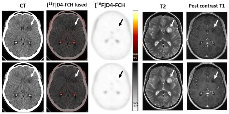
Figure 2.
[18F]D4–FCH PET/CT in a left thalamic low-grade glioma 6 months apart (top row, baseline; bottom row, 6 months later) showing minimal tracer uptake above background in the lesion, stable between scans, high signal on T2–weighted MRI imaging with no significant enhancement post-contrast. (Note physiological activity in the choroid plexus). Stable on continued follow-up 6 years later (not shown).

