ABSTRACT
Genes required for the lysosomal degradation pathway of autophagy play key roles in topologically distinct and physiologically important cellular processes. Some functions of ATG genes are independent of their role in degradative autophagy. One of the first described of these ATG gene-dependent, but degradative autophagy independent, processes is the requirement for a subset of ATG genes in interferon-γ (IFNγ)-induced inhibition of norovirus and Toxoplasma gondii replication. Herein, we identified additional genes that are required for, or that negatively regulate, this innate immune effector pathway. Enzymes in the UFMylation pathway negatively regulated IFNγ-induced inhibition of norovirus replication via effects of Ern1. IFNγ-induced inhibition of norovirus replication required Gate-16 (also termed GabarapL2), Wipi2b, Atg9a, Cul3, and Klhl9 but not Becn1 (encoding Beclin 1), Atg14, Uvrag, or Sqstm1. The phosphatidylinositol-3-phosphate and ATG16L1-binding domains of WIPI2B, as well as the ATG5-binding domain of ATG16L1, were required for IFNγ-induced inhibition of norovirus replication. Other members of the Cul3, Atg8, and Wipi2 gene families were not required, demonstrating exquisite specificity within these gene families for participation in IFNγ action. The generality of some aspects of this mechanism was demonstrated by a role for GATE-16 and WIPI2 in IFNγ-induced control of Toxoplasma gondii infection in human cells. These studies further delineate the genes and mechanisms of an ATG gene-dependent programmable form of cytokine-induced innate intracellular immunity.
IMPORTANCE
Interferon-γ (IFNγ) is a critical mediator of cell-intrinsic immunity to intracellular pathogens. Understanding the complex cellular mechanisms supporting robust interferon-γ-induced host defenses could aid in developing new therapeutics to treat infections. Here, we examined the impact of autophagy genes in the interferon-γ-induced host response. We demonstrate that genes within the autophagy pathway including Wipi2, Atg9, and Gate-16, as well as ubiquitin ligase complex genes Cul3 and Klhl9 are required for IFNγ-induced inhibition of murine norovirus (norovirus hereinafter) replication in mouse cells. WIPI2 and GATE-16 were also required for IFNγ-mediated restriction of parasite growth within the Toxoplasma gondii parasitophorous vacuole in human cells. Furthermore, we found that perturbation of UFMylation pathway components led to more robust IFNγ-induced inhibition of norovirus via regulation of endoplasmic reticulum (ER) stress. Enhancing or inhibiting these dynamic cellular components could serve as a strategy to control intracellular pathogens and maintain an effective immune response.
KEYWORDS: interferons, Toxoplasmsa gondii, autophagy, UFMylation, norovirus
INTRODUCTION
Macroautophagy (autophagy herein) requires formation of an isolation membrane that envelops cytoplasmic materials, organelles, or invading pathogens in a closed double membrane-bound autophagosome. Autophagosomes fuse with lysosomes to facilitate degradation of captured material (1, 2). Autophagy requires a series of autophagy genes (ATG genes), many of which are conserved broadly in evolution. It is now clear that these essential ATG genes are also required for additional topologically distinct cellular processes that have significant physiologic importance (3–5). Many of these autophagy-independent, but ATG gene-dependent, processes occur in myeloid cells. These include essential roles for ATG genes in the anti-microbial action of the key cytokine interferon-γ (IFNγ) (also referred to as type II IFN), the deposition of the ATG8-family proteins on the cytoplasmic surface of phagosomes (2, 6), the fusion of lysosomes to the polarized ruffled membrane of osteoclasts to secrete lysosomal proteases for extracellular degradation of bone (7), the regulation of neutrophilic inflammation during Mycobacterium tuberculosis infection (8), and the inhibition of inflammatory activation of tissue-resident macrophages (9). Of particular note, the role of ATG genes in the actions of IFNγ is fundamentally important to the survival of mammals because this cytokine plays an essential role in triggering cell-intrinsic immunity to intracellular bacteria, viruses, and parasites.
The fact that ATG genes play roles in both autophagy and topologically distinct cellular processes makes it imperative to further define the molecular machinery that is in common to, or distinguishes, these distinct cellular events (10). Here, we focus on IFNγ-induced ATG gene-dependent intracellular immunity to murine norovirus (norovirus herein) and Toxoplasma gondii (T. gondii). Inhibition of intracellular replication of these pathogens requires ATG genes Atg7, Atg5, Atg16l1, Atg12, Atg3, and Atg8 family members (11–19). ATG3, ATG7, and the ATG5-ATG12-ATG16L1 (herein the ATG5-12-16L1 complex) are required for the ubiquitin-like conjugation system of autophagy that conjugates phosphatidyl-ethanolamine (PE) to ATG8 proteins to localize them on membranes (1, 4). However, the topology of the intracellular events triggered by IFNγ is distinct for the two pathogens. T. gondii replicates in a parasitophorous vacuole (PV) sequestered away from the cytoplasm, while norovirus replicates on the cytoplasmic face of membranes. In mice, IFNγ-induced ATG gene-dependent clearance of T. gondii occurs by destruction of the parasitophorous vacuole (11, 14), while in human cells this pathway limits replication but may not eliminate the parasite (16, 20). In contrast, IFNγ inhibits norovirus replication by disrupting the formation and/or function of the membranous replication compartment upon which viral RNA synthesis occurs (12, 17).
Here, we show that an immortalized murine macrophage-like microglial cell line (BV-2 cells) (21) recapitulates ATG gene-dependent events in IFNγ-induced inhibition of norovirus replication as originally described in primary macrophages. We then identified additional positive and negative regulators of this process. Ufc1 and Uba5 negatively regulated IFNγ inhibition of norovirus replication. In contrast, Atg9a, Wipi2, Gate-16, Cul3, and Klhl9 were required for efficient IFNγ-induced inhibition of norovirus replication. The ATG16L1- and phosphatidylinositol 3-phosphate [PtdIns(3)P]-binding domains of WIPI2B, as well as the ATG5-binding domain of ATL16L1, were required for efficient IFNγ-induced inhibition of norovirus replication. WIPI2 and GATE-16 were also required for IFNγ-induced inhibition of T. gondii replication in human cells. In contrast to findings in the norovirus system, SQSTM1 (herein P62) was required for IFNγ-induced inhibition of T. gondii replication, but not for control of replication of norovirus in murine cells. Thus, components of the autophagy machinery outside of the ubiquitin-like conjugation systems involving the ATG5-12-16L1 complex are important components of a cellular immune mechanism used by IFNγ to block intracellular pathogen replication, potentially identifying targets for modulation of this type of immunity. The selective requirement for Gate-16, Wipi2, Cul3, Atg9a, and Klhl9, but not other members of their respective families, demonstrates the specificity of this form of intracellular immunity.
RESULTS
IFNγ inhibits norovirus replication in BV-2 cells in a Stat1- and Irf1-dependent manner
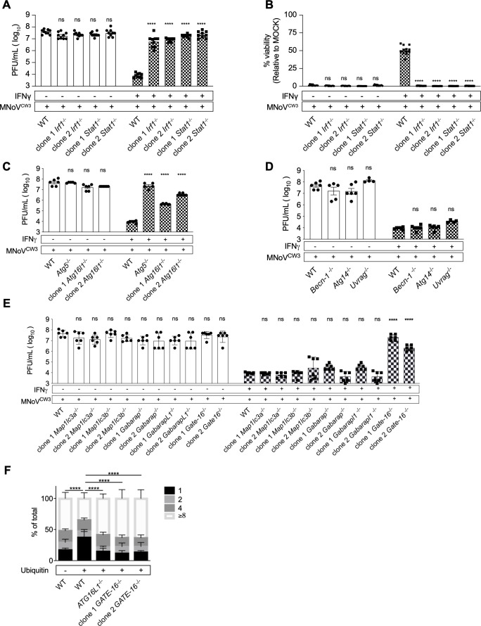
BV-2 cells are permissive for replication of murine norovirus strain MNoVCW3 (22), allowing us to test whether, in these cells, norovirus replication was inhibited by recombinant IFNγ as measured by plaque assay and cellular ATP content as a proxy for cell viability (Fig. S1). Pretreatment of BV-2 cells with IFNγ decreased MNoVCW3 (norovirus hereinafter) replication ~5,000-fold (Fig. 1A) and cytopathicity by 50% (Fig. 1B). Stat1 and Irf1 encode transcription factors essential for IFNγ responses (23) and are required for robust IFNγ-induced inhibition of norovirus replication both in vitro in primary macrophages and in vivo (24–26). To determine whether these transcription factors were required for IFNγ-induced inhibition of norovirus replication in BV-2 cells we generated two independent clonal Stat1 or Irf1 knockout BV-2 cell lines (Stat1−/−, Irf1−/−) (Fig. 1A and B). Throughout this work, we confirmed deletion of genes in cell lines using next generation sequencing (Table S1). IFNγ failed to efficiently inhibit norovirus replication and cytopathicity in either Stat1−/− or Irf1−/− cells (Fig. 1A and B), faithfully replicating the requirement for these genes in primary cells (26).
Fig 1.
Stat1, Irf1 Atg5, Atg16l1, and Gate-16 are required for IFNγ-induced inhibition of norovirus replication in BV-2 cells, Atg14, Becn1, and Uvrag are not; GATE-16 is required for IFNγ-induced growth restriction of T. gondii in HeLa cells. (A) Plaque assay of WT, Irf1−/−, and Stat1−/− BV-2 cells as described in Fig. S1. (B) Viability assay of WT, Irf1−/−, and Stat1−/− BV-2 cells as described in Fig. S1. (C–E) Plaque assay of WT, Atg5−/−, Ag16l1−/−, Atg14−/−, Becn1−/−, Uvrag−/−, and Atg8 family members in BV-2 cells. (F) WT, ATG16L1−/−, and GATE16−/− were treated with IFNγ and infected with T. gondii. Parasites within ubiquitin-positive (+) and ubiquitin-negative (−) vacuoles were counted by immunofluorescence microscopy. Average data pooled from 2 to 3 independent experiments are represented as means ± standard error of the mean (SEM). *P ≤ 0.05, **P ≤ 0.01, ***P ≤ 0.001, and ****P ≤ 0.0001 were considered statistically significant. ns, not significant. P value was determined by two-way analysis of variance (ANOVA) with Dunnett’s multiple comparison test; for T. gondii assay, P value was determined by two-way ANOVA with Tukey’s multiple comparison test.
The ATG5-12-16L1 complex is required for IFNγ-induced inhibition of norovirus replication in BV-2 cells
The ATG5-12-16L1 complex is crucial for IFNγ-induced control of norovirus in primary bone marrow-derived murine macrophages (12). Using Atg5 knockout BV-2 cells (Atg5−/−) (27, 28), we confirmed that Atg5 was required for IFNγ-induced inhibition of norovirus replication (Fig. 1C). To further define the role of the ATG5-12-16L1 complex we generated two independent clonal knockout BV-2 cell lines for Atg16l1 (Atg16l1−/−) (Table S1) (27). IFNγ failed to efficiently inhibit norovirus replication in cells lacking Atg16l1 (Fig. 1C). Deep sequencing confirmed saturating indel frequencies in Atg16l1 clonal knockout cells (Table S1) supporting a role for Atg16l1 in IFNγ inhibition of norovirus replication in these cells.
The ATG genes Becn1, Atg14, and Uvrag are not required for IFNγ-induced inhibition of norovirus replication
Certain upstream components of the autophagy pathway are not required for IFNγ-induced inhibition of norovirus or T. gondii replication (11, 12, 16, 17). To confirm these observations in BV-2 cells, we determined whether IFNγ efficiently inhibits norovirus replication in clonal Atg14−/− BV-2 cells (27, 28) and clonal Becn1−/− cell lines (28) (Fig. S3). Atg14, Becn1, and Uvrag were not required for IFNγ-induced inhibition of norovirus replication (Fig. 1D). We sought to extend these findings using cells lacking the VPS34 kinase which is responsible for PI3P generation by these Class II PI3 kinase complexes, but were unable to isolate viable cells lacking this protein. Additionally, in the setting of viral infection and IFNγ treatment, the drugs wortmannin (29), apilimod (30), and alpelisib (31) proved too toxic to provide interpretable data related to phosphatidylinositol involvement (not shown). A recent report using murine embryonic fibroblasts depleted for VPS34 expression found no evidence of a role for VPS34 in this form of intracellular immunity (32). Together with the data on the role of Stat1, Irf1, Atg5, and Atg16l1, these data support the validity of BV-2 cells as a model to further define mechanisms of IFNγ-induced ATG gene-dependent immunity to norovirus and indicate that the Class III PI3 kinase complexes that include Beclin 1 together with either ATG14 or UVRAG and that are required for degradative autophagy, are not required for this type of intracellular immunity.
Role of Atg8 family members in IFNγ-induced inhibition of norovirus and T. gondii infection
The ATG8 family of proteins plays a key role in multiple aspects of autophagy and ATG gene functions that are independent of degradative autophagy. Evidence has accumulated that individual family members can provide specific functions in different cell biology processes (18, 33). We therefore isolated two independent clones of cells lacking ATG8 family genes Map1lc3a, Map1lc3b, Gabarap, GabarapL1, or Gate-16. Only Gate-16 was required for IFNγ-induced inhibition of norovirus replication (Fig. 1E).
To assess the generality of this finding across pathogens, we analyzed the growth of T. gondii in the parasitophorous vacuole of human cells, using ATG16L1-deficient cells as a control for a gene known to be required for the activity of IFNγ against this pathogen (16). We quantitated T. gondii replication within the PV in parental and two independent GATE-16−/− human HeLa cells (Table S1), using a type III parasite that is susceptible to ATG gene-dependent IFNγ-induced growth control (16, 20). T. gondii multiplies by binary fission with a half-life of ~8 hours, generating vacuoles containing clusters of 1 to ≥8 parasites over 24 hours. A subset of parasitophorous vacuoles becomes labeled by ubiquitin in IFNγ-treated cells (16). Ubiquitin-positive vacuoles are targeted in an ATG gene-dependent manner that impairs replication resulting in decreased numbers of parasites per vacuole (16). We therefore counted parasites per ubiquitin-positive vacuole in wild type and GATE-16−/− HeLa cells (Fig. 1F) (16). In wild-type cells, we did not observe a growth restriction phenotype in PVs lacking ubiquitin (Fig. 1F, left); however, most ubiquitin-positive vacuoles contained only one parasite, indicative of IFNγ-induced growth restriction (Fig. 1F). In the absence of either ATG16L1 [as previously reported, (14, 16)] or GATE-16, IFNγ failed to efficiently restrict parasite growth, and fewer PVs containing one parasite were observed while the majority containing ≥8 parasites (Fig. 1F). Thus, the role of GATE-16 in IFNγ-induced inhibition of intracellular replication extends from a cytoplasmic RNA virus to an intravacuolar apicomplexan parasite.
CRISPR screen for identification of genes involved in IFNγ-induced inhibition of norovirus cytopathicity in BV-2 cells
We used CRISPR screening to identify candidate genes for a role in IFNγ action (Fig. S2). We designed an autophagy sgRNA library containing 1–4 independent guides targeting 695 candidate genes (Table S2). We included 300 guides with no known target as controls (2,979 guides total) (34). Genes were selected using three criteria: (i) present in the autophagy interaction landscape, including the baits used, as defined by Behrends et al. (35); (ii) murine genes related to autophagy using gene ontology (GO) (36, 37); and (iii) murine genes corresponding to human genes related to autophagy using GO (36, 37). The gene encoding CAPRIN1, recently shown to have a role in this form of immunity, was not included in the CRISPR screen (32). To enhance the sensitivity of this CRISPR screen, we targeted the generation of a cell pool with each guide represented by ~2,000 cells (38). After experimental selection (Fig. 2A), we quantified guides by DNA sequencing and determined the significance of differences in guide frequencies in different comparisons using STARS and negative binomial analysis (22, 27, 28, 34, 39) with a threshold of false discovery rate (FDR) < 0.1 to identify genes for further consideration (28).
Fig 2.
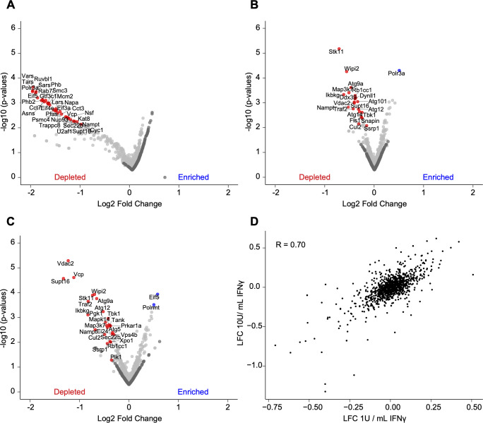
Identification of genes required for viability in passage or IFNγ-treated BV-2 cells. (A) Volcano plot of mock treatment guides enriched or depleted relative to 5 days post-puromycin selection. (B) Volcano plot of guides enriched or depleted after 1 U/mL IFNγ treatment relative to mock treatment. (C) As in (B) for 10 U/mL IFNγ treatment. (D) Average log2 fold change (LFC) of 1 U/mL IFNγ condition versus LFC of 10 U/mL IFNγ condition. Pearson correlations are indicated. For volcano plots, the LFC of all sgRNAs for each gene is plotted against the −log10(P value) for each gene. Blue and red highlighted genes in (A–C) represent a STARS score with FDR < 0.01 and dark gray genes represent non-targeting guides.
Genes required for cell survival in the absence of norovirus infection
The effects of genes required for cell survival or proliferation may obscure the identification of those required for a phenotype, especially when cell death is part of the biology being studied as is the case for norovirus replication and IFNγ treatment (22, 28, 34). We, therefore, identified differences in guide frequency between the original cell library and cells passaged under mock conditions (Fig. 2A; Table S3) as well as between cells passaged under mock conditions compared to those treated with IFNγ at two doses (Fig. 2B and C; Table S4). We analyzed both 1 U/mL and 10 U/mL because the lower dose is non-toxic, while the higher dose alters proliferation and/or cell survival of wild-type BV-2 cells (28, 34). The mock passage did not enrich guides for any gene but decreased guides for 52 genes, suggesting that these are essential genes for the growth or survival of BV-2 cells under these conditions. Among the targeted genes were Cdc37, Adsl, and Cct2. Mutations in ADSL result in the rare autosomal recessive disorder adenylosuccinate lyase deficiency (40), while CCT2 mutations evoke the rare disease Leber congenital amaurosis (41). We compared these candidate essential genes to those identified in Project Achilles (42–44). Three of 52 genes lacked a human homolog and one did not have specific guides in Project Achilles. Therefore, 48 candidate essential genes were considered for this comparison (Table S3). Forty-three of 48 (95%) depleted genes were considered common essential among immortalized cells (Table S3) (42–44), thus validating our screening approach via deprioritizing analysis of genes whose guides were depleted under control conditions.
Identification of genes enriched or depleted in the presence IFNγ
IFNγ treatment at 1 U/mL enriched guides for seven genes (Fig. 2B; Table S4). Escalation of the IFNγ dose to 10 U/mL enriched guides for 20 genes (Fig. 2C; Table S4). Among the targeted genes were Eif5, Polrmt, Eif3a, Polr3a, Eif4g2, and Eif4e (Fig. 2B and C; Table S4). These genes regulate mRNA translation and immune cell activation (45–47). IFNγ treatment at 1 U/mL depleted guides for 25 genes (Fig. 2B; Table S4), and at a dose of 10 U/mL depleted guides for 24 genes (Fig. 2C; Table S4). We observed agreement at the gene level between treatment with 1 U/mL or 10 U/mL IFNγ with a Pearson’s correlation of 0.70 (Fig. 2D). Among the targeted genes were Traf2, Tbk1, Ikbkg, Tank, Ei24, Map3k7, Atg5, and Atg14 (Fig. 2B and C; Table S4). Traf2, Tbk1, Ikbkg, Tank, Ei24, and Map3k7 regulate the tumor necrosis factor receptor signaling pathway (48–50). Consistent with these results, IFNγ-induced cell death in BV-2 cells is mediated by tumor necrosis factor (28). Our results confirmed the critical role of Atg14 and Atg5 in inhibiting IFNγ-induced cell death in BV-2 cells (Fig. 2B; Table S4) (28). Guides for Wipi2, Rb1cc1 (also known as Fip200), Atg9a, Atg101, and Atg12 were also depleted, though these genes were not reported to protect BV-2 cells from IFNγ-induced cell death (Fig. 2B and C; Table S4) (28).
Identification of genes that enhance IFNγ-induced inhibition of norovirus-induced cytopathicity
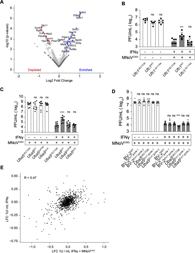
Guides for 18 genes (Fig. 3A; Table S4) were enriched in IFNγ-treated and norovirus-infected cells compared to mock conditions. We observed intermediate agreement of gene-level results, with a Pearson’s correlation of 0.47 between 1 U/mL IFNγ treatment alone compared to 1 U/mL IFNγ + norovirus-infected cells (Fig. 3B). Among the targeted genes were G3bp1, Sptcl1, and Sptcl2 which are required for efficient norovirus replication (22, 51–53). Among the genes identified as candidates for enhancing IFNγ-induced inhibition of norovirus replication were Iqgap1 and Ufc1 (Fig. 3A; Table S4). Deficiency of IQGAP1 in human monocytic cells results in hyperactive type I IFN responses to cytosolic nucleotides (54). Ufc1 and Uba5 encode enzymes in the ubiquitin-like system that covalently conjugates UFM1 to target proteins (UFMylation herein) (27, 55–57). UFMylation is required to maintain ER homeostasis, so in the absence of UFMylation consequent ER stress can lead to overactivation of IFNγ responses (27). We therefore quantified the effects of IFNγ on norovirus replication in cells lacking Ufc1 or Uba5 (Ufc1−/−, Uba5−/−) (27). IFNγ more potently inhibited norovirus replication in these cells (Fig. 3C and D) as shown by diminished potency of IFNγ upon expression of wild-type proteins (Ufc1WT or Uba5WT) but not the enzymatically dead versions of these proteins (Ufc1ΔC116A, Uba5ΔC248A) (Fig. 3C and D) (27). UBA5 contains a UFM1 interaction motif disrupted by the mutations of W340A/L344A (Fig. 3D, Uba5ΔUFIM) (27, 55, 56). Disruption of this motif prevented UBA5 from rescuing the increased IFNγ potency observed in Uba5−/− cells (Fig. 3C). However, deletion of UFM1 itself had no effect on IFNγ-induced control of norovirus infection (Fig. S4). As observed by Balce et al., Ern1 deletion reversed effects on IFNγ potency observed in Uba5−/− cells, consistent with a role for this aspect of the ER stress response in modulating IFNγ action (Fig. 3E) (27).
Fig 3.
Identification of genes required for IFNγ-induced norovirus cytopathicity in BV-2 cells. (A) Volcano plot of guides enriched or depleted after 1 U/mL IFNγ + norovirus infection relative to mock treatment. (B) Plaque assay of Ufc1VECTOR, Ufc1WT, and Ufc1ΔC116A BV-2 cells as described in Fig. S1. (C) Plaque assay of Uba5VECTOR, Uba5WT, Uba5ΔUFIM, and Uba5ΔCA BV-2 cells as described in Fig. S1. (D) Plaque assay of BV-2WT, BV-2Ern1 guide 1, BV-2Ern1 guide 2, Uba5VECTOR, Uba5Ern1 guide 1, and Uba5Ern1 guide 2 cells as described in Fig. S1. (E) Average LFC of 1 U/mL IFNγ condition versus LFC of 1 U/mL IFNγ + norovirus condition. For volcano plots, the average log2 fold change (LFC) of all sgRNAs for each gene is plotted against the −log10(P value) for each gene. Blue and red highlighted genes in (A) represent a STARS score with FDR < 0.01, and dark gray genes represent non-targeting guides. Values in (B–D) represent means ± SEM from 2 to 3 independent experiments. *P ≤ 0.05, **P ≤ 0.01, ***P ≤ 0.001 and ****P ≤ 0.0001 were considered statistically significant. ns, not significant. P value was determined by two-way ANOVA with Dunnett’s multiple comparison test.
Identification of candidate genes required for efficient inhibition of norovirus-induced cytopathicity by IFNγ
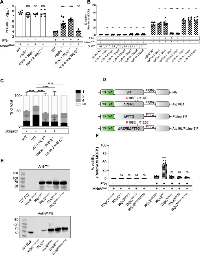
We next compared guide frequencies between mock and 1 U/mL IFNγ + norovirus infection, reasoning that guides decreased in cells surviving norovirus infection of IFNγ-treated cells might have a role in IFNγ-induced inhibition of norovirus replication (Fig. 3A). Guides for 32 genes (Fig. 3A; Table S4) were decreased in frequency in IFNγ + norovirus-infected cells. Among these, the autophagy-related genes Wipi2, Atg9a as well as the ubiquitin ligase Cul3 (below) were identified as candidates for a role in IFNγ-induced ATG gene-dependent innate immunity. To confirm screen findings, we generated one clonal BV-2 cell line lacking Wipi2 (Wipi2−/−) (Table S1) and one non-clonal cell line lacking Wipi2 (Wipi2−/+; cell line 2) (XX targeting of Wipi2, Table S1). We also examined BV-2 cells lacking Atg9a (Atg9a−/−) (Table S1) (27). Wipi2 and Atg9a were required for efficient IFNγ-induced inhibition of norovirus replication (Fig. 4A), indicating that they constitute key components of this form of intracellular innate immunity. This contrasts with a recent report that WIPI2 is not required for IFNγ-induced inhibition of norovirus replication in BV-2 cells (32). While we cannot explain this difference, we report results from multiple different methods to inhibit Wipi2 expression herein (Fig. 4A and B) and performed our studies using 1 U/mL, a non-toxic dose (28, 34) rather than 100 U/mL IFNγ.
Fig 4.
ATG16L1 and PtdIns(3)P-binding domains of WIPI2B are required for IFNγ-induced inhibition of norovirus cytopathicity in BV-2 cells; WIPI2 is required for IFNγ-induced growth restriction of T. gondii in HeLa cells. (A) Plaque assay of WT, Atg9a−/−, Wipi2−/− clonal, and Wipi2−/+ non-clonal knock-out cells as described in Fig. S1. (B) Viability assay of non-clonal BV-2-Cas9 knock-out cells transduced with non-targeting (NT) sgRNAs or sgRNAs targeting Wipi1, Wipi2, Wipi3, or Wipi4 as described in Fig. S1; percentage of the wild-type allele present in each non-clonal cell pool indicated. (C) WIPI2−/− HeLa cells were treated with IFNγ and infected with T. gondii. Parasites within ubiquitin positive (+) and ubiquitin negative (−) vacuoles were counted by immunofluorescence microscopy. (D) Schematic representation of WIPI2B complementation in clone 1 Wipi2−/− BV-2 cells. (E) Western blot detection of TY1 epitope or endogenous WIPI2 in complemented cells. (F) Viability assay of Wipi2VECTOR, Wipi2WT, Wipi2ΔRERE, Wipi2 ΔRERE, and Wipi2ΔRERE ΔFTTG complemented BV-2 cells as described in Fig. S1. Values in (A), (B), and (E) represent means ± SEM from 2 to 3 independent experiments. *P ≤ 0.05, **P ≤ 0.01, ***P ≤ 0.001, and ****P ≤ 0.0001 were considered statistically significant. ns, not significant. P value was determined by two-way ANOVA with Dunnett’s multiple comparison test; for T. gondii assay, P value was determined by two-way ANOVA with Tukey’s multiple comparison test.
To understand the possible specificity of Wipi2, as compared to related family members, in IFNγ-induced control of norovirus infection, we used two independent guide RNAs to generate non-clonal cell lines targeting each Wipi family member [greater than 90% of alleles targeted (Table S1), Fig. 4B]. IFNγ inhibited norovirus infection in Wipi1, Wipi3, and Wipi4 but not Wipi2, mutant cell lines. To determine the generality of the observation that WIPI2 plays a key role in IFNγ-induced intracellular immunity we examined control of T. gondii in two independent clonal cell lines lacking WIPI2. We found that WIPI2 is essential for efficient IFNγ-induced control of T. gondii replication within the PV (Fig. 4C).
Domains of WIPI2B and ATG16L1 required for IFNγ-induced inhibition of norovirus replication
We used mutagenesis to analyze the role of WIPI2 in greater detail because it interacts with ATG16L1 (58, 59), which plays a key role in IFNγ-induced inhibition of norovirus replication (Fig. 4D) (12, 17). Furthermore, it has been reported that the interaction between ATG16L1 and WIPI2 is important for the localization of LC3B to the norovirus replication complex in Hela cells (32). We defined the effects of the R108E/R125E (ΔRERE) mutations that inhibit binding to ATG16L1, the R224T/R225T (ΔFTTG) mutations that inhibit binding to PtdIns(3)P, and the double mutant ΔRERE/ΔFTTG (Fig. 4D) (58). We stably expressed these proteins and wild-type WIPI2B in Wipi2−/− clone 1 cells and assessed protein expression by western blot analysis (Fig. 4E). Probing for an N-terminal epitope tag or endogenous WIPI2 revealed equivalent expression of the different constructs (Fig. 4E). Expression of WIPI2B rescued the capacity of IFNγ to inhibit norovirus infection in Wipi2−/− cells (Fig. 4F). This confirms by complementation that the phenotype in this cell line is due to a lack of WIPI2B. In contrast, neither WIPI2BΔRERE nor WIPI2BDFTTG rescued inhibition of norovirus after IFNγ treatment (Fig. 4F). The double mutant WIPI2BDREREDFTTG protein also failed to restore IFNγ activity (Fig. 4F). Thus, WIPI2B binding to both ATG16L1 and PtdIns(3)P was necessary for immune control of norovirus by IFNγ.
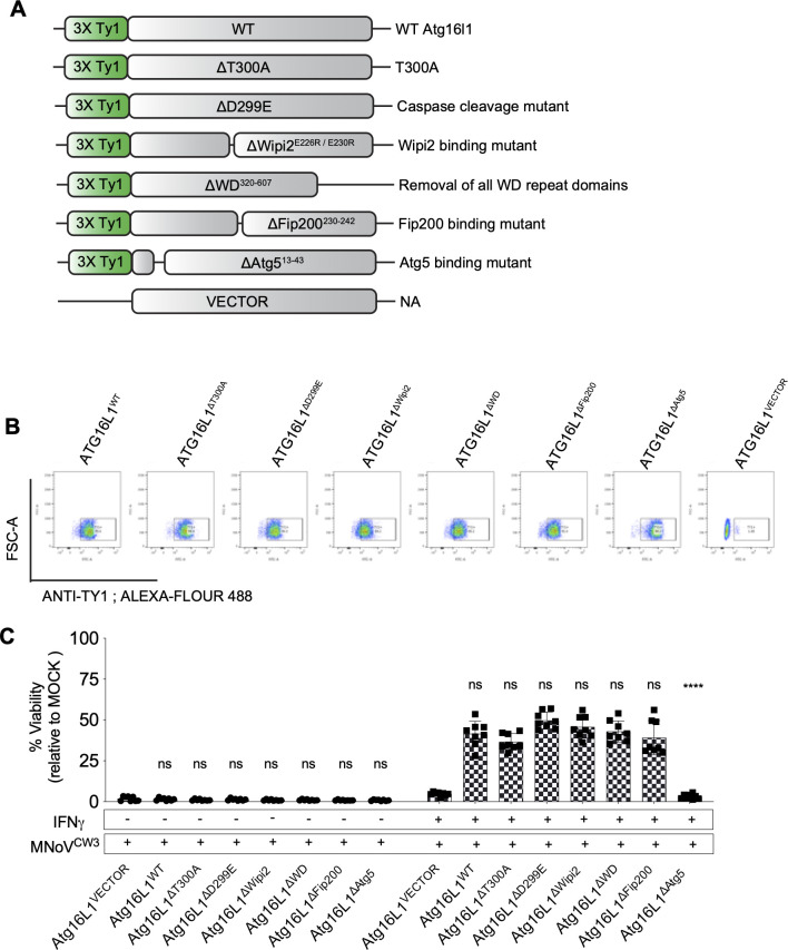
As WIPI2 binding to ATG16L1 was implicated in IFNγ control of norovirus infection, we mutagenized amino acids with known functions in ATG16L1 and expressed them in ATG16L1-deficient cells (Fig. 1C, Fig. 5A). The resulting mutant proteins were efficiently expressed (Fig. 5B). This analysis revealed that, uniquely among these known ATG16L1 functions, binding to ATG5 is important in control of norovirus infection (Fig. 5C). This interaction is likely of functional importance because mutation of ATG5 so that it cannot conjugate to ATG12 interferes with this form of intracellular immunity (12, 28).
Fig 5.
ATG5-binding domain of ATG16L1 is required for IFNγ-induced restriction of norovirus replication in BV-2 cells. (A) Schematic representation of ATG16L1 complementation in clone 1 Atg16l1−/− BV-2 cells. (B) Intracellular staining and detection of TY1 epitope in complemented cells by flow cytometry. (C) Viability assay of Atg16l1VECTOR, Atg16l1WT, Atg16l1ΔT300A, Atg16l1ΔD299E, Atg16l1ΔWipi2, Atg16l1ΔWD, Atg16l1ΔFip200, Atg16l1ΔAtg5, and Atg16l1VECTOR complemented BV-2 cells as described in Fig. S1. Values in (C) represent means ± SEM from three independent experiments. *P ≤ 0.05, **P ≤ 0.01, ***P ≤ 0.001, and ****P ≤ 0.0001 were considered statistically significant. ns, not significant. P value was determined by two-way ANOVA with Dunnett’s multiple comparison test.
P62 is required for IFNγ-induced inhibition of T. gondii in human HeLa cells but not required for IFNγ-induced inhibition of norovirus replication in BV-2 cells
Many functions of autophagy and ATG genes involve the P62 protein. In murine cells, the ubiquitin-binding protein P62 is recruited to vacuoles containing T. gondii in an IFNγ-dependent manner but is ultimately dispensable for inhibiting replication of the parasite (60). In human cells, P62 is also recruited to parasitophorous vacuoles in an IFNγ-dependent manner (16) but its requirement for the restriction of T. gondii replication is unknown. Therefore, we generated clonal P62−/− HeLa cells (Table S1) and observed that P62 was required for IFNγ-induced inhibition of T. gondii (Fig. S5A) but was not required for IFNγ-induced inhibition of norovirus replication in clonal p62−/− BV-2 cells (Fig. S5B; Table S1). Taken together, these data suggest the requirement for P62 in IFNγ-mediated control of intracellular pathogens can be species or cell type-specific.
Cul3 and Klhl9 are required for IFNγ-induced inhibition of norovirus replication
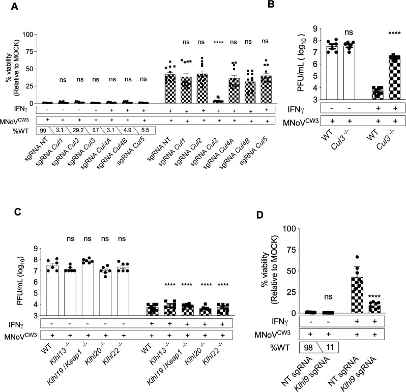
Given the important role of ubiquitin in autophagy, our attention was drawn to the screen finding suggesting that CUL3, a ubiquitin ligase involved in multiple cellular processes (61), was a candidate for a role in IFNγ-induced control of norovirus infection (Fig. 3A). We found that CRISPR guides targeting Cul3 inhibited IFNγ-induced control of norovirus infection (Fig. 6A), a finding confirmed in clonal Cul3−/− cell line (Fig. 6B). In contrast, targeting Cul1, Cul2, Cul4A, Cul4B, and Cul5 had no effect on control of norovirus infection (Fig. 6A). The specificity of CUL3-containing ubiquitin ligase complexes is conferred in some cases by assembly of CUL3 with various members of the KELCH domain family of proteins (61, 62). KlhlL7 and Klhl19 were the only KELCH domain protein-encoding genes included in our CRISPR library. Neither exhibited a meaningful effect on IFNγ-induced control of norovirus infection (Fig. 3A; Table S4). We therefore took a candidate approach to define a possible role for KELCH domain proteins in this form of intracellular immunity. Among KELCH domain proteins KLHL13 (63), KLHL19 (also Keap1) (64), KLHL20 (65), KLHL22 (66), and KLHL9 (63) are involved in cellular processes potentially related to the function of autophagy or ATG genes. Stable cell lines lacking Klhl13, Klhl19/Keap1, Klhl20, or Klhl22 were all able to control norovirus infection in the presence of IFNγ (Fig. 6C). We were unable to generate a clonal cell line mutated for Klhl9 (not shown). However, using Klhl9 specific guides we generated non-clonal cell populations in which ~88% of the Klhl9 alleles were disrupted (Fig. 6D). These cells were unable to effectively control norovirus infection in the presence of IFNγ, indicating that this KELCH domain protein is a candidate for involvement with CUL3 in IFNγ-induced control of norovirus infection.
Fig 6.
Cul3 and Klhl9 are required for IFNγ-induced inhibition of norovirus replication in BV-2 cells. (A) Viability assay of non-clonal BV-2-Cas9 knock-out cells transduced with non-targeting (NT) sgRNAs or sgRNAs targeting Cul1, Cul2, Cul3, Cul4A, Cul4B, and Cul5 as described in Fig. S1; percentage of the wild-type allele present in the non-clonal cell knock-out pool. (B and C) Plaque assay of WT, Cul3−/−, Klhl13−/−, Klhl19−/−, Klhl20−/−, and Klhl22−/− BV-2 clonal knock-out cells as described in Fig. S1. (D) Viability assay of non-clonal BV-2-Cas9 knock-out cells transduced with NT sgRNA or sgRNA targeting Klhl9 as described in Fig. S1; percentage of the wild-type allele present in the non-clonal cell pool provided. Values in (A–D) represent means ± SEM from 2 to 3 independent experiments. *P ≤ 0.05, **P ≤ 0.01, ***P ≤ 0.001, and ****P ≤ 0.0001 were considered statistically significant. ns, not significant. P value was determined by two-way ANOVA with Dunnett’s multiple comparison test.
DISCUSSION
Intracellular pathogens present diverse challenges to the immune system because they hijack different aspects of cell biology to their advantage. IFNγ is a central mediator of innate and adaptive immunity, in part via orchestrating the activities of populations of immune cells. However, IFNγ also induces cell-intrinsic immunity to intracellular pathogens via induction of transcriptional and other cellular pathways that create a cellular environment that is hostile to invading pathogens. Understanding the molecular and cellular mechanisms for this process is important for defining mechanisms of immunity to infection. We report herein cellular genes that regulate intracellular immunity and identify new components of the autophagy machinery including Gate16/GabarapL2, Atg9a, and Wipi2b that play an autophagy-independent role in IFNγ-induced intracellular immunity to T. gondii and/or norovirus. In addition, we found a previously unsuspected role of Cul3 and Klhl9, two components of a ubiquitin ligase complex, in this form of intracellular immunity. These data build on previous work on other ATG genes to elucidate a pathway that targets diverse pathogen-driven changes in intracellular membranes but that is not canonical degradative autophagy (11–19). The striking specificity of the genes Wipi2, Cul3, GATE-16, and Klhl9 within their respective large gene families for this form of immunity supports the previously proposed concept that cassettes of specific genes, some of which are also involved in degradative autophagy, provide a highly sophisticated programmable armamentarium for intracellular immunity (11, 14, 17).
ATG gene-dependent intracellular immunity
We and others have shown that IFNγ-induced intracellular immunity to norovirus and T. gondii does not require autophagy as a degradative function, but nevertheless requires essential ATG genes involved in the ubiquitin-like systems that conjugate ATG8 family proteins to PE during autophagy (11–14, 16, 17, 19, 67). The involved proteins include ATG7, which triggers the lipidation of ATG8 family proteins, ATG3 which complexes with ATG8 family proteins to foster their lipidation, and the E3-ligase-like ATG5-12-16L1 complex (11–14, 16, 17, 19, 67), which directs lipidation of ATG8 family proteins to appropriate sub-cellular locations. The ATG8 family comprises LC3 and GABARAP proteins involved in various aspects of autophagy and non-autophagic cellular processes (1, 4, 68). To further define the genetic requirements for IFNγ-induced inhibition of norovirus infection, we first validated the BV-2 cell system by demonstrating the expected roles for Stat1, Irf1, Atg5, and Ag16L1 (12, 17, 24, 26). CRISPR screening in mock- or IFNγ-treated cells revealed genes essential for cell survival in the absence of viral infection; these were not studied further. Analysis of cells treated with IFNγ and infected with norovirus also revealed genes known to be essential for efficient norovirus replication (22, 51–53) and genes that regulate responses to IFNγ such as Ufc1 and Uba5. In addition, this screen revealed new players in intracellular immunity including a single member of the murine ATG8 gene family. We found that GATE-16 is required for IFNγ-induced immunity to two very different intracellular pathogens in norovirus and T. gondii. This confirms and extends data indicating the murine GABARAPs, but not LC3 proteins, are required for IFNγ-induced intracellular immunity to T. gondii (18). The observation herein that a single ATG8 family member, GATE-16, is required for IFNγ to inhibit norovirus replication in this system is important for future studies since many studies use localization of LC3B as a marker for recruitment of ATG8 family proteins to autophagosomes or the norovirus replication complex. It may be that the mechanisms of recruitment to membranes are the same for all ATG8 family members, but in our system, only recruitment of GATE-16 is mechanistically important for cytokine control of viral replication.
It is notable that the cellular membranes targeted by these ATG genes are quite distinct for T. gondii and norovirus. T. gondii survives and replicates via binary fission inside a single membrane-bound vacuole created when it invades the cell. Replication occurs sequestered away from the cytoplasm. In contrast, norovirus replication occurs on the cytoplasmic face of intracellular membranes rearranged into a topologically complex replication complex responsible for orienting viral proteins and nucleic acids for efficient replication and assembly. Nevertheless, these two topologically distinct processes are interdicted by IFNγ in a manner dependent on a common set of ATG genes.
Not all essential ATG genes are required for this form of intracellular immunity
The ATG genes required for this form of IFNγ-induced intracellular immunity provide clues as to the mechanisms involved, but so do the ATG genes that are not required. We confirmed the previously reported lack of a role for Atg14 in IFNγ-induced inhibition of norovirus replication (17). This is a key observation because ATG14 is an essential component of the C1 form of the hetero-tetrameric Class III PI3Kinase complex required for the initiation of degradative autophagy (1, 3). Furthermore, ATG14 is not required for control of T. gondii in IFNγ-treated human HeLa cells (16). The C1 complex comprises ATG14, VPS15, VPS34, and Beclin 1, while the C2 complex substitutes UVRAG for ATG14 (1, 4, 5). We also show herein that UVRAG is not required for this form of intracellular immunity. These lipid kinase complexes generate PtdIns(3)P on membranes including the isolation membrane to initiate degradative autophagy. Beclin 1 is a core member of both autophagy-related Class III PI3Kinases. We report here that the Becn1 gene was not required for IFNγ-mediated inhibition of norovirus replication. This confirms that the autophagy-related complexes C1 and C2 are not required for this form of intracellular immunity. This conclusion is supported by the observation that the same cell lines used here were used to show that Becn1 and Atg14 are required for protection against IFNγ-induced cell death in BV-2 cells (28). Thus, these genes are functional in this cellular system via their expected role in autophagy. It is notable that there is overlap between the genes required to prevent cell death induced by IFNγ, and those required for IFNγ to inhibit norovirus replication. For example, both require Atg5 and Atg7. However, the mechanisms of IFNγ-induced inhibition of norovirus replication and prevention of cell death likely differ because prevention of IFNγ-induced cell death requires Atg14 and Becn1 (28), while prevention of norovirus replication does not. Together, these data show that the classical PI3K complexes involved in autophagy are not required for this form of intracellular immunity, despite the shared involvement of specific essential ATG genes that act downstream of the C1 and C2 complexes in degradative autophagy
The mechanism by which ATG proteins inhibit replication of both norovirus and T. gondii in IFNγ-treated cells involves the recruitment of IFN-inducible GTPases to the intracellular membranes responsible for supporting pathogen replication (15, 17, 67, 69–71). While the genes encoding sub-families of the interferon-γ-inducible immunity-related GTPase (IRGs) differ between humans and mice (67, 72), IRGs and guanylate-binding proteins (GBPs) in mice, and GBPs in humans (32, 73) are required for IFNγ-induced inhibition of the replication of both norovirus and T. gondii, as well as Chlamydia (71, 74, 75). In contrast to species variations in IFN-induced GTPases that act downstream of ATG proteins, the role of the ATG5-12-16L1 complex is conserved between humans and mice for this form of IFNγ-induced intracellular immunity. We speculate that there are conserved sequential events that result in targeting norovirus and T. gondii-related cellular membranes despite the striking differences in topology between the cellular compartments used by these two pathogens. First, the ATG5-12-16L1 complex is recruited to the relevant membranes followed by deposition of lipidated forms of the ATG8 family proteins. This is followed by recruitment of IFN-inducible GTPases to the target membranes [reviewed in reference (71)]. Less well-defined are events prior to ATG5-12-16L1 recruitment and those that occur after recruitment of IFNγ-induced immune effectors to pathogen-related membranes (71).
Role of Atg9a in IFNγ-induced ATG gene-dependent immunity
Atg9a is a lipid scramblase (76) that plays its role in autophagy via provision of lipids to developing autophagosomes via recruitment of ATG9A-positive small vesicles (58, 68, 77). While the role of Atg9a demonstrated herein could well involve these small vesicles, the regulation of Atg9a in IFNγ-induced intracellular immunity appears to differ from its regulation during autophagosome formation. In murine cells, neither the unc-5-like kinases ULK1 nor ULK2, which are required for initiation of autophagy, are required for IFNγ-mediated inhibition of norovirus replication (17, 68). This is particularly interesting since the role of ATG9A trafficking in degradative autophagy is tightly regulated by ULK1 kinase (68). This suggests that the regulation of Atg9a function in IFNγ-induced ATG gene-dependent immunity to norovirus is likely via a regulatory cascade distinct from that utilized to regulate the role of ATG9 in autophagy. Furthermore, recent work supports a role for an ATG9/ATG2 complex in autophagy (76), but we did not find a signal in our CRISPR screen for either Atg2a or Atg2b in our search for genes involved in the control of norovirus infection (Table S4). Lack of a signal in a genetic screen is suggestive only, and so this point will require further investigation.
Our data that Atg9a plays a key role in IFNγ-mediated inhibition of norovirus infection may be explained by the finding that deletion of ATG9 mRNA enhances induction of iNOS protein expression by IFNγ (27). Thus, the role of Atg9a might be either direct through action of this protein on pathogen-related intracellular membranes or indirect through changes in IFNγ signaling, or both. Our findings of a negative regulatory role for the UFMylation activity of the UFC1 and UBA5 enzymes are likely due to such an indirect effect on IFNγ signaling, mechanistically through the enhancement of IFNγ response via Ern1-dependent ER stress responses that occur when UFMylation is inhibited (27).
Role of WIPI2B in IFNγ-induced ATG gene-dependent immunity
The role of Wipi2b in our system may inform how the ATG5-12-16L1 complex is recruited to intracellular membranes during IFNγ-induced ATG gene-dependent intracellular immunity. WIPI2B binds to both PtdIns(3)P in cellular membranes and to ATG16L1 and is important for intracellular targeting and clearance of Salmonella enterica serovar Typhimurium via the recruitment of the Atg5-12-16L1 complex to cellular membranes followed by LC3 lipidation and the formation of autophagosomal membranes to engulf the bacteria (58). We found that amino acids required for WIPI2B to interact with both ATG16L1 and PtdIns(3)P were required for efficient IFNγ-induced inhibition of norovirus replication. One explanation for these findings is that WIPI2B is upstream of the effects of the ATG5-12-16L1 complex, perhaps by binding cellular lipids on target membranes and then recruitment of the ATG5-12-16L1 complex via binding to ATG16L1 as observed for its role in degradative autophagy (58). This concept is consistent with recent work showing that amino acids in ATG16L1 that confer binding of ATG16L1 to WIPI2B are important for IFNγ to inhibit norovirus replication (32). In contrast, we did not detect an effect of point mutations in murine ATG16L1 that inhibit binding of ATG6L1 to WIPI2B (58) on this form of immunity. These differences may relate to the fact that mutations used in the two studies differ in both types and in whether mutations were made in the full length versus a truncated form of ATG16L1 lacking the WD repeat region, or the possibility that the point mutations chosen here do not fully block binding of ATG16L1 to WIPI2B in the complex milieu of the viral replication complex. Further studies will be required to resolve this issue.
The finding herein that the PI3P-binding region of WIPI2B is important for IFNγ inhibition of norovirus replication presents an interesting conundrum because we found that Becn1, Atg14, and Uvrag, encoding proteins that are essential components of the Class III PI3Kinases to generate PtdIns(3)P required for autophagy, are not required for IFNγ-induced ATG gene-dependent inhibition of norovirus replication. Thus, the source of phospholipids that interact with WIPI2B in this form of immunity remains mysterious. Interestingly, activation of Beclin 1-dependent processes is not sufficient to target the parasitophorous vacuole inhabited by T. gondii for ATG gene-dependent immunity (16). It is interesting to speculate that this form of immunity may involve Beclin 2 as opposed to Beclin 1 (78–80). These observations open new questions on how pathogen-related membranes become labeled with specific phospholipids after IFNγ treatment. Resolving this question will require additional experimentation.
Species specificity of ATG gene-dependent immunity
Intriguingly, Atg9a and p62 were previously found to be dispensable for the localization of p62, GBPs, Irga6, and ubiquitin to the parasitophorous vacuole of T. gondii in mouse embryonic fibroblast cells (18, 60). However, we found herein that P62 was required for IFNγ-induced inhibition of T. gondii in HeLa cells, but not for norovirus replication in murine cells. Thus, while there are shared ATG genes involved in IFNγ-induced immunity to these two pathogens, there are proteins such as ATG9A and P62, which may play pathogen- or cell-type-specific roles in these topologically distinct cellular compartments in different experimental systems.
Role of CUL3 and KLHL9 in IFNγ-induced ATG gene-dependent immunity to norovirus infection
While the above studies shed light on some components of the pathway by which IFNγ controls intracellular pathogens, the mechanisms by which this process links to ubiquitin ligases have not been previously described. This is an important observation because the role of ATG8 family proteins, such as GATE-16 which we demonstrate herein is selectively required for this form of intracellular immunity, frequently requires ubiquitin ligase function. In this regard, we found that Cul3, but not other Cul gene family genes, plays a required role in this form of immunity to norovirus infection. The CUL proteins form ubiquitin ligases whose specificity is often conferred by the presence of specific KELCH domain-containing proteins in CUL protein-containing E3 ligase complexes. Based on our CRISPR screen data and subsequent confirmatory experiments, we found a role for KLHL9, leading us to speculate that the specificity of IFNγ-induced ATG gene-dependent immunity may rest on the function of E3 ligases such as CUL3 combined with specific KELCH domain proteins. It is particularly interesting in this regard that KLHL9 also plays a role in intracellular immunity to salmonella (63). Together, these data suggest that an ATG gene-dependent programmable intracellular system involved in the control of multiple different pathogens might rely on CUL/KELCH domain proteins in a combinatorial manner.
Summary
Data presented here, and substantial prior work, demonstrate the existence and physiological importance of a unique IFNγ-induced ATG gene-dependent process responsible for the creation of a cellular environment that is hostile to membrane-dependent replication of phylogenetically distinct pathogens. This mechanism appears programmable with the participation of a core machinery including WIPI2B, ATG9A, ATG7, ATG5-12-16L1, ATG3, and the ATG8 family member GATE-16 that is regulated by proteins in the UFMylation pathway and the involvement of specific ubiquitin ligases such as CUL3, and rendered specific by KELCH domain-containing proteins (12, 17, 71). These new data further support the previous conclusion that cassettes of ATG genes have been leveraged by the immune system to perform cytokine-induced tasks that block replication and clear pathogens (3, 11, 12). This system is programmable via the involvement of a variety of gene products and focuses on altered intracellular membranes created as diverse pathogens hijack and evade normal cellular functions. Since intracellular pathogens require rearrangement and hijacking of intracellular membranes as a lifestyle, this form of immunity is an important barrier to infectious diseases.
MATERIALS AND METHODS
Cells
BV-2, HEK293T, and HeLa cells were cultured in Dulbecco’s modified Eagle medium (Gibco) with 10% fetal bovine serum (FBS), and 1% HEPES. BV-2 Cas9 cell lines were generated using standard protocols (22). For selection, 5 µg/mL puromycin (Thermofisher) and 5 µg/mL blasticidin (Thermofisher) were added to BV-2 cells.
BV-2 and HeLa clonal knock-out cells were generated by transiently introducing Cas9 and gRNAs (Table S1) via nucleofection into wild type, and clones were screened for indels by sequencing the target region with Illumina MiSeq at approximately 500× coverage. Indel signature frequency was determined using an in-house algorithm (GEiC, Washington University School of Medicine, St. Louis, MO, USA). Non-clonal knock-out pools were generated in BV-2 Cas9 cells by transduction of lentivirus carrying sgRNA (Table S1) (21) followed by puromycin selection; 5 days post-puromycin selection indel frequency was determined by sequencing the target (Table S1).
For cDNA expression of ATG16L1 and WIPI2B, proteins cells were transduced with lentivirus carrying the gene of interest with an N-terminal 3× Ty1 tag (Table S5) on the pCDH-CMV-MCS-T2A-Puro backbone (CD522A-1, System Biosciences). cDNA expression of ATG proteins has been previously described (22, 27, 28).
Viruses and viral assays
MNoVCW3 (GenBank accession no. EF014462.1, termed norovirus in the main text) was generated by transfecting a molecular clone (81) into HEK293T cells (P0 stock), which was passaged on BV-2 cells. After two passages, infected cells were frozen at −80°C and thawed, cleared of cellular debris and virus was concentrated by tangential flow filtration. For infection, WT or knockout BV-2 cells were seeded at 104 cells/well of a 96-well plate. After 8 hours, cells were treated with 1 U/mL of IFNγ (BioLegend). After 16 hours, norovirus was added at a multiplicity of infction (MOI) of 5.0. Infected cells were harvested at 24 hpi and frozen at −80°C prior to plaque assay. For cell viability, CellTiter-Glo reagent (Promega) was added to wells of a 96-well plate, incubated for 10 minutes at room temperature, and then cellular ATP content was measured. Viral titers were determined in triplicate by plaque assay on BV-2 cells. BV-2 cells were seeded at 2 × 10⁶ cells/well of a six-well plate, and 24 hours later, 100 μl of 10-fold serially diluted samples was applied to each well for 1 hour with orbital rocking at room temperature. Viral inoculum was aspirated, and 2 mL of warmed MEM containing 10% FBS, 2 mM L-gluatmine, 10 mM HEPES, and 1% methylcellulose were added. Plates were incubated for 48–60 hours prior to visualization after staining with 0.2% crystal violet in 20% ethanol.
Antibodies and western blots
Cell lysates were run under reducing conditions on an Any kD Mini-PROTEAN TGX Stain-Free Protein Gel (BioRad) and imaged using a ChemiDoc imaging system (Biorad). WIPI2B and ATG16L1 proteins were tagged with three copies of Ty1 on the N terminus. Anti-Ty1 antibody (ThermoFisher #MA5-23513) was used at 1:1,000. Anti-Wipi2 (ThermoFisher # PA5-78465) was used at 1:1,000. Anti-beta-actin (R&D Systems #MAB8929) was used at 1:10,000. Anti-Uvrag (Cell-Singaling Technologies #5320) was used at 1:100. Anti-mouse horseradish peroxidase (HRP) (Jackson Immuno Research Laboratories #315-035-003) was used at 1:10,000. Anti-rabbit HRP (Jackson Immuno Research Laboratories #111-035-003) was used at 1:10,000.
Autophagy CRISPR-Cas9 subpool generation and screen
BV-2-Cas9 cells (1.4 × 10⁷) (22, 27, 28) were transduced with a lentivirus stock (transduction efficiency 40%) resulting in 6 × 10⁶ cells. After 36 hours, puromycin was added, and transduced cells were selected for 5 days. Five days later, 1 × 10⁷ cells were harvested, and DNA was for sequencing (QIAamp DNA Blood Midi Kit, Qiagen). For each experimental condition (mock, IFNγ, and IFNγ + norovirus), 1 × 10⁷ cells were seeded in a 15-cm2 dish. Eight hours later, cells were treated with media or 1 U/mL IFNγ. After 16 hours, cells were either mock infected or infected with norovirus at an MOI of 5.0. Cells were harvested 24 hours later for isolation of DNA for sequencing and assessment of cell viability (Trypan blue exclusion). Genomic DNA was sequenced and analyzed (22, 27, 28). Volcano plots were generated using the hypergeometric distribution method (https://github.com/mhegde/volcano_plots), and screen results were analyzed using STARS (https://portals.broadinstitute.org/gpp/public/software/stars).
T. gondii growth restriction assay
HeLa cells were treated with 100 U/mL IFNγ for 24 hours, infected with tachyzoites, and washed 2 hours later to remove extracellular parasites. Cells were fixed in 4% formaldehyde 24 hours later. T. gondii was localized using antibody against the RH strain tachyzoites (81), and ubiquitin was localized using mouse antibody clone FK2 (04-263; EMD Millipore Corporation). Parasites were enumerated per PV from 30 PV on three individual coverslips from three independent experiments.
Quantification and statistical analysis
Data were analyzed with Prism 7 software (GraphPad Software, San Diego, CA, USA). Volcano plot and Pearson visualizations were generated using R-studio (Integrated Development for R. RStudio, PBC, Boston, MA, USA; http://www.rstudio.com/).
ACKNOWLEDGMENTS
The authors would like to thank Rob Orchard, Craig Wilen, Yating Wang, Scott Handley, Tim Schaiff, Anne Paredes, and John Doench for discussion; Monica Sentmanat in the Genome Engineering and iPSC Center (GEiC) at Washington University School of Medicine for assistance with CRISPR cell line generation.
H.W.V. was supported by the National Institutes of Health (NIH) grant U19 AI109725. H.W.V., M.R.M., D.R.B., and S.H. were supported by U19 AI142784. M.R.M. was supported by U19 AI109725. A.P. was supported by T32 AI716340. J.B. and L.D.S. were supported by NIH grant AI 118426.
M.R.M. and H.W.V. designed the project and wrote the manuscript. A.O., A.P., M.E.S., J.B., L.D.S., D.R.B., and S.H. helped to edit and review the manuscript. M.R.M., A.O., A.P., M.E.S., J.B., L.D.S., D.R.B., and S.H. assisted with project design. M.R.M., J.B., A.O., A.P., and M.E.S. designed and/or assisted with experiments and data analysis. All the authors approved the manuscript.
Contributor Information
Michael R. McAllaster, Email: mmcallaster@vir.bio.
Herbert W. Virgin, Email: virgin@wustl.edu.
Stefan H. E. Kaufmann, Max Planck Institute for Infection Biology, Berlin, Germany
Eric Baehrecke, University of Massachusetts Medical School, Worcester, Massachusetts, USA.
SUPPLEMENTAL MATERIAL
The following material is available online at https://doi.org/10.1128/mbio.02332-23.
Schematic for seeding, treatment, and norovirus infection.
Autophagy CRISPR library screen design.
Uvrag-/- BV-2 cells lack UVRAG protein.
Ufm1 is not required for IFNγ-induced inhibition of norovirus replication in BV-2 cells.
p62 is not required for IFNγ-induced inhibition of norovirus replication in BV-2 cells; P62 is required for IFNγ-induced growth restriction of T. gondii in HeLa cells.
NGS validation data.
Autophagy CRISPR library.
Genes enriched and depleted for passage of BV-2 cells results; genes depleted during passage of BV-2 cells compared to Project Achilles.
Genes enriched and depleted in 1 U/mL IFNγ treated cells, 10 U/mL IFNγ treated cells, or 1 U/mL IFNγ + norovirus infected cells.
Supplemental sequences.
ASM does not own the copyrights to Supplemental Material that may be linked to, or accessed through, an article. The authors have granted ASM a non-exclusive, world-wide license to publish the Supplemental Material files. Please contact the corresponding author directly for reuse.
REFERENCES
- 1. Morishita H, Mizushima N. 2019. Diverse cellular roles of autophagy. Annu Rev Cell Dev Biol 35:453–475. doi: 10.1146/annurev-cellbio-100818-125300 [DOI] [PubMed] [Google Scholar]
- 2. Galluzzi L, Baehrecke EH, Ballabio A, Boya P, Bravo-San Pedro JM, Cecconi F, Choi AM, Chu CT, Codogno P, Colombo MI, Cuervo AM, Debnath J, Deretic V, Dikic I, Eskelinen E-L, Fimia GM, Fulda S, Gewirtz DA, Green DR, Hansen M, Harper JW, Jäättelä M, Johansen T, Juhasz G, Kimmelman AC, Kraft C, Ktistakis NT, Kumar S, Levine B, Lopez-Otin C, Madeo F, Martens S, Martinez J, Melendez A, Mizushima N, Münz C, Murphy LO, Penninger JM, Piacentini M, Reggiori F, Rubinsztein DC, Ryan KM, Santambrogio L, Scorrano L, Simon AK, Simon H-U, Simonsen A, Tavernarakis N, Tooze SA, Yoshimori T, Yuan J, Yue Z, Zhong Q, Kroemer G. 2017. Molecular definitions of autophagy and related processes. EMBO J 36:1811–1836. doi: 10.15252/embj.201796697 [DOI] [PMC free article] [PubMed] [Google Scholar]
- 3. Levine B, Mizushima N, Virgin HW. 2011. Autophagy in immunity and inflammation. Nature 469:323–335. doi: 10.1038/nature09782 [DOI] [PMC free article] [PubMed] [Google Scholar]
- 4. Cadwell K, Debnath J. 2018. Beyond self-eating: the control of nonautophagic functions and signaling pathways by autophagy-related proteins. J Cell Biol 217:813–822. doi: 10.1083/jcb.201706157 [DOI] [PMC free article] [PubMed] [Google Scholar]
- 5. Deretic V. 2021. Autophagy in inflammation, infection, and immunometabolism. Immunity 54:437–453. doi: 10.1016/j.immuni.2021.01.018 [DOI] [PMC free article] [PubMed] [Google Scholar]
- 6. Heckmann BL, Boada-Romero E, Cunha LD, Magne J, Green DR. 2017. LC3-associated phagocytosis and inflammation. J Mol Biol 429:3561–3576. doi: 10.1016/j.jmb.2017.08.012 [DOI] [PMC free article] [PubMed] [Google Scholar]
- 7. DeSelm CJ, Miller BC, Zou W, Beatty WL, van Meel E, Takahata Y, Klumperman J, Tooze SA, Teitelbaum SL, Virgin HW. 2011. Autophagy proteins regulate the secretory component of osteoclastic bone resorption. Dev Cell 21:966–974. doi: 10.1016/j.devcel.2011.08.016 [DOI] [PMC free article] [PubMed] [Google Scholar]
- 8. Kimmey JM, Huynh JP, Weiss LA, Park S, Kambal A, Debnath J, Virgin HW, Stallings CL. 2015. Unique role for ATG5 in neutrophil-mediated Immunopathology during M. tuberculosis infection. Nature 528:565–569. doi: 10.1038/nature16451 [DOI] [PMC free article] [PubMed] [Google Scholar]
- 9. Wang Y-T, Zaitsev K, Lu Q, Li S, Schaiff WT, Kim K-W, Droit L, Wilen CB, Desai C, Balce DR, Orchard RC, Orvedahl A, Park S, Kreamalmeyer D, Handley SA, Pfeifer JD, Baldridge MT, Artyomov MN, Stallings CL, Virgin HW. 2020. Select autophagy genes maintain quiescence of tissue-resident macrophages and increase susceptibility to Listeria monocytogenes. Nat Microbiol 5:272–281. doi: 10.1038/s41564-019-0633-0 [DOI] [PMC free article] [PubMed] [Google Scholar]
- 10. Mauthe M, Langereis M, Jung J, Zhou X, Jones A, Omta W, Tooze SA, Stork B, Paludan SR, Ahola T, Egan D, Behrends C, Mokry M, de Haan C, van Kuppeveld F, Reggiori F. 2016. An siRNA screen for ATG protein depletion reveals the extent of the unconventional functions of the autophagy proteome in virus replication. J Cell Biol 214:619–635. doi: 10.1083/jcb.201602046 [DOI] [PMC free article] [PubMed] [Google Scholar]
- 11. Zhao Z, Fux B, Goodwin M, Dunay IR, Strong D, Miller BC, Cadwell K, Delgado MA, Ponpuak M, Green KG, Schmidt RE, Mizushima N, Deretic V, Sibley LD, Virgin HW. 2008. Autophagosome-independent essential function for the autophagy protein ATG5 in cellular immunity to intracellular pathogens. Cell Host Microbe 4:458–469. doi: 10.1016/j.chom.2008.10.003 [DOI] [PMC free article] [PubMed] [Google Scholar]
- 12. Hwang S, Maloney NS, Bruinsma MW, Goel G, Duan E, Zhang L, Shrestha B, Diamond MS, Dani A, Sosnovtsev SV, Green KY, Lopez-Otin C, Xavier RJ, Thackray LB, Virgin HW. 2012. Nondegradative role of Atg5-Atg12/ Atg16L1 autophagy protein complex in antiviral activity of interferon gamma. Cell Host Microbe 11:397–409. doi: 10.1016/j.chom.2012.03.002 [DOI] [PMC free article] [PubMed] [Google Scholar]
- 13. Selleck EM, Fentress SJ, Beatty WL, Degrandi D, Pfeffer K, Virgin HW 4th, Macmicking JD, Sibley LD. 2013. Guanylate-binding protein 1 (Gbp1) contributes to cell-autonomous immunity against Toxoplasma gondii. PLoS Pathog 9:e1003320. doi: 10.1371/journal.ppat.1003320 [DOI] [PMC free article] [PubMed] [Google Scholar]
- 14. Choi J, Park S, Biering SB, Selleck E, Liu CY, Zhang X, Fujita N, Saitoh T, Akira S, Yoshimori T, Sibley LD, Hwang S, Virgin HW. 2014. The parasitophorous vacuole membrane of Toxoplasma gondii is targeted for disruption by Ubiquitin-like conjugation systems of autophagy. Immunity 40:924–935. doi: 10.1016/j.immuni.2014.05.006 [DOI] [PMC free article] [PubMed] [Google Scholar]
- 15. Haldar AK, Piro AS, Pilla DM, Yamamoto M, Coers J. 2014. The E2-like conjugation enzyme Atg3 promotes binding of IRG and Gbp proteins to chlamydia- and toxoplasma-containing vacuoles and host resistance. PLoS One 9:e86684. doi: 10.1371/journal.pone.0086684 [DOI] [PMC free article] [PubMed] [Google Scholar]
- 16. Selleck EM, Orchard RC, Lassen KG, Beatty WL, Xavier RJ, Levine B, Virgin HW, Sibley LD. 2015. A Noncanonical autophagy pathway restricts Toxoplasma gondii growth in a strain-specific manner in IFN-γ-activated human cells. mBio 6:e01157–15. doi: 10.1128/mBio.01157-15 [DOI] [PMC free article] [PubMed] [Google Scholar]
- 17. Biering SB, Choi J, Halstrom RA, Brown HM, Beatty WL, Lee S, McCune BT, Dominici E, Williams LE, Orchard RC, Wilen CB, Yamamoto M, Coers J, Taylor GA, Hwang S. 2017. Viral replication complexes are targeted by LC3-guided interferon-inducible GTPases. Cell Host Microbe 22:74–85. doi: 10.1016/j.chom.2017.06.005 [DOI] [PMC free article] [PubMed] [Google Scholar]
- 18. Sasai M, Sakaguchi N, Ma JS, Nakamura S, Kawabata T, Bando H, Lee Y, Saitoh T, Akira S, Iwasaki A, Standley DM, Yoshimori T, Yamamoto M. 2017. Essential role for GABARAP autophagy proteins in interferon-inducible GTPase-mediated host defense. Nat Immunol 18:899–910. doi: 10.1038/ni.3767 [DOI] [PubMed] [Google Scholar]
- 19. Bhushan J, Radke JB, Perng Y-C, Mcallaster M, Lenschow DJ, Virgin HW, Sibley LD. 2020. ISG15 connects autophagy and IFN-γ-dependent control of Toxoplasma gondii infection in human cells. mBio 11:e00852-20. doi: 10.1128/mBio.00852-20 [DOI] [PMC free article] [PubMed] [Google Scholar]
- 20. Krishnamurthy S, Konstantinou EK, Young LH, Gold DA, Saeij JPJ. 2017. The human immune response to Toxoplasma: autophagy versus cell death. PLoS Pathog 13:e1006176. doi: 10.1371/journal.ppat.1006176 [DOI] [PMC free article] [PubMed] [Google Scholar]
- 21. Blasi E, Barluzzi R, Bocchini V, Mazzolla R, Bistoni F. 1990. Immortalization of murine microglial cells by a v-raf / v-myc carrying retrovirus. J Neuroimmunol 27:229–237. doi: 10.1016/0165-5728(90)90073-v [DOI] [PubMed] [Google Scholar]
- 22. Orchard RC, Wilen CB, Doench JG, Baldridge MT, McCune BT, Lee Y-C, Lee S, Pruett-Miller SM, Nelson CA, Fremont DH, Virgin HW. 2016. Discovery of a proteinaceous cellular receptor for a norovirus. Science 353:933–936. doi: 10.1126/science.aaf1220 [DOI] [PMC free article] [PubMed] [Google Scholar]
- 23. Stark GR, Kerr IM, Williams BRG, Silverman RH, Schreiber RD. 1998. How cells respond to Interferons. Annu Rev Biochem 67:227–264. doi: 10.1146/annurev.biochem.67.1.227 [DOI] [PubMed] [Google Scholar]
- 24. Mumphrey SM, Changotra H, Moore TN, Heimann-Nichols ER, Wobus CE, Reilly MJ, Moghadamfalahi M, Shukla D, Karst SM. 2007. Murine norovirus 1 infection is associated with histopathological changes in immunocompetent hosts, but clinical disease is prevented by STAT1-dependent interferon responses▿. J Virol 81:3251–3263. doi: 10.1128/JVI.02096-06 [DOI] [PMC free article] [PubMed] [Google Scholar]
- 25. Karst SM, Wobus CE, Lay M, Davidson J, Virgin HW. 2003. STAT1-dependent innate immunity to a norwalk-like virus. Science 299:1575–1578. doi: 10.1126/science.1077905 [DOI] [PubMed] [Google Scholar]
- 26. Maloney NS, Thackray LB, Goel G, Hwang S, Duan E, Vachharajani P, Xavier R, Virgin HW. 2012. Essential cell-autonomous role for interferon (IFN) regulatory factor 1 in IFN-γ-mediated inhibition of norovirus replication in macrophages. J Virol 86:12655–12664. doi: 10.1128/JVI.01564-12 [DOI] [PMC free article] [PubMed] [Google Scholar]
- 27. Balce DR, Wang Y-T, McAllaster MR, Dunlap BF, Orvedahl A, Hykes BL, Droit L, Handley SA, Wilen CB, Doench JG, Orchard RC, Stallings CL, Virgin HW. 2021. UFMylation inhibits the proinflammatory capacity of interferon-γ–activated macrophages. Proc Natl Acad Sci U S A 118:e2011763118. doi: 10.1073/pnas.2011763118 [DOI] [PMC free article] [PubMed] [Google Scholar]
- 28. Orvedahl A, McAllaster MR, Sansone A, Dunlap BF, Desai C, Wang Y-T, Balce DR, Luke CJ, Lee S, Orchard RC, Artyomov MN, Handley SA, Doench JG, Silverman GA, Virgin HW. 2019. Autophagy genes in myeloid cells counteract IFNγ-induced TNF-mediated cell death and fatal TNF-induced shock. Proc Natl Acad Sci U S A 116:16497–16506. doi: 10.1073/pnas.1822157116 [DOI] [PMC free article] [PubMed] [Google Scholar]
- 29. Wymann MP, Bulgarelli-Leva G, Zvelebil MJ, Pirola L, Vanhaesebroeck B, Waterfield MD, Panayotou G. 1996. Wortmannin inactivates phosphoinositide 3-kinase by covalent modification of Lys-802, a residue involved in the phosphate transfer reaction. Mol Cell Biol 16:1722–1733. doi: 10.1128/MCB.16.4.1722 [DOI] [PMC free article] [PubMed] [Google Scholar]
- 30. Cai X, Xu Y, Cheung AK, Tomlinson RC, Alcázar-Román A, Murphy L, Billich A, Zhang B, Feng Y, Klumpp M, Rondeau J-M, Fazal AN, Wilson CJ, Myer V, Joberty G, Bouwmeester T, Labow MA, Finan PM, Porter JA, Ploegh HL, Baird D, De Camilli P, Tallarico JA, Huang Q. 2013. PIKfyve, a class III PI kinase, is the target of the small molecular IL-12/IL-23 inhibitor apilimod and a player in toll-like receptor signaling. Chem Biol 20:912–921. doi: 10.1016/j.chembiol.2013.05.010 [DOI] [PMC free article] [PubMed] [Google Scholar]
- 31. Wilhoit, PharmD T, M. Patrick, PharmD, BCOP J, B. May, PharmD, BCOP M. 2020. Alpelisib: A novel therapy for patients with PIK3Ca-Mutated metastatic breast cancer. JADPRO 11:768–775. doi: 10.6004/jadpro.2020.11.7.9 [DOI] [PMC free article] [PubMed] [Google Scholar]
- 32. Kurhade C, Kang S, Biering SB, Hwang S, Randall G. 2023. Caprin1 is required for control of viral replication complexes by interferon gamma. mBio 14:e0017223. doi: 10.1128/mbio.00172-23 [DOI] [PMC free article] [PubMed] [Google Scholar]
- 33. Weidberg H, Shvets E, Shpilka T, Shimron F, Shinder V, Elazar Z. 2010. LC3 and GATE‐16/GABARAP subfamilies are both essential yet act differently in autophagosome biogenesis. EMBO J 29:1792–1802. doi: 10.1038/emboj.2010.74 [DOI] [PMC free article] [PubMed] [Google Scholar]
- 34. Doench JG, Fusi N, Sullender M, Hegde M, Vaimberg EW, Donovan KF, Smith I, Tothova Z, Wilen C, Orchard R, Virgin HW, Listgarten J, Root DE. 2016. Optimized sgRNA design to maximize activity and minimize off-target effects of CRISPR-Cas9. Nat Biotechnol 34:184–191. doi: 10.1038/nbt.3437 [DOI] [PMC free article] [PubMed] [Google Scholar]
- 35. Behrends C, Sowa ME, Gygi SP, Harper JW. 2010. Network organization of the human autophagy system. Nature 466:68–76. doi: 10.1038/nature09204 [DOI] [PMC free article] [PubMed] [Google Scholar]
- 36. Ashburner M, Ball CA, Blake JA, Botstein D, Butler H, Cherry JM, Davis AP, Dolinski K, Dwight SS, Eppig JT, Harris MA, Hill DP, Issel-Tarver L, Kasarskis A, Lewis S, Matese JC, Richardson JE, Ringwald M, Rubin GM, Sherlock G. 2000. Gene ontology: tool for the unification of biology. Nat Genet 25:25–29. doi: 10.1038/75556 [DOI] [PMC free article] [PubMed] [Google Scholar]
- 37. Consortium TGO, Carbon S, Douglass E, Good BM, Unni DR, Harris NL, Mungall CJ, Basu S, Chisholm RL, Dodson RJ, et al. 2021. The gene ontology resource: enriching a gold mine. Nucleic Acids Res 49:D325–D334. doi: 10.1093/nar/gkaa1113 [DOI] [PMC free article] [PubMed] [Google Scholar]
- 38. Haney MS, Bohlen CJ, Morgens DW, Ousey JA, Barkal AA, Tsui CK, Ego BK, Levin R, Kamber RA, Collins H, Tucker A, Li A, Vorselen D, Labitigan L, Crane E, Boyle E, Jiang L, Chan J, Rincón E, Greenleaf WJ, Li B, Snyder MP, Weissman IL, Theriot JA, Collins SR, Barres BA, Bassik MC. 2018. Identification of phagocytosis regulators using magnetic genome-wide CRISPR screens. Nat Genet 50:1716–1727. doi: 10.1038/s41588-018-0254-1 [DOI] [PMC free article] [PubMed] [Google Scholar]
- 39. Brestoff JR, Wilen CB, Moley JR, Li Y, Zou W, Malvin NP, Rowen MN, Saunders BT, Ma H, Mack MR, Hykes BL, Balce DR, Orvedahl A, Williams JW, Rohatgi N, Wang X, McAllaster MR, Handley SA, Kim BS, Doench JG, Zinselmeyer BH, Diamond MS, Virgin HW, Gelman AE, Teitelbaum SL. 2021. Intercellular mitochondria transfer to macrophages regulates white adipose tissue homeostasis and is impaired in obesity. Cell Metab 33:270–282. doi: 10.1016/j.cmet.2020.11.008 [DOI] [PMC free article] [PubMed] [Google Scholar]
- 40. Jurecka A, Zikanova M, Kmoch S, Tylki-Szymańska A. 2015. Adenylosuccinate lyase deficiency. J Inherit Metab Dis 38:231–242. doi: 10.1007/s10545-014-9755-y [DOI] [PMC free article] [PubMed] [Google Scholar]
- 41. Minegishi Y, Ota N, Soga S, Shimotoyodome A. 2016. Effects of nutritional supplementation with milk fat globule membrane on physical and muscle function in healthy adults aged 60 and over with semiweekly light exercise: a randomized double-blind, placebo-controlled pilot trial. J Nutr Sci Vitaminol (Tokyo) 62:409–415. doi: 10.3177/jnsv.62.409 [DOI] [PubMed] [Google Scholar]
- 42. Dempster JM, Rossen J, Kazachkova M, Pan J, Kugener G, Root DE, Tsherniak A. 2019. Extracting biological insights from the project achilles genome-scale CRISPR screens in cancer cell lines. Cancer Biology. doi: 10.1101/720243 [DOI]
- 43. Meyers RM, Bryan JG, McFarland JM, Weir BA, Sizemore AE, Xu H, Dharia NV, Montgomery PG, Cowley GS, Pantel S, Goodale A, Lee Y, Ali LD, Jiang G, Lubonja R, Harrington WF, Strickland M, Wu T, Hawes DC, Zhivich VA, Wyatt MR, Kalani Z, Chang JJ, Okamoto M, Stegmaier K, Golub TR, Boehm JS, Vazquez F, Root DE, Hahn WC, Tsherniak A. 2017. Computational correction of copy number effect improves specificity of CRISPR–Cas9 essentiality screens in cancer cells. Nat Genet 49:1779–1784. doi: 10.1038/ng.3984 [DOI] [PMC free article] [PubMed] [Google Scholar]
- 44. Kroczynska B, Mehrotra S, Arslan AD, Kaur S, Platanias LC. 2014. Regulation of interferon-dependent mRNA translation of target genes. J Interferon Cytokine Res 34:289–296. doi: 10.1089/jir.2013.0148 [DOI] [PMC free article] [PubMed] [Google Scholar]
- 45. Su X, Yu Y, Zhong Y, Giannopoulou EG, Hu X, Liu H, Cross JR, Rätsch G, Rice CM, Ivashkiv LB. 2015. Interferon-γ regulates cellular metabolism and mRNA translation to potentiate macrophage activation. Nat Immunol 16:838–849. doi: 10.1038/ni.3205 [DOI] [PMC free article] [PubMed] [Google Scholar]
- 46. Ogunjimi B, Zhang S-Y, Sørensen KB, Skipper KA, Carter-Timofte M, Kerner G, Luecke S, Prabakaran T, Cai Y, Meester J, Bartholomeus E, Bolar NA, Vandeweyer G, Claes C, Sillis Y, Lorenzo L, Fiorenza RA, Boucherit S, Dielman C, Heynderickx S, Elias G, Kurotova A, Auwera AV, Verstraete L, Lagae L, Verhelst H, Jansen A, Ramet J, Suls A, Smits E, Ceulemans B, Van Laer L, Plat Wilson G, Kreth J, Picard C, Von Bernuth H, Fluss J, Chabrier S, Abel L, Mortier G, Fribourg S, Mikkelsen JG, Casanova J-L, Paludan SR, Mogensen TH. 2017. Inborn errors in RNA polymerase III underlie severe varicella zoster virus infections. J Clin Invest 127:3543–3556. doi: 10.1172/JCI92280 [DOI] [PMC free article] [PubMed] [Google Scholar]
- 47. Choi J-M, Devkota S, Sung YH, Lee H-W. 2013. EI24 regulates epithelial-to-mesenchymal transition and tumor progression by suppressing TRAF2-mediated NF-κB activity. Oncotarget 4:2383–2396. doi: 10.18632/oncotarget.1434 [DOI] [PMC free article] [PubMed] [Google Scholar]
- 48. Wajant H, Siegmund D. 2019. TNFR1 and TNFR2 in the control of the life and death balance of macrophages. Front Cell Dev Biol 7:91. doi: 10.3389/fcell.2019.00091 [DOI] [PMC free article] [PubMed] [Google Scholar]
- 49. Sanjo H, Nakayama J, Yoshizawa T, Fehling HJ, Akira S, Taki S. 2019. Cutting edge: TAK1 safeguards macrophages against proinflammatory cell death. J Immunol 203:783–788. doi: 10.4049/jimmunol.1900202 [DOI] [PubMed] [Google Scholar]
- 50. Orchard RC, Wilen CB, Virgin HW. 2018. Sphingolipid biosynthesis induces a conformational change in the murine norovirus receptor and facilitates viral infection. Nat Microbiol 3:1109–1114. doi: 10.1038/s41564-018-0221-8 [DOI] [PMC free article] [PubMed] [Google Scholar]
- 51. Fritzlar S, Aktepe TE, Chao Y-W, Kenney ND, McAllaster MR, Wilen CB, White PA, Mackenzie JM. 2019. Mouse norovirus infection arrests host cell translation uncoupled from the stress granule-PKR-eIF2α axis. mBio 10:e00960-19. doi: 10.1128/mBio.00960-19 [DOI] [PMC free article] [PubMed] [Google Scholar]
- 52. Hosmillo M, Lu J, McAllaster MR, Eaglesham JB, Wang X, Emmott E, Domingues P, Chaudhry Y, Fitzmaurice TJ, Tung MK, Panas MD, McInerney G, Locker N, Wilen CB, Goodfellow IG. 2019. Noroviruses subvert the core stress granule component G3Bp1 to promote viral VPg-dependent translation. Elife 8:e46681. doi: 10.7554/eLife.46681 [DOI] [PMC free article] [PubMed] [Google Scholar]
- 53. Tocker AM, Durocher E, Jacob KD, Trieschman KE, Talento SM, Rechnitzer AA, Roberts DM, Davis BK. 2017. The scaffolding protein IQGAP1 interacts with NLRC3 and inhibits type I IFN production. J Immunol 199:2896–2909. doi: 10.4049/jimmunol.1601370 [DOI] [PMC free article] [PubMed] [Google Scholar]
- 54. Habisov S, Huber J, Ichimura Y, Akutsu M, Rogova N, Loehr F, McEwan DG, Johansen T, Dikic I, Doetsch V, Komatsu M, Rogov VV, Kirkin V. 2016. Structural and functional analysis of a novel interaction motif within UFM1-activating enzyme 5 (UBA5) required for binding to Ubiquitin-like proteins and Ufmylation. J Biol Chem 291:9025–9041. doi: 10.1074/jbc.M116.715474 [DOI] [PMC free article] [PubMed] [Google Scholar]
- 55. Huber J, Obata M, Gruber J, Akutsu M, Löhr F, Rogova N, Güntert P, Dikic I, Kirkin V, Komatsu M, Dötsch V, Rogov VV. 2020. An atypical LIR motif within UBA5 (Ubiquitin like modifier activating enzyme 5) interacts with GABARAP proteins and mediates membrane localization of UBA5. Autophagy 16:256–270. doi: 10.1080/15548627.2019.1606637 [DOI] [PMC free article] [PubMed] [Google Scholar]
- 56. Komatsu M, Chiba T, Tatsumi K, Iemura S, Tanida I, Okazaki N, Ueno T, Kominami E, Natsume T, Tanaka K. 2004. A novel protein‐conjugating system for Ufm1, a Ubiquitin‐fold modifier. EMBO J 23:1977–1986. doi: 10.1038/sj.emboj.7600205 [DOI] [PMC free article] [PubMed] [Google Scholar]
- 57. Dooley HC, Razi M, Polson HEJ, Girardin SE, Wilson MI, Tooze SA. 2014. WIPI2 links LC3 conjugation with PI3P, autophagosome formation, and pathogen clearance by recruiting Atg12–5-16L1. Mol Cell 55:238–252. doi: 10.1016/j.molcel.2014.05.021 [DOI] [PMC free article] [PubMed] [Google Scholar]
- 58. Lystad AH, Carlsson SR, de la Ballina LR, Kauffman KJ, Nag S, Yoshimori T, Melia TJ, Simonsen A. 2019. Distinct functions of ATG16L1 Isoforms in membrane binding and LC3B lipidation in autophagy-related processes. Nat Cell Biol 21:372–383. doi: 10.1038/s41556-019-0274-9 [DOI] [PMC free article] [PubMed] [Google Scholar]
- 59. Lee Y, Sasai M, Ma JS, Sakaguchi N, Ohshima J, Bando H, Saitoh T, Akira S, Yamamoto M. 2015. P62 plays a specific role in interferon-γ-induced presentation of a Toxoplasma vacuolar antigen. Cell Rep 13:223–233. doi: 10.1016/j.celrep.2015.09.005 [DOI] [PubMed] [Google Scholar]
- 60. Genschik P, Sumara I, Lechner E. 2013. The emerging family of CULLIN3‐RING Ubiquitin Ligases (CRL3s): Cellular functions and disease implications. EMBO J 32:2307–2320. doi: 10.1038/emboj.2013.173 [DOI] [PMC free article] [PubMed] [Google Scholar]
- 61. Prag S, Adams JC. 2003. Molecular phylogeny of the kelch-repeat superfamily reveals an expansion of BTB/Kelch proteins in animals. BMC Bioinformatics 4:42. doi: 10.1186/1471-2105-4-42 [DOI] [PMC free article] [PubMed] [Google Scholar]
- 62. Begun J, Lassen KG, Jijon HB, Baxt LA, Goel G, Heath RJ, Ng A, Tam JM, Kuo S-Y, Villablanca EJ, Fagbami L, Oosting M, Kumar V, Schenone M, Carr SA, Joosten LAB, Vyas JM, Daly MJ, Netea MG, Brown GD, Wijmenga C, Xavier RJ. 2015. Integrated genomics of crohn’s disease risk variant identifies a role for CLEC12A in antibacterial autophagy. Cell Rep 11:1905–1918. doi: 10.1016/j.celrep.2015.05.045 [DOI] [PMC free article] [PubMed] [Google Scholar]
- 63. Thimmulappa RK, Lee H, Rangasamy T, Reddy SP, Yamamoto M, Kensler TW, Biswal S. 2006. Nrf2 is a critical regulator of the innate immune response and survival during experimental sepsis. J Clin Invest 116:984–995. doi: 10.1172/JCI25790 [DOI] [PMC free article] [PubMed] [Google Scholar]
- 64. Lee Y-R, Yuan W-C, Ho H-C, Chen C-H, Shih H-M, Chen R-H. 2010. The cullin 3 substrate adaptor KLHL20 mediates DAPK Ubiquitination to control interferon responses. EMBO J 29:1748–1761. doi: 10.1038/emboj.2010.62 [DOI] [PMC free article] [PubMed] [Google Scholar]
- 65. Chen J, Ou Y, Yang Y, Li W, Xu Y, Xie Y, Liu Y. 2018. KLHL22 activates amino-acid-dependent mTORC1 signalling to promote tumorigenesis and ageing. Nature 557:585–589. doi: 10.1038/s41586-018-0128-9 [DOI] [PubMed] [Google Scholar]
- 66. Park S, Choi J, Biering SB, Dominici E, Williams LE, Hwang S. 2016. Targeting by autophagy proteins (TAG): targeting of IFNG-inducible GTPases to membranes by the LC3 conjugation system of autophagy. Autophagy 12:1153–1167. doi: 10.1080/15548627.2016.1178447 [DOI] [PMC free article] [PubMed] [Google Scholar]
- 67. Mercer TJ, Gubas A, Tooze SA. 2018. A molecular perspective of mammalian autophagosome biogenesis. J Biol Chem 293:5386–5395. doi: 10.1074/jbc.R117.810366 [DOI] [PMC free article] [PubMed] [Google Scholar]
- 68. Ling YM, Shaw MH, Ayala C, Coppens I, Taylor GA, Ferguson DJP, Yap GS. 2006. Vacuolar and plasma membrane stripping and autophagic elimination of Toxoplasma gondii in primed effector macrophages. J Exp Med 203:2063–2071. doi: 10.1084/jem.20061318 [DOI] [PMC free article] [PubMed] [Google Scholar]
- 69. Ohshima J, Lee Y, Sasai M, Saitoh T, Su Ma J, Kamiyama N, Matsuura Y, Pann-Ghill S, Hayashi M, Ebisu S, Takeda K, Akira S, Yamamoto M. 2014. Role of mouse and human autophagy proteins in IFN-γ–induced cell-autonomous responses against Toxoplasma gondii. J Immunol 192:3328–3335. doi: 10.4049/jimmunol.1302822 [DOI] [PubMed] [Google Scholar]
- 70. Brown HM, Biering SB, Zhu A, Choi J, Hwang S. 2018. Demarcation of viral shelters results in destruction by membranolytic GTPases: antiviral function of autophagy proteins and interferon‐inducible GTPases. Bioessays 40:e1700231. doi: 10.1002/bies.201700231 [DOI] [PMC free article] [PubMed] [Google Scholar]
- 71. Pilla-Moffett D, Barber MF, Taylor GA, Coers J. 2016. Interferon-inducible GTPases in host resistance, inflammation and disease. J Mol Biol 428:3495–3513. doi: 10.1016/j.jmb.2016.04.032 [DOI] [PMC free article] [PubMed] [Google Scholar]
- 72. Fisch D, Bando H, Clough B, Hornung V, Yamamoto M, Shenoy AR, Frickel E-M. 2019. Human GBP1 is a microbe‐specific gatekeeper of macrophage apoptosis and pyroptosis. EMBO J 38:e100926. doi: 10.15252/embj.2018100926 [DOI] [PMC free article] [PubMed] [Google Scholar]
- 73. Piro AS, Hernandez D, Luoma S, Feeley EM, Finethy R, Yirga A, Frickel EM, Lesser CF, Coers J. 2017. Detection of cytosolic Shigella flexneri via a C-terminal triple-arginine motif of GBP1 inhibits actin-based motility. mBio 8:e01979-17. doi: 10.1128/mBio.01979-17 [DOI] [PMC free article] [PubMed] [Google Scholar]
- 74. Kutsch M, Sistemich L, Lesser CF, Goldberg MB, Herrmann C, Coers J. 2020. Direct binding of polymeric GBP1 to LPS disrupts bacterial cell envelope functions. EMBO J 39:e104926. doi: 10.15252/embj.2020104926 [DOI] [PMC free article] [PubMed] [Google Scholar]
- 75. van Vliet AR, Chiduza GN, Maslen SL, Pye VE, Joshi D, De Tito S, Jefferies HBJ, Christodoulou E, Roustan C, Punch E, Hervás JH, O’Reilly N, Skehel JM, Cherepanov P, Tooze SA. 2022. ATG9A and ATG2A form a heteromeric complex essential for autophagosome formation. Mol Cell 82:4324–4339. doi: 10.1016/j.molcel.2022.10.017 [DOI] [PubMed] [Google Scholar]
- 76. Nishimura T, Tooze SA. 2020. Emerging roles of ATG proteins and membrane lipids in autophagosome formation. Cell Discov 6:32. doi: 10.1038/s41421-020-0161-3 [DOI] [PMC free article] [PubMed] [Google Scholar]
- 77. He C, Wei Y, Sun K, Li B, Dong X, Zou Z, Liu Y, Kinch LN, Khan S, Sinha S, Xavier RJ, Grishin NV, Xiao G, Eskelinen E-L, Scherer PE, Whistler JL, Levine B. 2013. Beclin 2 functions in Autophagy, degradation of G protein-coupled receptors, and metabolism. Cell 154:1085–1099. doi: 10.1016/j.cell.2013.07.035 [DOI] [PMC free article] [PubMed] [Google Scholar]
- 78. Zhu M, Deng G, Tan P, Xing C, Guan C, Jiang C, Zhang Y, Ning B, Li C, Yin B, Chen K, Zhao Y, Wang HY, Levine B, Nie G, Wang R-F. 2020. Beclin 2 negatively regulates innate immune signaling and tumor development. J Clin Invest 130:5349–5369. doi: 10.1172/JCI133283 [DOI] [PMC free article] [PubMed] [Google Scholar]
- 79. Deng G, Li C, Chen L, Xing C, Fu C, Qian C, Liu X, Wang HY, Zhu M, Wang R-F. 2022. Negatively regulates Inflammasome sensors through ATG9A-dependent but ATG16L1- and LC3-independent non-canonical autophagy. Autophagy 18:340–356. doi: 10.1080/15548627.2021.1934270 [DOI] [PMC free article] [PubMed] [Google Scholar]
- 80. Strong DW, Thackray LB, Smith TJ, Virgin HW. 2012. Protruding domain of capsid protein is necessary and sufficient to determine murine norovirus replication and pathogenesis in vivo. J Virol 86:2950–2958. doi: 10.1128/JVI.07038-11 [DOI] [PMC free article] [PubMed] [Google Scholar]
- 81. Mordue DG, Monroy F, La Regina M, Dinarello CA, Sibley LD. 2001. Acute toxoplasmosis leads to lethal overproduction of Th1 Cytokines. J Immunol 167:4574–4584. doi: 10.4049/jimmunol.167.8.4574 [DOI] [PubMed] [Google Scholar]
Associated Data
This section collects any data citations, data availability statements, or supplementary materials included in this article.
Supplementary Materials
Schematic for seeding, treatment, and norovirus infection.
Autophagy CRISPR library screen design.
Uvrag-/- BV-2 cells lack UVRAG protein.
Ufm1 is not required for IFNγ-induced inhibition of norovirus replication in BV-2 cells.
p62 is not required for IFNγ-induced inhibition of norovirus replication in BV-2 cells; P62 is required for IFNγ-induced growth restriction of T. gondii in HeLa cells.
NGS validation data.
Autophagy CRISPR library.
Genes enriched and depleted for passage of BV-2 cells results; genes depleted during passage of BV-2 cells compared to Project Achilles.
Genes enriched and depleted in 1 U/mL IFNγ treated cells, 10 U/mL IFNγ treated cells, or 1 U/mL IFNγ + norovirus infected cells.
Supplemental sequences.


