



Abstract
Androgenetic alopecia (AGA) is a common and benign cause of chronic hair loss that affects both males and females. Platelet-rich plasma (PRP) is a safe and minimally invasive technique with promising outcomes in patients with AGA, alongside other therapeutics use. The currently available data in the literature assures that the rate of side effects is low but includes infection and localized reaction (Stevens and Khetarpal, Feb. 2019) [1]. This article describes a case of herpes zoster ophthalmicus (HZO) following PRP treatment for androgenic alopecia, while shedding light on the importance of respecting the guidelines when injecting PRP therapy to ensure a safe outcome with no complications.
Keywords: AGA: Androgenetic Alopecia, PRP: platelet-rich plasma, HZO: herpes zoster ophthalmicus
1. Introduction
Platelet-rich plasma (PRP), also known as platelet-rich fibrin (PRF), platelet-rich growth factors (GFs), matrix, and platelet concentrate, is a technique with multiple applications in medicine [1,2]. In the 1970s, this procedure was created by hematologists to treat patients with thrombocytopenia. Ten years later, PRP was used in multiple medical departments starting with maxillofacial surgeries then progressively other medical fields. Recently, PRP has been widely applied in dermatology, i.e., wound healing, scar revision, tissue regeneration, skin rejuvenation, and alopecia [2]. With PRP being minimally invasive, the literature described side effects like immune rejection and infection as rare [1]. This technique is based on preparing platelet-concentrated plasma via cell separation by centrifugation of the patient's whole blood. When applied in alopecia, platelets get activated once injected sub- or intradermally into androgen-dependent areas of the scalp, releasing growth factors and cytokines that aid in hair growth [1]. Androgenetic alopecia (AGA) is a common and benign cause of chronic hair loss that affects both males and females but with different patterns [3].
Studies have shown that PRP is a safe and effective approach for hair loss in AGA compared with other commonly used treatments such as Minoxidil, Finasteride, and Dutasteride [4]. This technique can be used alone or as a pretreatment to preserve hair grafts before transplantation, which showed promising results compared to saline-preserved hair grafts [5].
The procedure is easy and non-painful and consists of subcutaneous small injections; the choice of the injecting sites is not random but follows each patients’ affected scalp area which in certain cases might be close to the trigeminal nerve innervation branches.
Which leads us to this case report discussing a case of herpes zoster ophthalmicus (HZO), a trigeminal nerve targeting virus, in a female patient after receiving PRP therapy.
2. Case presentation
A 71-year-old female patient, previously healthy, presented for left-sided facial erythema, edema, and facial paresthesia of 5 days duration. Her symptoms were associated with ipsilateral periorbital edema, purulent discharge, and lacrimation. Seven days before her presentation, the patient underwent PRP injections into her scalp for hair growth restoration. Within 24 hours, the patient started gradually developing a rash extending over her scalp, left forehead, and nasal bridge. She also reported ipsilateral periorbital edema and new onset of left blurry vision. She denied any associated photosensitivity, rhinorrhea, anosmia, or hearing impairment. The patient does not take any medication and has no allergies. She denied tobacco, alcohol, or drug use. She also denied environmental exposures and previous surgeries. Upon presentation, her vital signs were within normal ranges. Her physical examination was remarkable for left-sided facial erythema and edema with a vesicular rash in a dermatomal distribution that involved the ophthalmic (V1) branch of the trigeminal nerve and was respecting the midline (Fig. 1). The rash involved the root and side of the nose, also known as the Hutchinson sign, which implies the involvement of the nasociliary nerve. The rash also extends to the left eyelid. It was accompanied by ipsilateral periorbital edema indicating blepharitis and conjunctival injection indicating conjunctivitis (Fig. 2, Fig. 3), one of the most common complications of HZO. Visual acuity was not affected, and no corneal ulcer or abrasion was noted. Her initial blood work-up was unremarkable. (WBC of 10.2 x10³/μL with 64 % neutrophils and 24 % lymphocytes, C reactive protein (CRP) of 1.2 mg/dL. An eye swab culture was taken on admission and identified coagulase-negative staphylococci, no Polymerease Chain Reaction to detect the virus was available and the diagnosis was made clinically.
Fig. 1.

Hair loss pattern in both male and female.
Fig. 2.
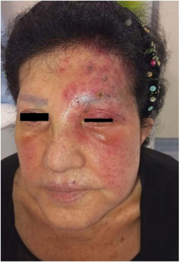
Vesicular Rash following V1 dermatomal distribution.
Fig. 3.

Blepharoconjunctivitis.
The admitting diagnosis was HZO with superimposed bacterial infection. The patient was started on Clindamycin (600 mg intravenously every 8 hours), Acyclovir (500 mg intravenously every 8 hours), and Ofloxacin (1 drop every 6 hours).
After two days, the patient improved clinically and was discharged on oral Clindamycin, oral Valaciclovir, and topical Ofloxacin.
3. Discussion
Many treatments are commonly used to manage androgenic alopecia (AA), whether in males or females. Although it is a benign condition, depression, anxiety, and psychosocial impacts are expected consequences of AA [6,7]. The United States Food and Drug Administration (FDA) only approved topical minoxidil, but other treatments like oral minoxidil, acetate, and finasteride are wildly present in the market. These treatments can be time-consuming, and the symptoms can return to medication nonadherence [8]. Subsequently, many patients seek alternative, long-lasting treatment such as the Platelet-Rich Plasma injection [8,9]. Studies are being performed on PRP efficacy for hair restoration, bone, cartilage regeneration, and wound repair. With this minimally invasive approach, local infection is one of the common complications, mainly if not performed in a sterile technique. This patient developed HZO with superimposed bacterial infection following PRP use; this condition needed intravenous antibiotics and hospitalization for monitoring. Herpes Zoster Ophtalmicus is an ophthalmic emergency as it can be sight-threatening if not treated appropriately and promptly. HZO is defined as an infection of the ophthalmic division of the 5th cranial nerve by Herpes zoster. Direct ocular involvement can be present, such as in this case where the patient complained of blurry vision along with systemic symptoms such as fever, malaise, and headaches. The infection can spread and manifest as, conjunctivitis and blepharitis like in this case, but also uveitis, keratitis, or rashes. Early diagnosis and treatment are essential, and the medical treatment requires intravenous or oral antivirals (acyclovir, valacyclovir, famciclovir) to limit the viral replication. Steroids might be added to the regimen to reduce the inflammatory response. This patient was also started on clindamycin to cover a possible superimposed infection, later confirmed by the eye swab culture.
Furthermore, the Naranjo scale for determining the likelihood of the adverse drug effect was calculated, and the score was 6, meaning the adverse event is probably due to the drug in question (Appendix 1).
Trying to establish a causality between HZO and the PRP treatment, we looked at other risk factors or triggers that could explain the reactivation of the virus. Of which we can eliminate in this case a weakened immune system as the patient was not on any steroid or anti-inflammatory treatment and as a negative personal and family medical history for autoimmune diseases and hasn't have any other procedures done before receiving this treatment. She did not receive any recent vaccination, which leaves the PRP therapy the most probable cause of stress that by consequence lead to the eruption of varicella [10].
Finally, to ensure that the patient received adequate care, we asked for follow up pictures (Fig. 4, Fig. 5). In these pictures we can clearly see that no vesicles, rash or scars are present 1.5 months after the episode.
Fig. 4.
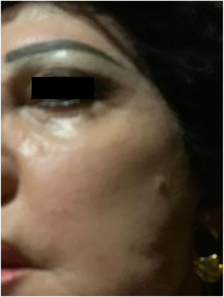
Follow up pictures after a successful treatment.
Fig. 5.
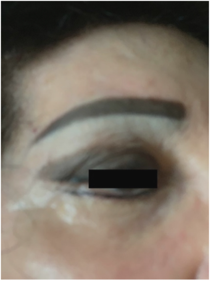
Follow up pictures after a successful treatment.
4. Limitations
We lack enough data on the steps applied during PRP and the sterility of the products used, making us unable to link this complication to a particular stage in the procedure. We also did not test for the viral load in blood or body fluid. The managelent was based on clinical exam and improvement after antiviral instauration.
Nonetheless, other potential risk factors could have contributed to the development of HZO in this patient, such as the patient's age,immune system function, or previous exposure to the varicella-zoster virus that was not taken into account during patients' interview.
5. Conclusion
To sum up, the bried article discusses the case of a female who suffered unfortunate skin infection after PRP treatment. Although it is understandable that alopecia can have psychological and emotional burden especially to a female patient, adhering to the guidelines and safe protocols is important to avoid adverse outcomes. PRP is a minimally invasive but it can have serious side effects and can cause acute stress if not executed adequately. Early diagnosis and prompt management of HZO is crucial as it can have detrimental effects on the patient, the most severe being vision loss.
Data availability statement
No new data were created or analyzed in this study. Data sharing is not applicable to this article.
CRediT authorship contribution statement
Rawan Zeineddine: Writing – review & editing, Writing – original draft. Danielle Bou Khater: Supervision, Writing – original draft, Writing – review & editing. Yara Mouawad: Writing – original draft. Cima Hamieh: Writing – review & editing. Mahmoud El-Hussein: Writing – review & editing, Writing – original draft, Supervision, Methodology.
Declaration of competing interest
The authors declare that they have no known competing financial interests or personal relationships that could have appeared to influence the work reported in this paper.
Appendix.
Are there previous conclusive reports on this reaction?
Yes (+1)
2. Did the adverse events appear after the suspected drug was given?
Yes (+2)
3. Did the adverse reaction improve when the drug was discontinued or a specific antagonist was given?
Yes (+1)
4. Did the adverse reaction appear when the drug was readministered?
Do not know or not done (0)
5. Are there alternative causes that could have caused the reaction?
NO (+2)
6. Did the reaction reappear when a placebo was given?
Do not know or not done (0)
7. Was the drug detected in any body fluid in toxic concentrations?
Do not know or not done (0)
8. Was the reaction more severe when the dose was increased, or less severe when the dose was decreased?
Do not know or not done (0)
9. Did the patient have a similar reaction to the same or similar drugs in any previous exposure?
No (0)
10. Was the adverse event confirmed by any objective evidence?
No (0)
Scoring
-
•
≥ 9 = definite ADR
-
•
5–8 = probable ADR
-
•
1–4 = possible ADR
-
•
0 = doubtful ADR
References
- 1.Stevens J., Khetarpal S. Platelet-rich plasma for androgenetic alopecia: a review of the literature and proposed treatment protocol. International Journal of Women’s Dermatology. Feb. 2019;5(1):46–51. doi: 10.1016/j.ijwd.2018.08.004. [DOI] [PMC free article] [PubMed] [Google Scholar]
- 2.https://www.freepik.com/premium-vector/hair-loss-stages-male-female-alopecia-circle-infographic-steps-baldness_27523843.htm
- 3.Alves R., Grimalt R. A review of platelet-rich plasma: history, biology, mechanism of action, and classification. Skin Appendage Disord. 2018;4(1):18–24. doi: 10.1159/000477353. [DOI] [PMC free article] [PubMed] [Google Scholar]
- 4.Gentile P., Garcovich S. Systematic review of platelet-rich plasma use in androgenetic alopecia compared with Minoxidil®, Finasteride®, and adult stem cell-based therapy. Indian J. Manag. Sci. Apr. 2020;21(8):2702. doi: 10.3390/ijms21082702. [DOI] [PMC free article] [PubMed] [Google Scholar]
- 5.Abdelkader R., Abdalbary S., Naguib I., Makarem K. Effect of platelet rich plasma versus saline solution as a preservation solution for hair transplantation. Plastic and Reconstructive Surgery - Global Open. Jun. 2020;8(6):e2875. doi: 10.1097/GOX.0000000000002875. [DOI] [PMC free article] [PubMed] [Google Scholar]
- 6.Starace M., Orlando G., Alessandrini A., Piraccini B.M. Female androgenetic alopecia: an update on diagnosis and management. Am. J. Clin. Dermatol. Feb. 2020;21(1):69–84. doi: 10.1007/s40257-019-00479-x. [DOI] [PubMed] [Google Scholar]
- 7.Russo P.M., Fino E., Mancini C., Mazzetti M., Starace M., Piraccini B.M. HrQoL in hair loss‐affected patients with alopecia areata, androgenetic alopecia and telogen effluvium: the role of personality traits and psychosocial anxiety. J. Eur. Acad. Dermatol. Venereol. Mar. 2019;33(3):608–611. doi: 10.1111/jdv.15327. [DOI] [PubMed] [Google Scholar]
- 8.Kanti V., et al. Evidence-based (S3) guideline for the treatment of androgenetic alopecia in women and in men - short version. J. Eur. Acad. Dermatol. Venereol. Jan. 2018;32(1):11–22. doi: 10.1111/jdv.14624. [DOI] [PubMed] [Google Scholar]
- 9.Gentile P., Dionisi L., Pizzicannella J., de Angelis B., de Fazio D., Garcovich S. A randomized blinded retrospective study: the combined use of micro-needling technique, low-level laser therapy and autologous non-activated platelet-rich plasma improves hair re-growth in patients with androgenic alopecia. Expet Opin. Biol. Ther. Sep. 2020;20(9):1099–1109. doi: 10.1080/14712598.2020.1797676. [DOI] [PubMed] [Google Scholar]
- 10.https://www.healthline.com/health/what-causes-shingles-to-activate#causes
Associated Data
This section collects any data citations, data availability statements, or supplementary materials included in this article.
Data Availability Statement
No new data were created or analyzed in this study. Data sharing is not applicable to this article.