

Introduction
Amniotic band syndrome (ABS) is an isolated or a group of fetal structural malformations detected at birth. This is an uncommon and poorly understood embryo-fetopathy. The estimated incidence of amniotic band syndrome ranges between 1 in 1200 and 1 in 15,000 live births [1]. Fetal malformations could be one of the three main categories: (A) limb defects, (B) craniofacial defects and (C) visceral defects. The common presentations are: constriction bands, acrosyndactyly, clubbed feet, rarely auto-amputations of the limbs and digits. Origin of these malformations is thought to be multifactorial [2]. Early diagnosis is essential for its management. Presently, prenatal screening and diagnosis is widely used in obstetric practice. The aim of this presentation is to create awareness about this problem among the obstetricians, so that early diagnosis could be made. The parents need to be counseled for the options available for the updated management.
Case Presentation
Mrs AL, a 25-year-old primigravida, non-consanguineous marriage, no history of teratogenic drug intake, at 37 + weeks of gestation, (corresponding to early trimester ultrasonography scan), presented at the obstetric emergency of RL JALAPPA Hospital, Karnataka, with the complaints of pain abdomen and leakage of liquor per vaginam for a duration of 4 h. She was an unbooked case (level 1) and had no history of exanthematous illnesses. There was no clinical evidence of chorioamnionitis. General physical examination was normal. Obstetric examination revealed: uterus 34 weeks’ size with ‘fetus hugging the uterus’ suggestive of gross oligohydramnios. There was persistent fetal bradycardia of 90 to 100 beats per minute over a period of 20 min assessment. Mild uterine contractions were there. Pelvic examination (speculum) revealed cervix: 2 cm long, Os: 2 cm dilated, leakage of clear liquor was seen through the Os. Routine hematological investigations as done were within normal limits. Patient underwent emergency cesarean delivery (CD) in view of grossly reduced liquor and fetal bradycardia. Intraoperatively, patient was diagnosed with bicornuate uterus. A live baby girl was born with birth weight of 2062gm. Baby cried immediately after birth. Apgar at 1 min.:6 and at 5 min. 8. Baby was identified to have multiple congenital malformations suggestive of amniotic band syndrome (ABS). Thorough neonatal examination revealed: left lower limb had a pterygium extending from the hip to the ankle (Fig. 1). At birth knee was found flexed at 45 degrees.
Fig. 1.
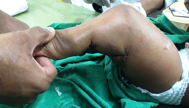
Left lower limb following Z plasty
Club foot of the left side along with right calcaneovalgus deformity were observed. Multiple missing digits and syndactyly in the upper limbs (Fig. 2) with small volume of muscles were observed.
Fig. 2.
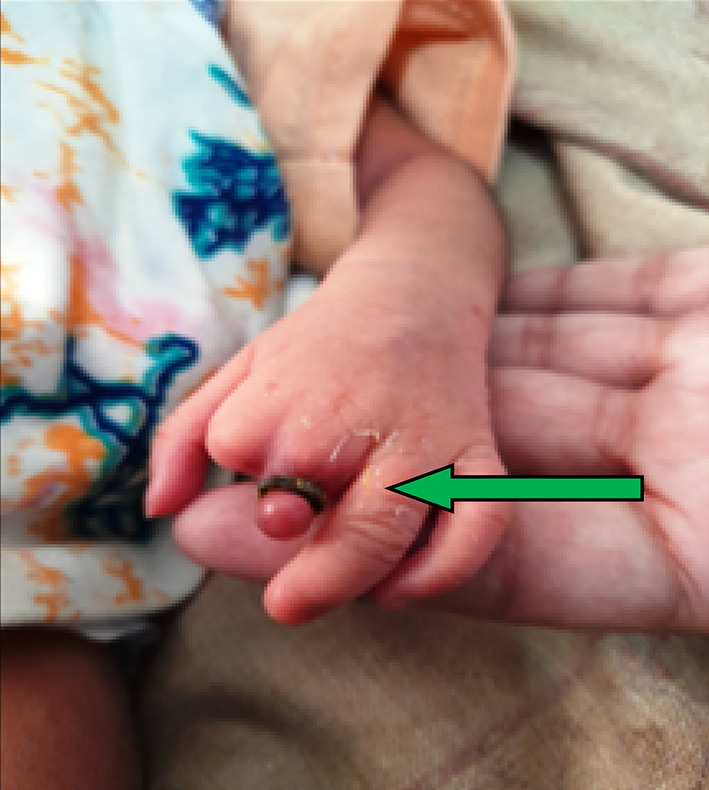
Left hand of the neonate showing amputation of the digits of the index and middle finger. The constricting band on the digit is seen (arrow)
The neonate was managed initially in the neonatal intensive care unit (NICU). Karyotype revealed normal pattern. The existing constriction bands were removed. On the 5th day, baby was shifted to a tertiary care center for surgical corrections. At the age of 6 months, baby underwent Zplasty along with tendoachilles lengthening (Fig. 1). Baby is doing well and waiting for next step of reconstructive surgery.
Management Options
Management of ABS varies significantly depending upon the site and severity of affection However, involvement of the multidisciplinary team management is important. Diagnosis was missed in the antenatal period. Early prenatal targeted ultrasonography using 3D/ 4D machine by an expert can detect amniotic bands [3]. Doppler study can assess the vascular supply of the affected parts. MRI can be used for confirmation of prenatal diagnosis and also for the details of information.
Fetoscopic laser interruption of the amniotic bands has been done successfully [4]. Medical Termination of Pregnancy (MTP) should be offered for cases with the presence of severe craniofacial and visceral malformations. Surgery is not needed for shallow constriction bands that are not circumferential or without any distal part swelling.
Discussion
In our case, multiple abnormalities were detected involving both the lower and the upper limbs as described above. Amniotic bands were detected around both the limbs. However, the presence of an amniotic membrane in contact with these injuries is not essential for the diagnosis [2].
Exact etiology of ABS is poorly understood. Amniotic band syndrome is the result of fibrous amniotic bands or floating amniotic strands entangling the body parts of the fetus. In most cases, it involves the limbs. It may encircle the digits. Cases of limb-body wall complex associated with amniotic band syndrome have been observed [5].
The origin of this malformation is multifactorial, involving multiple pathological processes (genetic, infectious or environmental). Researchers have developed many theories (exogenous, endogenous, vascular or genetic) based on clinical findings or animal experiments, but none has been proven so far [2]. ABS has been classified with extrinsic and intrinsic theories. Both explained the pathology being the germ plasm defect or the mesodermal germ cell defect, leading to either sloughing of the superficial skin or abnormal creases of the skin forming constricting bands to cause avascularity. Depending upon the severity, this results in limb or digital maldevelopment or even amputation. These bands are difficult to see by ultrasonography as in our case. Based upon the extrinsic theory, there is lack of a complete amniotic lining in the placenta of neonates with ABS. It is accepted that intrauterine trauma led to premature rupture of the membranes and ultimately led to the formation of strands that encircle the body parts to cause the pathology. Studies often correlate with several high risk factors as mentioned: •Use of recreational drugs •Trauma •Prematurity •History of amniocentesis •Chorion villous sampling •Use of Misoprostol for the 1st trimester abortion. •Uterine malformations or placental abnormalities. But none is conclusively established. Our patient had bicornuate uterus. She presented in early labor following premature rupture of membranes (PROM).
Conclusion
ABS is an uncommon embryo-fetopathy of multifactorial etiology. Antenatal early diagnosis is possible. Interventions are tailored according to an individual fetus/neonate. Multidisciplinary team approach is needed for the management. The aim of this paper is to create increase in awareness about this abnormality. Parents need to be counseled about the need of surgery.
Acknowledgements
We acknowledge the contribution of the surgical team in the management of this case. We thank Md Jakir Hossain for his assistance in manuscript preparation, and typing the manuscript.
Funding
This study received no grant from any funding agency.
Declarations
Conflict of interest
We declare that we have no conflicts of interest relevant to this article.
Ethical approval
All procedures followed were in accordance with the ethical standards of the responsible committee on the human participants (institutional and national) and with the Helsinki Declaration of 1975, as revised in 2008.
Informed consent
Informed consent was obtained from the participant of the study.
Footnotes
Publisher's Note
Springer Nature remains neutral with regard to jurisdictional claims in published maps and institutional affiliations.
References
- 1.Sentilhes L, Verspyck E, Patrier S, Eurin D, Lechevallier J, Marpeau L. Maladie des brides amniotiques: etiopathogenie, diagnostic antenatal et prise en charge neonatale. J Gynecol Obstet Biol Reprod (Paris) 2003;32:693–704. [PubMed] [Google Scholar]
- 2.Mourabbih LM, Ouajih O, Jalal M, Fichtali K, Bouhya S. Amniotic band syndrome: a case report. Int J Surg Case Rep. 2022;95:1–4. doi: 10.1016/j.ijscr.2022.107096. [DOI] [PMC free article] [PubMed] [Google Scholar]
- 3.Clarke JC, Salonen DC. Amniotic band syndrome. Can Assoc J Radiol. 2000;51(2):134–136. [PubMed] [Google Scholar]
- 4.Gueneuc A, Chalouhi GE, Borali D, Mediouni I, Stirnemann J, Ville Y (2019) Fetoscopic release of amniotic bands causing limb constriction: case series and review of the literature. Fetal Diagnosis Therapy. [DOI] [PubMed]
- 5.Mekonen E, Muhidin AB, Abiye H, et al. Amniotic band syndrome associated with extremely severe atypical clefts of the orofacial region. Eur J Plast Surg. 2022;45:509–513. doi: 10.1007/s00238-021-01860-y. [DOI] [Google Scholar]