Abstract
Calculus in the urethra of the female is very unusual. The patient remains asymptomatic or uncommonly presents with symptoms of dysuria, post-void urinary dribbling, and dyspareunia. If asymptomatic, it can be diagnosed incidentally on gynecological examination. Being hard in consistency, it may mimic metastatic lesion. We present a case of a female who presented to us for management of ovarian mass. On routine examination there was a hard mass in her vagina which was suspected to be a metastatic lesion. This mass on evaluation came out to be a urethral diverticulum with a large calculus. Very large urethral calculus are a very rare presentation in a female.
Introduction
Urethral diverticulum (UD) is infrequently found in women. The presence of calculus within the urethral diverticulum is even more uncommon. Due to shorter and straighter urethra, only a few cases of UD are reported in females. The incidence of UD in adult women varies between 0.6 and 6% [1]. Calculus is reported in 1.5–10% of UD [1]. The quality of life of patients having diverticulum (especially with calculi) is significantly disturbed many a time. The classic presentation of the urethral diverticulum is described historically as the three Ds: dyspareunia, dribbling (post-void), and dysuria which is seen only in 5% of patients [2]. As many as 20% of patients are asymptomatic. Urethral diverticular cancer and calculi can mimic metastatic deposits [3]. Clinically, it occurs concomitantly with other urogynaecological cancers. We present a case of a female having a complex ovarian mass with suspected vaginal metastasis, which was diagnosed as a giant urethral calculus on evaluation. This was successfully managed with diverticulectomy and calculi extraction by vaginal route.
Case Summary
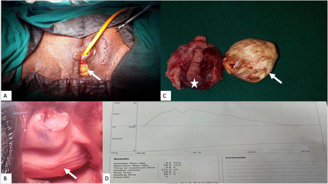
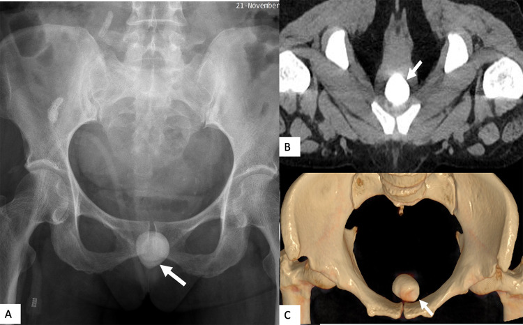
A 64-year-old postmenopausal female known case of hypertension and diabetes mellitus was referred from a private hospital with chief complaints of pain in the lower abdomen, abdominal lump, constipation and bloating from the past 20 to 25 days in the Department of Obstetrics and Gynecology. The pain was insidious in onset gradually progressive with no aggravating factor. This was associated with feeling of some lump in lower abdomen with a gradual increase in size. The patient also had a decreased frequency of micturition from the past week along with hesitancy and urgency. There was no history of weight loss or loss of appetite and postmenopausal bleeding. She was suspected of having ovarian malignancy on ultrasound and was referred for management of the same. On evaluation and physical examination, the patient’s general condition was fair. There were no palpable cervical, axillary, or inguinal lymph nodes. On per abdominal examination, there was an abdominal mass in the right iliac fossa of size 9 × 10 cm which was vaguely defined, non-tender with restricted mobility. On per speculum examination, a sub-urethral bulge was noted of 4 × 5 cm, about 1 cm behind the urethral meatus, with a grossly healthy-looking cervix and vagina (Fig. 1a). A stony hard mass was felt at the anterior vaginal wall on per vaginal examination. Presence of a firm to cystic non-tender mass of 10 × 9 cm with restricted mobility felt in the right adnexa was confirmed, along with a normal size uterus felt separate from it. Thus, a diagnosis of suspected right ovarian malignancy with unusual vaginal metastasis or bone growth was made. A radiograph of the pelvis and CT scan were performed, which revealed a large solid cystic right ovarian tumour with papillary projections and septations likely to be malignant. In addition, a posterior urethral diverticulum with impacted large hyperdense calculus was also seen (Fig. 2a–c). Her serum Ca 125 was 72.4 U/ml, CA-19.9 was 0.3 U/ml, and CEA was 2.343 U/ml. The RMI score calculated was 651.6.
Fig. 1.
On examination a sub-urethral bulge (A) was noted (arrow), about 1 cm behind the urethral meatus (B), Specimen of urethral diverticulum (asterisk) and 4 × 4 cm calculus (arrow) (C). Uroflowmetry showed an unobstructed curve with maximum flow rate of 18.5ml/sec (D)
Fig. 2.
Radiograph of pelvis anteroposterior view (A), non-contrast computed tomography axial image (B) and 3D volume rendered image (C) showing a large hyperdense calculus in the urethral diverticulum (white arrow)
The diagnosis of a malignant right ovarian mass with a large impacted calculus in UD was made. The patient was planned for staging laparotomy along with lithotripsy and diverticulum repair in conjunction with the urology department. Intraoperatively, a large solid cystic mass with intact capsule was seen in right ovary. Left adnexa was normal. Examination of abdominal organs in a clockwise direction showed no evidence of metastasis. The frozen section of ovarian mass came to be borderline serous cystadenoma. Decision of total abdominal hysterectomy, bilateral salpingo-oophorectomy, bilateral retroperitoneal pelvic lymph node dissection (RPLND), and infracolic omentectomy was taken looking at borderline nature of cyst and postmenopausal state of women. Cystourethroscopy showed UD with impacted calculus. Transvaginal urethral diverticulum excision was done, and a 4 × 4 cm calculus was retrieved from UD (Fig. 1c). This was followed by repair with Martius flap interposition.
The patient was discharged on postoperative day (POD) five. No wound-related complications were observed. The Foleys catheter was removed on POD 21. Histopathology of the ovarian mass showed serous cystadenofibroma. No tumour deposits were noted in a total of 20 retrieved lymph nodes. The urethral diverticulum showed inflammatory changes without evidence of any malignancy.
In follow-up, the patient is voiding satisfactorily without any symptoms. Uroflowmetry showed an unobstructed curve with a maximum flow rate of 18.5 ml/s (Fig. 1d).
Discussion
The presentation of urethral diverticulum is highly variable, ranging from incidental detection during physical examination or imaging to storage LUTS, dyspareunia, urinary incontinence, or tumour [4]. The possible underlying mechanism for diverticulum formation is a urethral abscess secondary to instrumentation, trauma, or infection, which ruptures gradually in the urethra, leading to diverticulum formation [2]. The quality of life of those with a diverticulum (especially with stones) may be significantly disturbed because of varied symptomatology. The main complications of UD are inflammation, stone formation, and neoplastic changes. Urethral stones are rare and generally more common in men with urethral stricture or diverticulum. Patients with lower urinary tract symptoms that are unresponsive to traditional treatment should be suspected of having UD. Stasis of infected urine with deposition of salts and occasional mucoid desquamation of the epithelial lining is known to be the causal factor for stone formation. It is associated with pyuria and intermittent dysuria [2]. Distal urethral obstruction also contributes to this process [1, 2]. UD calculi are usually composed of triple phosphate (struvite). A hard anterior vaginal mass indicates calculus or cancer within UD, requiring urgent investigation. Radiographs, CT scan, and cystourethrography helps in confirming the presence of a urethral diverticulum and calculus. However, these are sometimes insufficient because of the possibility of impacted calculus or no space in the diverticula where magnetic resonance imaging can be a good alternative [2, 4].
The calculus (4cm) in this case report is one of the largest calculus removed from UD. Up to 10% of patients with UD show atypical pathology even after normal imaging findings. The incidence of malignancy is 1–6% in UD. The most common are adenocarcinoma, transitional cell carcinoma, and squamous cell carcinoma. Allen et al. noted isolated urethral metastasis in the case of primary ovarian malignancy [3]. Ovarian cancer is the second most common gynaecological cancer (9.4/100,000) [3]. It primarily disseminates through transperitoneal mode in the peritoneal cavity. Malignant ovarian mass can occasionally metastasize to the urethral region. So, the differential diagnosis of hard anterior vaginal masses in patients with ovarian masses can be UD with calculus, UD with urethral cancer, or metastasis.
The management of UD with calculus depends on the size and site of the calculus, with no clear guidelines due to its rarity [1]. Agwu et al., in their large retrospective study of ten years, have found only one woman with urethral calculi. They have discussed possible management options for calculi [4]. Small stones (less than 10 mm) can be left to pass on their own. With any surgical approach, there is risk of incontinence and stricture formation. If calculus is proximal near the bladder, it can be manipulated back into bladder and extracted with cystolitholapaxy. For distal calculus, extraction with urethral meatotomy is an option. Large anterior calculi are taken out with open surgery like urethroplasty. The associated complications are urinary tract infection, fistula formation, urinary retention, incontinence, and strictures [2, 4]. Other endoscopic techniques like pneumatic lithotripsy combined with ultrasound lithotripsy (PLCUL) have also been tried by surgeons depending on size and site. Manual extraction is usually avoided because of the risk of abrasive injury and stenosis. We performed diverticulectomy along with calculus extraction from the vaginal route.
Conclusion
This case highlights the importance of thorough clinical examination. Large UD with stone can mimic a metastatic deposit in periurethral region in cases of known pelvic tumours.
Funding
This research did not receive any specific grant from funding agencies in the public, commercial, or not-for-profit sectors.
Declarations
Conflict of interest
No potential conflict of interest to this article was reported.
Informed Consent
Written informed consent was obtained from the patient for publication of this case report.
Footnotes
Neha Agrawal: Senior Resident, MD, DNB, FMAS; Shashank Tripathi: Senior Resident, MBBS, MS; Priyanka Kathuria: Associate professor, MBBS, DGO, MS, FMAS, DipMAS; Shashank Shekhar: Professor, MD, MRCOG (UK), DNB, FMAS; Deepak Prakash Bhirud: Assistant Professor, MS, MCh; Taruna Yadav: Additional Professor, MD; Mahendra Singh: Associate Professor.
Publisher's Note
Springer Nature remains neutral with regard to jurisdictional claims in published maps and institutional affiliations.
References
- 1.Ati N, Chakroun M, Boussaffa H, et al. Female urethral diverticulum containing calculi: a rare and tricky condition. Urol Case Rep. 2018;20(21):101–103. doi: 10.1016/j.eucr.2018.09.012. [DOI] [PMC free article] [PubMed] [Google Scholar]
- 2.Greiman AK, Rolef J, Rovner ES. Urethral diverticulum: a systematic review. Arab J Urol. 2019;17(1):49–57. doi: 10.1080/2090598X.2019.1589748. [DOI] [PMC free article] [PubMed] [Google Scholar]
- 3.Allan M, Tailor A, Butler-Manuel S, et al. Isolated urethral metastasis from a primary ovarian malignancy. J Obstet Gynaecol Can. 2018;40(12):1632–1634. doi: 10.1016/j.jogc.2018.05.012. [DOI] [PubMed] [Google Scholar]
- 4.Agwu NP, Abdulwahab-Ahmed A, Sadiq AM, et al. Management of impacted urethral calculi: an uncommon cause of acute urine retention in North-western Nigeria. Int J Clin Urol. 2020;4(1):1. doi: 10.11648/j.ijcu.20200401.11. [DOI] [Google Scholar]


