Dear Editor,
We present a case of combined application of CIRCLE software (Carl Zeiss Meditec AG, Germany) and topography-guided laser-assisted in situ keratomileusis (Topo-LASIK) for small-incision lenticule extraction (SMILE) enhancement. SMILE is a safe, minimally invasive corneal laser surgery using a femtosecond laser to create an extractable stromal lenticule. However, potential complications include black spots and suction loss[1].
Suction loss in SMILE surgery is most common mainly due to excessive liquid on the cornea, poor eye fixation, sudden eye movements, or conjunctiva embedded in the gap between the suction ring and cornea during scanning. Loss of suction may lead to scan termination. Incorrect realignment or separation during a second attempt can cause postoperative refractive errors and visual impairments[2]. This case describes a new enhancement technique for suction loss during SMILE. Written informed consent was obtained from the patient according to the protocol approved by the ethics review board of Eye and ENT hospital of Fudan University (approve number: 2019092). The study followed the tenets of the Declaration of Helsinki.
A 27-year-old man came to our hospital for bilateral SMILE surgery to correct myopia. The preoperative manifest refraction was -7.5 D/-0.75 D×175° in the right eye, with a corrected distance visual acuity (CDVA) of 20/20, and -3.5 D/-1.5 D×15° in the left, with a CDVA of 20/20. Surgery was performed with a corneal cap diameter of 7.6 mm and depth of 120 µm using the VisuMax femtosecond laser system (Carl Zeiss Meditec AG, Germany). Surgery on the right eye was successful. However, during surgery on the left eye, suction loss occurred when the corneal cap was scanned to a diameter of 0.51 mm (7%). Following realignment, the corneal cap was rescanned with the initial parameters, and the lenticule extraction was successful. There were no postoperative complications, such as infection or hazy opacity.
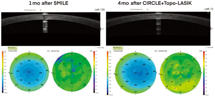
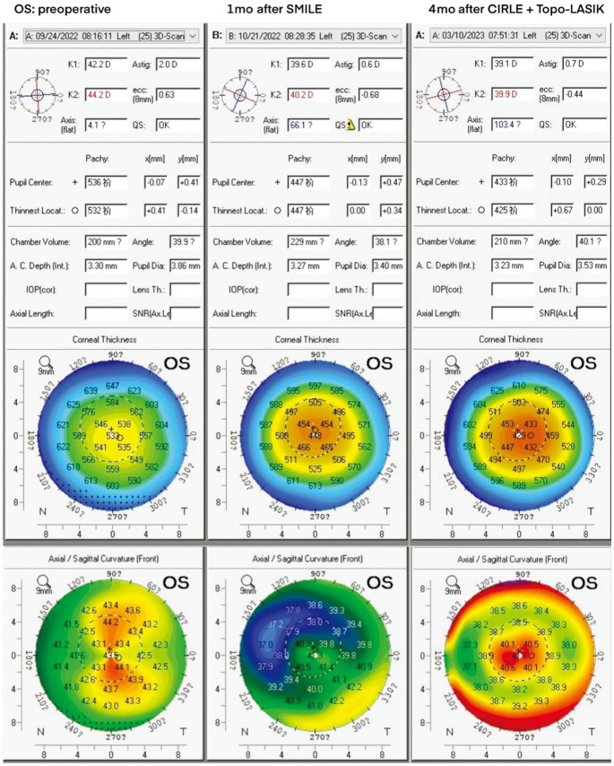
One day after surgery, good recovery was observed in the right eye, with a refractive error of -0.25 D and a best-corrected visual acuity of 20/20. In contrast, the left eye exhibited a manifest refraction of 0/-1.25 D×65° and a CDVA of 20/50. Twenty days after surgery, the manifest refraction in the left eye was -0.5 D/-1 D×65° with a CDVA of 20/100. Anterior segment optical coherence tomography (AS-OCT) using RTVue-100 (Optovue Inc., Fremont, CA, USA) revealed no significant abnormalities, and the corneal stroma was transparent (Figure 1). Postoperative corneal tomography using Pentacam HR (Oculus, Wetzlar, Germany) revealed an abnormally high corneal curvature in the temporal inferior central region of the left eye up to 41.7 D (Figure 2), indicating that the second corneal cap scan was not performed in the same plane as the first, resulting in a thinner central region of the separated lenticule, causing irregular astigmatism and decreased vision in the left eye (Figure 3).
Figure 1. AS-OCT results for the patient's left eye 1mo after SMILE surgery and 4mo after enhancement surgery.
AS-OCT: Anterior segment optical coherence tomography; SMILE: Small-incision lenticule extraction.
Figure 2. Pentacam results for the patient's left eye before surgery, 1mo after SMILE surgery, and 4mo after enhancement surgery.
SMILE: Small-incision lenticule extraction.
Figure 3. Illustration of irregular astigmatism in the patient's left eye caused by suction loss.
One month after SMILE, enhancement surgery was performed on the left eye using the CIRCLE procedure combined with Topo-LASIK. Prior to surgery, the five best corneal tomography scans obtained from the Topolyzer Vario (WaveLight, Erlangen, Germany) were used to plan the Topo-LASIK treatment. The CIRCLE procedure was programmed to create an 8.1 mm corneal flap at a depth of 120 µm using a Visumax femtosecond laser in “junction up and down” mode to replace the corneal flap required by LASIK. Corneal flap hinge was placed on the nasal side. After lifting the corneal flap, a Mel-90 excimer laser system (Carl Zeiss Meditec AG, Germany) was used for stromal ablation, with a 6 mm optical zone, a corrective spherical power of -0.5 D, and an ablation depth of 45 µm. A soft contact lens was placed on the left eye after surgery, and fluorometholone and levofloxacin eye drops were administered four times a day, with gradual reductions in frequency over the course of 2wk.
One day after enhancement surgery, the contact lens was removed, and manifest refraction in the left eye was -0.25 DS, with a CDVA of 20/25. Pentacam results revealed decreased central corneal curvature and increased symmetry (Figure 2). No complications, such as displacement of the corneal cap or epithelial implantation, occurred after surgery. Four months after enhancement surgery, the left eye exhibited no refractive error, with an uncorrected visual acuity reaching 20/20.
The incidence of suction loss during SMILE ranges from 0.17% to 0.93%, with the majority of cases (>50%) occurring during corneal cap scanning[3]. The most common approach to the latter is rescanning the corneal cap using the same scan parameters after careful central alignment[2]. Suction loss of up to 7% during corneal cap scanning has not been reported. The risk of creating a different layer during re-scanning is worthy of attention. In addition, SMILE procedures, during which suction loss occurred, that were discontinued and rescheduled for secondary SMILE surgery (re-SMILE) or LASIK have also been reported, with patients experiencing satisfactory visual outcomes[4].
Enhancement surgery following SMILE is required for cases of overcorrection, undercorrection, or irregular astigmatism in 1%-4% of cases. The main risk factors include high refractive error, high astigmatism, suction loss, and patient age over 35 years[3]. Such procedures include re-SMILE, photorefractive keratectomy (PRK), thin-flap LASIK, and cap-to-flap conversion treatment using CIRCLE software. In this case, identifying residual stromal tissue on the corneal cap after re-scanning was challenging on AS-OCT, making removal difficult during re-SMILE, with the possibility of entering the same false plane caused by the initial SMILE procedure[5]. In addition, PRK has an approximately 2.5% risk of developing haze, while thin-flap LASIK is more suitable in cases of corneal cap ≥160 µm[5]. Therefore, CIRCLE enhancement plan was chosen.
CIRCLE software uses a Visumax femtosecond laser for side cuts, converting the SMILE cap into a LASIK flap, which is suitable for residual stromal tissue caused by SMILE in complex situations, such as interface irregularities not visible on AS-OCT or residual lenticules on the stromal bed due to the formation of false planes[6]. Kostin et al[7] reported a case of post-SMILE refractive regression treatment using CIRCLE software combined with a MEL-80 excimer laser; after surgery, the patient showed no refractive error. In a matched comparative study by Siedlecki et al[8], the safety and effectiveness of CIRCLE for post-SMILE enhancement surgery were investigated, and it was found to produce better visual outcomes than PRK.
At the same time, since patients' corneal curvature in the central temporal inferior region of the cornea increased abnormally due to suction loss, topography-guided corneal ablation plan is more suitable for its excellent ability to correct congenital and surgery-induced corneal irregularities. Therefore, a combined CIRCLE and Topo-LASIK enhancement plan was chosen. Farooqui and Al-Muammar[9] suggested that Topo-LASIK decreases the likelihood of postoperative spherical aberrations and coma compared with traditional LASIK, resulting in better night vision quality. Alió et al[10] demonstrated that Topo-LASIK exerts significant therapeutic effects on irregular astigmatism induced after refractive surgery. These studies demonstrate the significance of customized ablation in treating irregular astigmatism after SMILE surgery.
This study provides preliminary evidence for the feasibility and effectiveness of using CIRCLE software combined with Topo-LASIK for enhancement surgery in patients with suction loss after SMILE. Further research with larger sample and longer follow-up periods is required.
Acknowledgments
Authors' contributions: Conception and design of the study: Zhou XT. Illustrations: Sun BQ. Administrative, technical, or material support: Li MY, and Xu HP. Drafting of the manuscript: Sun BQ. Critical revision of the manuscript: Li MY.
Foundations: Supported by the Shanghai Rising-Star Program (No.21QA1401500); Shanghai Natural Science Foundation (No.23ZR1409200).
Conflicts of Interest: Sun BQ, None; Xu HP, None; Zhou XT, None; Li MY, None.
REFERENCES
- 1.Tian M, Jian WJ, Miao HM, Li M, Xia F, Zhou XT. Five-year follow-up of visual outcomes and optical quality after small incision lenticule extraction for moderate and high myopia. Ophthalmol Ther. 2022;11(1):355–363. doi: 10.1007/s40123-021-00436-0. [DOI] [PMC free article] [PubMed] [Google Scholar]
- 2.Wan KH, Lin TPH, Lai KHW, Liu S, Lam DSC. Options and results in managing suction loss during small-incision lenticule extraction. J Cataract Refract Surg. 2021;47(7):933–941. doi: 10.1097/j.jcrs.0000000000000546. [DOI] [PubMed] [Google Scholar]
- 3.Sharma N, Asif M, Bafna R, Mehta J, Reddy J, Titiyal J, Maharana P. Complications of small incision lenticule extraction. Indian J Ophthalmol. 2020;68(12):2711. doi: 10.4103/ijo.IJO_3258_20. [DOI] [PMC free article] [PubMed] [Google Scholar]
- 4.Wong CW, Chan C, Tan D, Mehta JS. Incidence and management of suction loss in refractive lenticule extraction. J Cataract Refract Surg. 2014;40(12):2002–2010. doi: 10.1016/j.jcrs.2014.04.031. [DOI] [PubMed] [Google Scholar]
- 5.Siedlecki J, Luft N, Priglinger SG, Dirisamer M. Enhancement options after myopic small-incision lenticule extraction (SMILE): a review. Asia Pac J Ophthalmol (Phila) 2019;8(5):406–411. doi: 10.1097/APO.0000000000000259. [DOI] [PMC free article] [PubMed] [Google Scholar]
- 6.Ganesh S, Brar S, K V M. CIRCLE software for the management of retained lenticule tissue following complicated SMILE surgery. J Refract Surg. 2019;35(1):60–65. doi: 10.3928/1081597X-20181120-01. [DOI] [PubMed] [Google Scholar]
- 7.Kostin, Rebrikov SV, Ovchinnikov АI, Stepanov, Takhchidi KP. Results of residual ametropia correction using CIRCLE technology after femtosecond laser SMILE surgery. Vestn Oftal'mol. 2017;133(1):55. doi: 10.17116/oftalma2017133155-59. [DOI] [PubMed] [Google Scholar]
- 8.Siedlecki J, Siedlecki M, Luft N, Kook D, Meyer B, Bechmann M, Wiltfang R, Sekundo W, Priglinger SG, Dirisamer M. Surface ablation versus CIRCLE for myopic enhancement after SMILE: a matched comparative study. J Refract Surg. 2019;35(5):294–300. doi: 10.3928/1081597X-20190416-02. [DOI] [PubMed] [Google Scholar]
- 9.Farooqui MA, Al-Muammar AR. Topography-guided CATz versus conventional LASIK for myopia with the NIDEK EC-5000: a bilateral eye study. J Refract Surg. 2006;22(8):741–745. doi: 10.3928/1081-597X-20061001-03. [DOI] [PubMed] [Google Scholar]
- 10.Alió JL, Belda JI, Osman AA, Shalaby AMM. Topography-guided laser in situ keratomileusis (TOPOLINK) to correct irregular astigmatism after previous refractive surgery. J Refract Surg. 2003;19(5):516–527. doi: 10.3928/1081-597X-20030901-06. [DOI] [PubMed] [Google Scholar]


