Abstract
URA5 genes encode orotidine-5′-monophosphate pyrophosphorylase (OMPpase), an enzyme involved in pyrimidine biosynthesis. We cloned the Histoplasma capsulatum URA5 gene (URA5Hc) by using a probe generated by PCR with inosine-rich primers based on relatively conserved sequences in OMPpases from other organisms. Transformation with this gene restored uracil prototrophy and OMPpase activity to UV-mutagenized ura5 strains of H. capsulatum. We attempted to target the genomic URA5 locus in this haploid organism to demonstrate homologous allelic replacement with transforming DNA, which has not been previously done in H. capsulatum and has been challenging in some other pathogenic fungi. Several strategies commonly used in Saccharomyces cerevisiae and other eukaryotes were unsuccessful, due to the frequent occurrence of ectopic integration, linear plasmid formation, and spontaneous resistance to 5-fluoroorotic acid, which is a selective agent for URA5 gene inactivation. Recent development of an efficient electrotransformation system and of a second selectable marker (hph, conferring hygromycin B resistance) for this fungus enabled us to achieve allelic replacement by using transformation with an insertionally inactivated Δura5Hc::hph plasmid, followed by dual selection with hygromycin B and 5-fluoroorotic acid, or by screening hygromycin B-resistant transformants for uracil auxotrophy. The relative frequency of homologous gene targeting was approximately one allelic replacement event per thousand transformants. This work demonstrates the feasibility but also the potential challenge of gene disruption in this organism. To our knowledge, it represents the first example of experimentally directed allelic replacement in H. capsulatum, or in any dimorphic systemic fungal pathogen of humans.
A number of molecular genetic tools and techniques have recently been developed for the dimorphic pathogenic fungus Histoplasma capsulatum (15, 18, 28, 41–43, 45, 46), as for other pathogenic fungi that are of increasing clinical significance and scientific interest (6, 10, 13, 14, 17–19, 22, 29, 32, 39, 40). We have described genetic transformation following chemical treatment or by electrotransformation, and two selection systems using either the Podospora anserina URA5 gene in H. capsulatum ura5 mutants generated by UV mutagenesis or the hph gene encoding hygromycin phosphotransferase, which is also efficacious in nonmutagenized strains (41–43, 46). We have also described the appearance of transforming DNA either integrated apparently randomly in the genome or on de novo-generated linear plasmids, and telomeric shuttle plasmids for efficient DNA delivery to and recovery from H. capsulatum (41–43, 46). Available protocols thus allow delivery of exogenous DNA either episomally or by apparently random genomic integration, but the cardinal requirement of reverse genetics—replacing a genomic locus with a disrupted or otherwise modified copy of a cloned gene—has not previously been demonstrated in this organism. In this study, we report cloning of the native H. capsulatum URA5 gene (URA5Hc), encoding orotidine-5′-monophosphate pyrophosphorylase (OMPpase), and disruption of the genomic locus by allelic replacement with an insertionally inactivated copy supplied by transformation, at a frequency of about one homologously targeted event per thousand transformants.
(A preliminary report of some of this work has appeared elsewhere [42a].)
MATERIALS AND METHODS
Fungal strains.
H. capsulatum G184AS, G184ASura5-11, and G217Bura5-21 have been described previously (41). The parental strains G184A (ATCC 26027) and G217B (ATCC 26032) are clinical isolates of restriction fragment length polymorphism (RFLP) classes 3 and 2, respectively. G184ASura5-11 and G217Bura5-21 were isolated after UV mutagenesis and selection using 5-fluoroorotic acid (FOA) (41, 45). Experiments were done with H. capsulatum grown as yeast cells at 37°C.
Bacterial strains.
Plasmids were propagated in Escherichia coli HB101 (supE44 hsdS20 recA13 ara-14 proA2 lacY1 galK2 rpsL20 xyl-5 mtl-1), JM109 (supE44 hsdR17 recA1 mcrA endA1 thi gyrA96 relA1 Δ[lac proAB] [F′ traD36 proAB lacIqZΔM15]), and SURE (e14− [mcrA]Δ[mcrCB-hsdSMR-mrr]171 endA1 supE44 thi-1 gyrA96 relA1 lac recB recJ sbsC umuC::Tn5 uvrC [F′ proAB lacIqZΔM15Tn10]) (obtained from Stratagene Cloning Systems, La Jolla, Calif.).
Media.
H. capsulatum was grown in broth in rich defined medium HMM or minimal defined medium 3M (44), supplemented with uracil (100 μg/ml) for nonselective growth of ura5 strains. Growth in these media without exogenous uracil was used as the criterion of uracil prototrophy and of complementation of auxotrophic ura5 strains. Solid medium also contained 0.5% (wt/vol) agarose (SeaKem LE agarose, lot no. 634592; FMC BioProducts, Rockland, Maine) and was supplemented with an additional 10 μM FeSO4. For hph selection, hygromycin B (Boehringer Mannheim Biochemicals, Indianapolis, Ind.) was added to solid or liquid medium at a concentration of 200 μg/ml. Penicillin and streptomycin were added to broth, and gentamicin was added to agarose. For selection of uracil auxotrophs, FOA (American Biorganics, Niagra Falls, N.Y.) was added to 3 M at a concentration of 2 mg/ml, together with uracil (100 μg/ml). E. coli was grown in LB broth (3, 31) or on LB–1.5% agarose solid medium, supplemented with appropriate antibiotics.
Nucleic acid preparation.
Plasmids were prepared from E. coli by using an alkaline lysis miniprep protocol (47) or using pZ523 columns (5 Prime→3 Prime, Boulder, Colo.) according to the manufacturer’s recommendations. The procedures for genomic Histoplasma DNA preparations using enzymatic spheroplasting, alkaline-sodium dodecyl sulfate lysis, RNase A and proteinase K treatments, and organic extractions have been described previously (41). Histoplasma total RNA was prepared with an RNeasy RNA isolation kit (Qiagen, Santa Clarita, Calif.). Briefly, log-phase yeasts were pelleted by centrifugation, resuspended in RLT lysis buffer as specified in the kit, mixed with an equal volume of acid-washed glass beads, and disrupted in a Mini-Beadbeater-8 cell disruption apparatus (Biospec Products, Bartlesville, Okla.) for four 30-s intervals at high speed, separated by cooling on ice for 30 s, after which RNA was isolated according to the manufacturer’s recommendations. Histoplasma first-strand cDNA was synthesized from total RNA by using the primer dT16 (Boehringer Mannheim Biochemicals) and Superscript II reverse transcriptase (Gibco BRL, Gaithersburg, Md.) according to the manufacturers’ recommendations.
Plasmids.
DNA fragments were cloned in pBR328 or pBluescript SK+. Both vectors were used in plasmids for transformation of H. capsulatum, for which purpose we have observed no difference between them (43). Vector pBluescript derivatives were also used for DNA sequencing. Two partial libraries were prepared by size fractionation of G184AS genomic DNA fragments on agarose gels after complete restriction digestion with the endonucleases indicated below and then screened by colony hybridization with radiolabeled probes (3, 31). Plasmid pWU10 consists of a 4-kb G184AS genomic DNA XhoI fragment cloned into XhoI-cut pBluescript that hybridized with the PCR-generated URA5Hc probe (see Results) but did not complement the auxotrophy of Histoplasma ura5 strains; sequence analysis showed the fragment to contain an incomplete URA5Hc gene, lacking the 5′ flanking region and start of the coding sequence. Plasmid pWU16 consists of a pWU10 insert-overlapping 0.9-kb G184AS genomic DNA HindIII/BamHI fragment cloned into the large HindIII/BamHI pBluescript fragment; the pWU16 insert includes about 0.4 kb of genomic sequence 5′ to the terminus of the pWU10 insert sequence. The pWU16 insert containing the URA5Hc 5′ end was unified with subclones of pWU10 in the appropriate orientation to generate plasmids containing the entire URA5Hc gene that were shown to confer uracil prototrophy on Histoplasma ura5 mutants. Southern blotting was used to confirm that the restriction endonuclease map of the cloned locus matches that of the strain G184AS genomic locus for all enzymes tested (data not shown). Additional subclones with 5′ or 3′ truncations were constructed and tested for complementation to delineate a minimal complementing fragment that corresponded with the gene predicted from nucleotide sequencing (see Results).
The Δura5Hc::hph disruption plasmid pMAD10 was constructed from pWU90 (43), which contains the 2.9-kb hph selectable marker (including an upstream Aspergillus promoter region) in the polylinker of pBluescript. First, a 1.1-kb ClaI (vector polylinker site)/SmaI (URA5Hc internal site) fragment containing the upstream flanking and 5′ coding region of the URA5Hc gene was cloned into pWU90 digested in the polylinker with ClaI and EcoRV (on one side of the hph marker). This plasmid was then digested in the polylinker on the other side of the hph marker with XbaI and NotI and ligated to a 3-kb AvrII (internal site)/NotI (vector polylinker site) fragment containing the downstream flanking region of the URA5Hc gene. Plasmid pMAD10 thus contains the maximal flanking sequences of the URA5Hc locus that have been cloned, with the 2.9-kb hph marker replacing an internal 0.4-kb SmaI/AvrI fragment including a 3′ part of the coding sequence. The hph marker in pMAD10 is flanked on the URA5Hc 5′ side by 1.1 kb and on the 3′ side by 3 kb of URA5Hc sequence.
Chemicals and molecular biology reagents and methods.
Unless otherwise indicated, chemicals were obtained from Sigma Chemical Company (St. Louis, Mo.). Radionuclides were obtained from Amersham Pharmacia Biotech (Arlington Heights, Ill.) ([α-35S]dATP, [α-32P]dATP, [α-33P]dATP, and [γ-32P]ATP) or New England Nuclear (Boston, Mass.) ([14C-orotic acid]). Restriction endonucleases, T4 DNA ligase, and T4 DNA kinase were obtained from Boehringer Mannheim Biochemicals or New England BioLabs (Beverly, Mass.) and used according to the manufacturer’s recommendations. Amplitaq thermostable DNA polymerase was obtained from Perkin-Elmer (Foster City, Calif.) and used according to the manufacturer’s recommendations. Methods for DNA electrophoresis, staining, band purification from agarose gels, Southern blotting, and random-primed radiolabeling of DNA probes have been described previously (41).
Oligodeoxyribonucleotides used in DNA sequencing, primer extension, or PCR were synthesized at the Washington University Protein Chemistry Laboratory or obtained from Gibco BRL. Nucleotide sequencing was performed on genomic DNA and cDNA clones by the dideoxy-chain termination method (3, 31) by using Sequenase (Amersham Pharmacia Biotech) or cycle sequencing kits (fmol [Promega, Madison, Wis.] or Amplitaq [Perkin-Elmer]), with radiolabeling by internal incorporation or by using an end-labeled primer. The genomic DNA sequence reported here was confirmed on both strands. DNA sequence analyses were performed using Genetics Computer Group (Madison, Wis.) and MacVector (Eastman Kodak, Rochester, N.Y.) software applications.
For determination of the 5′ end of the URA5Hc transcript, primer extension was performed with 0.2 pmol of oligonucleotide 5′ GAAGGGGAGAGCTTGTGGAC 3′ (corresponding to nucleotides 34 to 15), end labeled by using [γ-32P]ATP and T4 DNA kinase, mixed with 10 to 50 μg of G184AS total RNA, incubated at 70°C for 1 min and then at 42°C for 2 min, and reverse transcribed with Superscript II at 42°C. The extension product was precipitated with 1/20 volume of 3 M sodium acetate and 2 volumes of ethanol, resuspended in 4 μl of sequence stop solution (Promega), and electrophoresed on a 6% Long Ranger (FMC BioProducts) gel. Comparison was made to a sequencing ladder prepared from the URA5Hc gene by using the same primer included on the same gel.
The conditions used for PCR amplification of the 131-bp URA5Hc product from G184AS genomic DNA with inosine-rich primers (see Results) were 1 μg of template DNA, 200 pmol of each primer, 200 μM deoxynucleoside triphosphates, 1× buffer, and 2.5 U of Amplitaq in 100 μl of reaction mixture, and PCR for 30 cycles of 90°C for 1 min, 45°C for 1 min, and 72°C for 3 min.
For confirmation of putative intron sequences, PCR was performed with H. capsulatum G184AS genomic DNA and cDNA, using primers that bind to sites outside the boundaries of the two putative introns (5′ ACGACTTTCCTCGAGTCCTG 3′ [corresponding to nucleotides 46 to 65] and 5′ ACGGGGATCCCACGATGCTCCC 3′ [corresponding to nucleotides 548 to 527]) and 25 cycles of 94°C for 30 s, 57°C for 30 s, and 72°C for 30 s, followed by 72°C extension for 10 min. Genomic DNA and cDNA PCR products were electrophoresed in a 1.8% agarose gel for size comparison. A restriction fragment of the cDNA product was cloned into pBluescript and sequenced for comparison to the corresponding genomic DNA sequence.
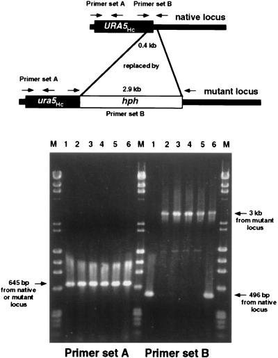
For PCR confirmation of URA5Hc gene disruption by using plasmid pMAD10 in strain G184AS, parental or transformant genomic DNAs were used as templates in two reactions (both for 30 cycles of 94°C for 1 min, 68°C for 1 min, and 72°C for 2 min, followed by 72°C extension for 10 min): a control reaction with URA5Hc primers (5′ ACCAACACCCCTCTCCCCTCG 3′ [corresponding to nucleotides −97 to −77] and 5′ ACGGGGATCCCACGATGCTCCC 3′ [corresponding to nucleotides 548 to 527]) amplifying a 645-bp product predicted to be present in all samples, and a test reaction with URA5Hc primers (5′ GAAGGCGGCAAAGTGGTGGGC 3′ [corresponding to nucleotides 632 to 652] and 5′ CCCGTGTCTTCTCATTCCGAGAG 3′ [corresponding to nucleotides 1127 to 1105]) targeted to sites that flank the site of hph insertion and predicted to amplify a 496-bp product from the native locus and a 3-kb product from the disruption construct.
Genetic transformation.
E. coli was transformed by standard protocols (3, 31). For H. capsulatum, chemical transformation using treatment with lithium acetate and polyethylene glycol (41, 46) and electrotransformation (43) were performed as described previously. Selection for uracil prototrophy was done by plating on HMM agarose without exogenous uracil; selection for FOA resistance (and theoretically uracil auxotrophy) was done by plating on 3 M–FOA–uracil agarose. In the gene disruption experiments with pMAD10, 20 μg of ClaI-digested plasmid DNA was used per electroporation cuvette; aliquots of the electrotransformed G184AS yeast suspensions were plated on Nytran filters on 3 M–uracil agarose or on 3 M–FOA–uracil agarose and incubated at 37°C for 24 to 48 h to allow expression time for the hph marker, and then the filters were transferred to solid medium with the same constituents plus hygromycin B. Colonies arising on 3 M–uracil–hygromycin B were toothpicked to duplicate HMM-hygromycin B agarose plates with or without exogenous uracil, and uracil auxotrophs were further examined by PCR and Southern blotting. Colonies arising on 3 M–FOA–uracil–hygromycin B were considered presumptively to be uracil auxotrophs; this phenotype was confirmed by comparing growth on HMM-hygromycin B agarose with or without uracil, and the fate of the transforming DNA was examined by PCR and Southern blotting. For these experiments, the total number of hygromycin B-resistant transformants was calculated based on the number of colonies arising on a 3 M–uracil–hygromycin B agarose plate (without FOA).
Measurement of OMPpase activity.
Generation of 14CO2 from [14C]orotic acid by yeast cell extracts was performed as described previously (45).
Image photodocumentation.
Ethidium bromide-stained gels were transilluminated with UV light and photographed or were imaged by using a Bio-Rad (Hercules, Calif.) Gel Doc 1000 and Molecular Analyst software. Radioactively labeled filters were exposed to film or phosporimaged with a Bio-Rad GS-525 molecular imager system and Molecular Analyst software. Digital image files were obtained from autoradiographs or photographs by using a scanner and Photoshop software (Adobe Systems, Grove City, Ohio). Figures were constructed from the image files using Canvas software (Deneba, Miami, Fla.).
Nucleotide sequence accession number.
The URA5Hc genomic DNA sequence reported here is available from GenBank under accession no. AF070928.
RESULTS AND DISCUSSION
Cloning and characterization of the URA5Hc gene from strain G184AS.
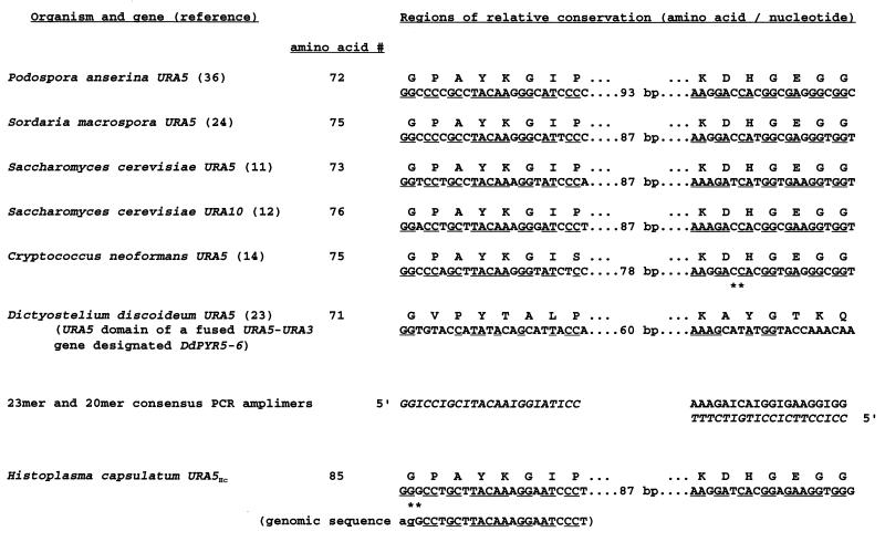
We have previously used a heterologous URA5 gene from P. anserina as a selectable marker in H. capsulatum (41–43, 46). We found this gene to have no detectable hybridization homology with Histoplasma genomic DNA, even under conditions of very low stringency (data not shown). Therefore, we examined the predicted protein sequences of OMPpase from other organisms and identified two short regions that were highly conserved, although the encoding genes showed substantial nucleotide divergence, mainly at the third position of codons (Fig. 1). Based on these regions, we designed PCR primers that contained inosines at most of these wobble positions and used them to amplify a 131-bp product from G184AS genomic DNA that was of appropriate size in comparison to the other genes and had a nucleotide sequence consistent with those of other OMPpase-encoding genes. This fragment was used as a probe to screen plasmid libraries of G184AS genomic DNA. The entire putative URA5Hc gene was cloned on two overlapping fragments, followed by construction of plasmids containing the entire gene, as described in Materials and Methods.
FIG. 1.
Relatively conserved regions of reported OMPpase proteins and the genes encoding them, used to design inosine-rich PCR primers and amplify a 131-bp URA5Hc gene fragment from strain G184AS. Nucleotides that exactly match the primer sequences are underlined. ∗∗, nucleotides separated by an intron in the Cryptococcus and Histoplasma genomic sequences.
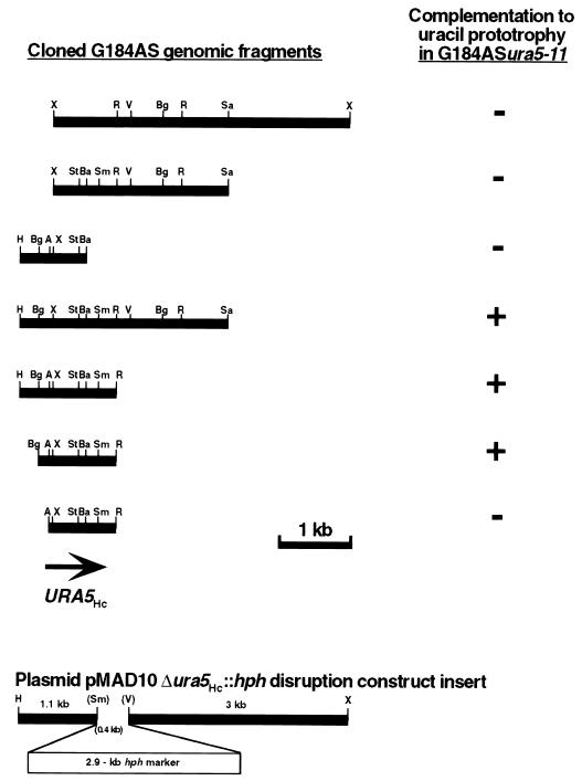
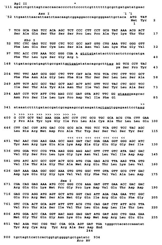
Subclones of URA5Hc DNA were tested for the ability to complement the uracil auxotrophy of strain G184ASura5-11 by transformation (Fig. 2). The nucleotide sequence of the minimal complementing 1.05-kb BglII/EcoRV fragment (Fig. 3) revealed a coding sequence bounded by start and stop codons and containing two putative introns (74 bp from nucleotides 119 to 192; 71 bp from nucleotides 329 to 399) with consensus 5′ splice, branch point, and 3′ splice sites (4). One of these putative introns overlapped slightly one of the PCR primers used to amplify the original product from genomic DNA, but fortuitously this overlap occurred near the 5′ end of the primer and did not interfere with the PCR. Excision of these putative introns would result in a predicted 26.6-kDa, 244-amino-acid protein (Fig. 3) with relatively high amino acid conservation and good alignment with other OMPpases (data not shown). To confirm these introns, PCR was performed on G184AS genomic DNA and cDNA by using primers flanking the region containing both putative introns, corresponding to nucleotides 46 to 65 and 548 to 527. The amplified products showed the sizes (503 bp for genomic DNA; 358 bp for cDNA) predicted to result from the presence or absence of these introns, and nucleotide sequencing confirmed their excision from the cDNA (data not shown). The predicted protein shows 53% identity and 68% similarity with the P. anserina URA5 gene product, and there is 61% homology between the genes, consistent with our failure to detect hybridization between these sequences.
FIG. 2.
Complementation to uracil prototrophy in strain G184ASura5-11 conferred by URA5Hc DNA fragments, and diagram of plasmid pMAD10 Δura5Hc::hph disruption construct insert. Restriction endonuclease sites: A, AsnI; Ba, BamHI; Bg, BglII; H, HindIII; R, EcoRV; Sa, SalI; Sm, SmaI; St, StuI; V, AvrII; X, XhoI. Only mapped sites are shown for each fragment, and not all fragments were mapped with each enzyme.
FIG. 3.
DNA and predicted protein sequences of the minimal complementing URA5Hc fragment (accession no. AF070928). In the DNA sequence (upper lines, with position numbers at the left and at the end of the sequence), protein-coding and stop codon nucleotides are uppercase, and upstream and downstream flanking and intron sequences are lowercase. The probable transcriptional start site (nucleotide −84 T) is marked by an asterisk. The two introns have 5′ splice, branch point, and 3′ splice sites (underlined) that match well the consensus sequences (4) (intron 1 gtctgt…ctgat…tag, intron 2 gtaagc…ctgat…aag, consensus gt[nngy]…ctray…[y]ag [n = any nucleotide; y = pyrimidine C or T; r = purine A or G]). The binding sites for the inosine-rich primers used to amplify a product used as a probe for cloning the entire gene (nucleotides 398 to 420 and 528 to 509) are indicated by inverted arrowheads. 5′ truncation to the indicated AsnI site ablated functional complementation in strain G184ASura5-11. The indicated XhoI site near the start of the coding sequence is the location of a gene fusion with lacZ reported previously (43). The indicated SmaI site marks the beginning of the 0.4-kb fragment deleted and replaced by the hph marker in the plasmid pMAD10 Δ ura5Hc::hph disruption construct; the deletion extends to an AvrII site beyond the 3′ terminus of this fragment. The predicted protein sequence is indicated on the lower lines, with amino acid numbers on the right and below the first and last amino acids.
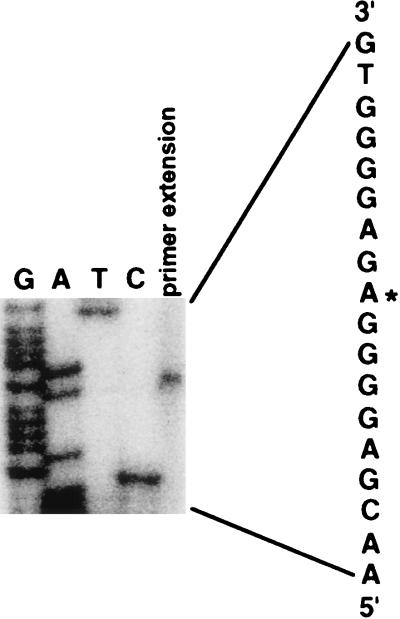
The minimal complementing fragment contains only 111 bp upstream of the start codon and 54 bp downstream of the stop codon. Primer extension analysis performed with strain G184AS RNA and a primer corresponding to nucleotides 34 to 15 localized the 5′ end of the transcript (likely the transcriptional start site) to nucleotide −84 T (Fig. 4). Further 5′ truncation of the upstream region by 70 bp to the AsnI site, which removes the probable transcriptional start site and leaves 41 bp upstream of the start codon, ablated functional complementation in G184ASura5-11 (Fig. 2). Although we have not examined this gene’s regulatory sequences in depth, it is clear that effective function for transformation to uracil prototrophy requires relatively short upstream and downstream flanking sequences. In addition to driving expression of the native gene, this presumptive promoter region has also been used to direct expression of a URA5Hc-lacZ fusion gene (a translational fusion constructed at the XhoI site near the start of the coding sequence), resulting in production of enzymatically active β-galactosidase in H. capsulatum (43).
FIG. 4.
Primer extension analysis of the URA5Hc gene. The first four lanes show a sequencing ladder (reverse strand, sequence indicated to the right), and the fifth lane shows the primer extension product, both generated by using the same primer as described in the text. This image is representative of three experiments, all indicating the 5′ end of the transcript at nucleotide −84 T (denoted by reverse-strand A*), which probably represents the transcriptional start site unless posttranscriptional processing has occurred.
As further confirmation that the gene described here is the structural gene encoding OMPpase, we measured the encoded enzyme activity in yeast cell extracts. We used for transformation plasmid pWU51 (43), a telomeric shuttle plasmid containing a 1.5-kb HindIII/EcoRV URA5Hc fragment (Fig. 2), but selected a transformant that contained the transforming DNA chromosomally integrated and not on a linear plasmid. OMPpase activities expressed as nanomoles of CO2 produced from orotic acid per minute per milligram of protein were 3.68 for Ura+ G184AS, <0.001 for transformation recipient G184ASura5-11, and 4.10 for G184ASura5-11 [integrated pWU51]. Thus, the URA5Hc transformant of an OMPpase-lacking uracil auxotroph showed over 100% of the enzyme activity of its naturally uracil-prototrophic ancestor. Additionally, plasmids containing the URA5Hc gene conferred uracil prototrophy on strain G217Bura5-21 as well as strain G184ASura5-11 after transformation.
Fates of transforming DNA containing an intact URA5Hc gene.
The native URA5Hc gene does not show any apparent increase in transformation efficiency compared to the Podospora gene (43). We examined whether the fate of the transforming DNA is influenced by inclusion of a native gene, particularly whether homologous targeting to the genomic locus readily occurs. This phenomenon, which provides a critical molecular genetic tool to accomplish reverse genetic manipulations, has not been demonstrated in this organism, and its relative frequency varies widely in different eukaryotes. We used plasmids containing an intact, functional URA5Hc gene (circular or linearized outside the URA5Hc sequence) to transform strain G184ASura5-11, selected for uracil prototrophy, and used Southern blotting to examine whether the genomic locus was targeted by insertion or replacement with the transforming DNA. Although the mutation in this nonreverting recipient strain is not known, it is presumed to be a point mutation since it was generated by UV mutagenesis (45) and since we have not observed any gross deletions or rearrangements by Southern blot RFLP analysis using several restriction endonucleases (data not shown). Transformant genomic DNAs were electrophoresed uncut or digested with restriction enzymes that did not cut within the URA5Hc fragment and then probed with URA5Hc or vector sequences. In over 100 transformants, there were invariably one or more hybridizing bands in addition to the native genomic locus seen in untransformed yeasts, resulting from ectopic chromosomal integration or linear plasmid formation (data not shown). Both of these outcomes were also associated frequently with multimerization of the transforming DNA and, at least occasionally, with rearrangement. All of these phenomena (random integration, linear plasmid formation, multimerization, and rearrangement) are the same as may occur after transformation with the heterologous Podospora URA5 gene (41–43, 46). We found no evidence for the desired single-crossover homologous insertion or double-crossover allelic replacement events, in spite of the obvious homology of the transforming DNA to a genomic locus.
Fates of transforming DNA examined by double-strand break and gapping approaches.
Other approaches (16, 21, 27, 33, 35) commonly used in the model yeast Saccharomyces cerevisiae, bacteria, and some other eukaryotes were unsuccessful in achieving homologous gene targeting. First, we attempted a double-strand break approach (16, 21, 27, 33) for homologous insertion using plasmids linearized at several different sites in different transformations within the URA5Hc coding sequence. The transforming DNA did not contain a contiguous functional URA5Hc gene (here both the target gene and the selectable marker) for complementation in the recipient strain G184ASura5-11, and we hypothesized that uracil prototrophy would be achieved only in transformants in which the entire plasmid had integrated at the native genomic locus, resulting in functional and mutant copies separated by the vector sequence. Uracil-prototrophic transformants were readily obtained, but all contained linear plasmids of different sizes, generally about twice the size of the transforming plasmid or somewhat smaller. Southern blotting of uncut and restriction enzyme-digested transformant genomic DNAs indicated that in all cases, exact ligation of two plasmid molecules had occurred to regenerate a contiguous functional URA5Hc gene in the middle of a linear plasmid (data not shown). Some variable removal of DNA from one or both termini of the doublet plasmid had generally occurred before the addition of telomeric sequences. Interestingly, this in vivo ligation phenomenon occurred whether the restriction digestion-generated termini of the original transforming plasmid had complementary cohesive termini or were blunt. Although we have not pursued this phenomenon extensively, it is clear that under our experimental conditions, this event occurred much more readily than the intended single-crossover homologous insertion event for which the protocol was designed, in marked contrast to results for S. cerevisiae.
Second, we used a more stringent extension of the homologous insertion approach, transforming with gel-purified, gapped plasmids that had been digested with two different restriction enzymes, each with a single site in the plasmid within the URA5Hc gene. The resulting linear molecule lacked an internal fragment of the URA5Hc gene and thus could not provide functional complementation on its own or by self-ligation. Uracil prototrophy should result only from homologous insertion at the native genomic locus, with repair of the gap in the transforming plasmid DNA from the target genomic sequence and correction or complementation of the undetermined genomic UV mutation by the transforming plasmid sequence. Since we do not know the location of the genomic UV mutation, we performed this experiment with a plasmid gapped with two different sets of restriction enzymes in different transformations, excising nonoverlapping internal fragments from the URA5Hc gene. In both cases, we obtained no uracil-prototrophic transformants, indicating no evidence for the desired single-crossover homologous insertion event including gap repair. We cannot exclude the theoretical possibility that G184ASura5-11 contains two different mutations in the URA5Hc gene, one in each of the regions excluded from the transforming plasmids in our two sets of experiments, which would also result in inability of both constructs to provide complementation by this approach.
Using phenotypic selection or screening for targeted URA5Hc gene inactivation.
Next, we attempted allelic replacement of the intact functional URA5Hc gene in strain G184AS by transformation with a mutant copy, taking advantage of the ability to select for uracil auxotrophs (including ura5 mutants) by using FOA, a toxic precursor analog in the pyrimidine biosynthetic pathway (5). We have previously used such selection to isolate UV-generated H. capsulatum ura5 mutants (41, 45), and FOA is commonly used with other fungi such as S. cerevisiae and Candida albicans for counterselection against a functional URA3 gene (1, 5, 17). Initially lacking another uracil-independent selectable marker, we used a plasmid with an internal segment of the URA5Hc gene deleted by restriction digestion and religation and with no other fungal selectable marker on the plasmid. After prolonged incubation following transformation, FOA-resistant colonies arose, but further examination showed them all to be prototrophic for uracil. Their FOA resistance presumably resulted from mutations elsewhere that were spontaneous or conceivably induced by the transformation protocol.
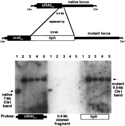
Our final approach exploited the recent demonstration of the utility of a hygromycin B resistance marker (hph) in H. capsulatum (43), which could provide a mechanism for selection of transformants independent of FOA selection for URA5Hc gene inactivation resulting from homologous gene targeting and allow determination of the relative frequency of allelic replacement events. We replaced a 0.4-kb internal segment of the URA5Hc gene including 3′ coding sequence with the 2.9-kb hph marker to construct plasmid pMAD10. This plasmid includes the greatest extent of URA5Hc locus flanking sequences available: 1.1 kb on the 5′ side and 3 kb on the 3′ side (Fig. 2). We selected hygromycin B-resistant H. capsulatum transformants following delivery of the restriction-digested deletion construct as a linear molecule. Two protocols were used, both of which resulted in the desired allelic replacement events (Table 1), as confirmed by PCR (Fig. 5) and Southern blotting (Fig. 6) as well as demonstration of uracil auxotrophy. In the first, we initially selected transformants solely for resistance to hygromycin B and then screened for uracil auxotrophy. In the second, we imposed direct selection for resistance to both hygromycin B and FOA on 3 M–uracil agarose. Interestingly, in addition to four genuine URA5Hc gene-disrupted transformants, we obtained a hygromycin B- and FOA-resistant colony that was uracil prototrophic and had no genomic alteration at the URA5Hc locus detectable by PCR (Fig. 5, lanes 6), presumably resulting from spontaneous or transformation-induced mutation to FOA resistance in a yeast transformed to hygromycin B resistance due to ectopic integration or linear plasmid formation by the transforming DNA. In different experiments, the homologous targeting frequency ranged from 1.0 × 10⁻³ to 2.6 × 10⁻³ allelic replacement events per hygromycin B-resistant transformant, for a cumulative frequency of 1.4 × 10⁻³. Resulting URA5Hc disruptants were auxotrophic for uracil and were confirmed by two analyses of genomic DNA. PCR using flanking primers showed loss of the native product and the appearance of a product appropriately 2.5 kb larger (Fig. 5). Southern blotting revealed loss of the native genomic band and the appearance of an approximately 2.5-kb-larger band when probed with URA5Hc sequence that also hybridized to an hph probe but not to the 0.4-kb fragment deleted from the disruption construct pMAD10 (Fig. 6). Subsequently, we were able to transform these disruptants back to uracil prototrophy by using plasmids carrying the Podospora URA5 gene.
TABLE 1.
Transformation of H. capsulatum G184AS with ClaI-cut pMAD10, containing the Δura5Hc::hph mutant locus
| Expt | No. of hygromycin B-resistant transformants | Selectionb | Screening | No. of uracil auxotrophs |
|---|---|---|---|---|
| 1 | 379 | Hygromycin B | Uracil requirement tested by growth on medium with and without exogenous uracil | 1 |
| 1,516 (calculateda) | Hygromycin B, FOA | Uracil requirement confirmed by growth on medium with and without exogenous uracil | 2c | |
| 2 | 980 (calculateda) | Hygromycin B, FOA | Uracil requirement confirmed by growth on medium with and without exogenous uracil | 1 |
Based on the number of colonies arising on a 3 M–uracil–hygromycin B agarose plate (without FOA).
Exogenous uracil was always provided.
Another isolate from the original transformation plates was uracil prototrophic, and this hygromycin B-resistant transformant was presumably a spontaneous or transformation-induced FOA-resistant mutant.
FIG. 5.
PCR evidence for pMAD10-mediated URA5Hc gene disruption in strain G184AS. Genomic DNAs from transformation recipient strain G184AS (lanes 1), the four hygromycin B-resistant, uracil-auxotrophic transformants (lanes 2 to 5), and a hygromycin B-resistant, uracil-prototrophic isolate (lanes 6) were subjected to PCR using primers targeted to a URA5Hc region upstream of the site of hph insertion (primer set A) or primers targeted to sites that flank the site of hph insertion (primer set B). The four uracil auxotrophs (lanes 2 to 5) have lost the native product and gained a new larger product due to hph insertion with primer set B. The uracil prototroph (lanes 6) shows both the unchanged native product and the larger product with primer set B, due to ectopic integration or linear plasmid formation by the transforming DNA. Lanes M, molecular size standards (λ/HindIII and φX/HaeIII).
FIG. 6.
Southern blot evidence for pMAD10-mediated URA5Hc gene disruption in strain G184AS. Genomic DNAs from transformation recipient strain G184AS (lanes 1) and the four hygromycin B-resistant, uracil-auxotrophic transformants (lanes 2 to 5) were digested with ClaI, electrophoresed, Southern blotted, and probed with radiolabeled fragments of the URA5Hc gene, the 0.4-kb internal URA5Hc fragment deleted from pMAD10, and the inserted selectable marker hph. The four uracil auxotrophs (lanes 2 to 5) have lost the native genomic band that hybridizes with the URA5Hc probe and gained a new, larger band due to hph insertion that hybridizes with both the URA5Hc and hph probes, as well as failing to show any hybridization with the 0.4-kb fragment deleted from the disruption construct pMAD10. Fragment sizes were estimated by comparison to molecular size standards in the ethidium bromide-stained gel.
H. capsulatum is far from unique in being a eukaryotic pathogen for which reverse genetics has proved technically challenging. Targeted gene disruption is a crucial experimental step in fulfilling Koch’s postulates on a molecular genetic level to establish the biological function or pathogenic role of a gene and gene product. In contrast to S. cerevisiae, the model yeast in which homologous gene targeting essentially occurs invariably in appropriately designed experiments (16, 21, 27, 33), other fungi (particularly those pathogenic for humans) have generally revealed one or more complications that hinder facile accomplishment of this process. Although C. albicans undergoes homologous gene targeting with efficiency similar to that of the closely related S. cerevisiae, it is an obligate diploid necessitating disruption of two alleles for each gene, and only the development of the “ura-blaster” or similar techniques and appropriate strains has made this endeavor less daunting (1, 17, 19). Haploid strains of pathogenic Aspergillus species and Cryptococcus neoformans both show relatively frequent ectopic integration of homologous transforming DNA, necessitating screening of many transformants for gene disruptants or developing selection or enrichment protocols for targeted events (8, 9, 14, 25, 30, 32, 37). In addition to ectopic integration, the basidiomycete C. neoformans also displays frequent generation of linear plasmids (which thus are not homologously targeted to the chromosomal locus) (13, 37, 38), and both of these phenomena are also observed in the ascomycete H. capsulatum. Several cryptococcal targeted gene disruptants have been constructed, at a relative frequency varying from approximately 10−1 to 10−4 per transformant (8, 9, 25, 30). These accomplishments were facilitated in some cases by the use of experimental designs for enrichment of targeted events, such as positive-negative selection (8, 9), a technique earlier used with mammalian embryonic stems cells (2, 20, 26). In fact, the URA5Hc gene described here can theoretically be used in a positive-negative selection strategy as a counterselectable marker flanking a target gene insertionally disrupted with another selectable marker (e.g., hph), although this approach may be complicated by the occurrence of spontaneous or transformation-induced FOA resistance that we have observed in H. capsulatum, necessitating confirmation of or even initial screening for uracil auxotrophy. Mammalian cells, of course, provide the dual challenges of diploidy and low frequency of homologous integration, but extensive work and development of new strategies have resulted in numerous gene disruptions in mice (2, 7, 20, 26, 34). Similar advances have increased the manipulability of other pathogenic fungi such as C. albicans and C. neoformans. We have now demonstrated the occurrence of homologous gene targeting in H. capsulatum, and we and others are continuing to work to increase the efficiency of this experimental technique and extend it to other target genes that may be important biologically and/or for virulence.
ACKNOWLEDGMENTS
This work was supported by Public Health Service grants HL55949 from the National Heart, Lung, and Blood Institute to J.P.W. and AI25584 from the National Institute of Allergy and Infectious Diseases to W.E.G. D.M.R. was supported by National Research Service Award F32 AI09720 from the National Institute of Allergy and Infectious Diseases. J.P.W. is a Lucille P. Markey Scholar, and this work was supported in part by Scholar Award 94-21 from the Lucille P. Markey Charitable Trust. W.E.G. is a recipient of the Burroughs Wellcome Fund Scholar Award in Pathogenic Mycology.
REFERENCES
- 1.Alani E, Cao L, Kleckner N. A method for gene disruption that allows repeated use of URA3 selection in the construction of multiply disrupted yeast strains. Genetics. 1987;116:541–545. doi: 10.1534/genetics.112.541.test. [DOI] [PMC free article] [PubMed] [Google Scholar]
- 2.Askew G R, Doetschman T, Lingrel J B. Site-directed point mutations in embryonic stem cells: a gene-targeting tag-and-exchange strategy. Mol Cell Biol. 1993;13:4115–4124. doi: 10.1128/mcb.13.7.4115. [DOI] [PMC free article] [PubMed] [Google Scholar]
- 3.Ausubel F M, Brent R, Kingston R E, Moore D D, Seidman J G, Smith J A, Struhl K, editors. Current protocols in molecular biology. New York, N.Y: John Wiley & Sons, Inc.; 1994. . (CD-ROM version). [Google Scholar]
- 4.Ballance D J. Sequences important for gene expression in filamentous fungi. Yeast. 1986;2:229–236. doi: 10.1002/yea.320020404. [DOI] [PubMed] [Google Scholar]
- 5.Boeke J D, LaCroute F, Fink G R. A positive selection for mutants lacking orotidine-5′-phosphate decarboxylase activity in yeast: 5-fluoro-orotic acid resistance. Mol Gen Genet. 1984;197:345–346. doi: 10.1007/BF00330984. [DOI] [PubMed] [Google Scholar]
- 6.Brown D H, Jr, Slobodkin I V, Kumamoto C A. Stable transformation and regulated expression of an inducible reporter construct in Candida albicans using restriction enzyme-mediated integration. Mol Gen Genet. 1996;251:75–80. doi: 10.1007/BF02174347. [DOI] [PubMed] [Google Scholar]
- 7.Capecchi M R. Altering the genome by homologous recombination. Science. 1989;244:1288–1292. doi: 10.1126/science.2660260. [DOI] [PubMed] [Google Scholar]
- 8.Chang Y C, Kwon-Chung K J. Complementation of a capsule-deficient mutation of Cryptococcus neoformans restores its virulence. Mol Cell Biol. 1994;14:4912–4919. doi: 10.1128/mcb.14.7.4912. [DOI] [PMC free article] [PubMed] [Google Scholar]
- 9.Chang Y C, Penoyer L A, Kwon-Chung K J. The second capsule gene of Cryptococcus neoformans, CAP64, is essential for virulence. Infect Immun. 1996;64:1977–1983. doi: 10.1128/iai.64.6.1977-1983.1996. [DOI] [PMC free article] [PubMed] [Google Scholar]
- 10.Cox G M, Toffaletti D L, Perfect J R. Dominant selection system for use in Cryptococcus neoformans. J Med Vet Mycol. 1996;34:385–391. [PubMed] [Google Scholar]
- 11.de Montigny J, Belarbi A, Hubert J-C, LaCroute F. Structure and expression of the URA5 gene of Saccharomyces cerevisiae. Mol Gen Genet. 1989;215:455–462. doi: 10.1007/BF00427043. [DOI] [PubMed] [Google Scholar]
- 12.de Montigny J, Kern L, Hubert J-C, LaCroute F. Cloning and sequencing of URA10, a second gene encoding orotate phosphoribosyl transferase in Saccharomyces cerevisiae. Curr Genet. 1990;17:105–111. doi: 10.1007/BF00312853. [DOI] [PubMed] [Google Scholar]
- 13.Edman J C. Isolation of telomerelike sequences from Cryptococcus neoformans and their use in high-efficiency transformation. Mol Cell Biol. 1992;12:2777–2783. doi: 10.1128/mcb.12.6.2777. [DOI] [PMC free article] [PubMed] [Google Scholar]
- 14.Edman J C, Kwon-Chung K J. Isolation of the URA5 gene from Cryptococcus neoformans var. neoformans and its use as a selective marker for transformation. Mol Cell Biol. 1990;10:4538–4544. doi: 10.1128/mcb.10.9.4538. [DOI] [PMC free article] [PubMed] [Google Scholar]
- 15.Eissenberg L G, Goldman W E. Histoplasma variation and adaptive strategies for parasitism: new perspectives on histoplasmosis. Clin Microbiol Rev. 1991;4:411–421. doi: 10.1128/cmr.4.4.411. [DOI] [PMC free article] [PubMed] [Google Scholar]
- 16.Fincham J R S. Transformation in fungi. Microbiol Rev. 1989;53:148–170. doi: 10.1128/mr.53.1.148-170.1989. [DOI] [PMC free article] [PubMed] [Google Scholar]
- 17.Fonzi W A, Irwin M Y. Isogenic strain construction and gene mapping in Candida albicans. Genetics. 1993;134:717–728. doi: 10.1093/genetics/134.3.717. [DOI] [PMC free article] [PubMed] [Google Scholar]
- 18.Goldman W E. Molecular genetic technology transfer to pathogenic fungi. Arch Med Res. 1995;26:437–440. [PubMed] [Google Scholar]
- 19.Gorman J A, Chan W, Gorman J W. Repeated use of GAL1 for gene disruption in Candida albicans. Genetics. 1991;129:19–24. doi: 10.1093/genetics/129.1.19. [DOI] [PMC free article] [PubMed] [Google Scholar]
- 20.Hasty P, Ramírez-Solis R, Krumlauf R, Bradley A. Introduction of a subtle mutation into the Hox-2.6 locus in embryonic stem cells. Nature. 1991;350:243–246. doi: 10.1038/350243a0. [DOI] [PubMed] [Google Scholar]
- 21.Hinnen A, Hicks J B, Fink G R. Transformation of yeast. Proc Natl Acad Sci USA. 1978;75:1929–1933. doi: 10.1073/pnas.75.4.1929. [DOI] [PMC free article] [PubMed] [Google Scholar]
- 22.Hogan L H, Klein B S. Transforming DNA integrates at multiple sites in the dimorphic fungal pathogen Blastomyces dermatitidis. Gene. 1997;186:219–226. doi: 10.1016/s0378-1119(96)00713-5. [DOI] [PubMed] [Google Scholar]
- 23.Jacquet M, Guilbaud R, Garreau H. Sequence analysis of the DdPYR5-6 gene coding for UMP synthase in Dictyostelium discoideum and comparison with orotate phosphoribosyl transferases and OMP decarboxylases. Mol Gen Genet. 1988;211:441–445. doi: 10.1007/BF00425698. [DOI] [PubMed] [Google Scholar]
- 24.Le Chevanton L, Leblon G. The ura5 gene of the ascomycete Sordaria macrospora: molecular cloning, characterization, and expression in Escherichia coli. Gene. 1989;77:39–49. doi: 10.1016/0378-1119(89)90357-0. [DOI] [PubMed] [Google Scholar]
- 25.Lodge J K, Jackson-Machelski E, Toffaletti D L, Perfect J R, Gordon J I. Targeted gene replacement demonstrates that myristoyl-CoA:protein N-myristoyltransferase is essential for viability of Cryptococcus neoformans. Proc Natl Acad Sci USA. 1994;91:12008–12012. doi: 10.1073/pnas.91.25.12008. [DOI] [PMC free article] [PubMed] [Google Scholar]
- 26.Mansour S L, Thomas K R, Capecchi M R. Disruption of the proto-oncogene int-2 in mouse embryo-derived stem cells: a general strategy for targeting mutations to non-selectable genes. Nature. 1988;336:348–352. doi: 10.1038/336348a0. [DOI] [PubMed] [Google Scholar]
- 27.Orr-Weaver T L, Szostak J W, Rothstein R J. Genetic applications of yeast transformation with linear and gapped plasmids. Methods Enzymol. 1983;101:228–244. doi: 10.1016/0076-6879(83)01017-4. [DOI] [PubMed] [Google Scholar]
- 28.Patel J B, Batanghari J W, Goldman W E. Probing the yeast phase-specific expression of the CBP1 gene in Histoplasma capsulatum. J Bacteriol. 1998;180:1786–1792. doi: 10.1128/jb.180.7.1786-1792.1998. [DOI] [PMC free article] [PubMed] [Google Scholar]
- 29.Peng M, Cooper C R, Jr, Szaniszlo P J. Genetic transformation of the pathogenic fungus Wangiella dermatitidis. Appl Microbiol Biotechnol. 1995;44:444–450. doi: 10.1007/BF00169942. [DOI] [PubMed] [Google Scholar]
- 30.Salas S D, Bennett J E, Kwon-Chung K J, Perfect J R, Williamson P R. Effect of the laccase gene, CNLAC1, on virulence of Cryptococcus neoformans. J Exp Med. 1996;184:377–386. doi: 10.1084/jem.184.2.377. [DOI] [PMC free article] [PubMed] [Google Scholar]
- 31.Sambrook J, Fritsch E F, Maniatis T. Molecular cloning: a laboratory manual. 2nd ed. Cold Spring Harbor, N.Y: Cold Spring Harbor Laboratory Press; 1989. [Google Scholar]
- 32.Smith J M, Tang C M, Noorden S V, Holden D W. Virulence of Aspergillus fumigatus double mutants lacking restricocin and an alkaline protease in a low-dose model of invasive pulmonary aspergillosis. Infect Immun. 1994;62:5247–5254. doi: 10.1128/iai.62.12.5247-5254.1994. [DOI] [PMC free article] [PubMed] [Google Scholar]
- 33.Struhl K. The new yeast genetics. Nature. 1983;305:391–397. doi: 10.1038/305391a0. [DOI] [PubMed] [Google Scholar]
- 34.te Riele H, Maandag E R, Clarke A, Hooper M, Berns A. Consecutive inactivation of both alleles of the pim-1 proto-oncogene by homologous recombination in embryonic stem cells. Nature. 1990;348:649–651. doi: 10.1038/348649a0. [DOI] [PubMed] [Google Scholar]
- 35.Timberlake W E, Marshall M A. Genetic engineering of filamentous fungi. Science. 1989;244:1313–1317. doi: 10.1126/science.2525275. [DOI] [PubMed] [Google Scholar]
- 36.Turcq B, Bégueret J. The ura5 gene of the filamentous fungus Podospora anserina: nucleotide sequence and expression in transformed strains. Gene. 1987;53:201–209. doi: 10.1016/0378-1119(87)90008-4. [DOI] [PubMed] [Google Scholar]
- 37.Varma A, Edman J C, Kwon-Chung K J. Molecular and genetic analysis of URA5 transformants of Cryptococcus neoformans. Infect Immun. 1992;60:1101–1108. doi: 10.1128/iai.60.3.1101-1108.1992. [DOI] [PMC free article] [PubMed] [Google Scholar]
- 38.Varma A, Kwon-Chung K J. Formation of a minichromosome in Cryptococcus neoformans as a result of electroporative transformation. Curr Genet. 1994;26:54–61. doi: 10.1007/BF00326305. [DOI] [PubMed] [Google Scholar]
- 39.Voisard C, Wang J, McEvoy J L, Xu P, Leong S A. urbs1, a gene regulating siderophore biosynthesis in Ustilago maydis, encodes a protein similar to the erythroid transcription factor GATA-1. Mol Cell Biol. 1993;13:7091–7100. doi: 10.1128/mcb.13.11.7091. [DOI] [PMC free article] [PubMed] [Google Scholar]
- 40.Wickes B L, Edman J C. The Cryptococcus neoformans GAL7 gene and its use as an inducible promoter. Mol Microbiol. 1995;16:1099–1109. doi: 10.1111/j.1365-2958.1995.tb02335.x. [DOI] [PubMed] [Google Scholar]
- 41.Woods J P, Goldman W E. In vivo generation of linear plasmids with addition of telomeric sequences by Histoplasma capsulatum. Mol Microbiol. 1992;6:3603–3610. doi: 10.1111/j.1365-2958.1992.tb01796.x. [DOI] [PubMed] [Google Scholar]
- 42.Woods J P, Goldman W E. Autonomous replication of foreign DNA in Histoplasma capsulatum: role of native telomeric sequences. J Bacteriol. 1993;175:636–641. doi: 10.1128/jb.175.3.636-641.1993. [DOI] [PMC free article] [PubMed] [Google Scholar]
- 42a.Woods J P, Heinecke E L. Abstracts of the 96th General Meeting of the American Society for Microbiology 1996. Washington, D.C: American Society for Microbiology; 1996. Homologous gene targeting in Histoplasma capsulatum: URA5 gene disruption using a hygromycin resistance marker, abstr. F-116; p. 94. [Google Scholar]
- 43.Woods J P, Heinecke E L, Goldman W E. Electrotransformation and expression of bacterial genes encoding hygromycin phosphotransferase and β-galactosidase in the pathogenic fungus Histoplasma capsulatum. Infect Immun. 1998;66:1697–1707. doi: 10.1128/iai.66.4.1697-1707.1998. [DOI] [PMC free article] [PubMed] [Google Scholar]
- 44.Worsham P L, Goldman W E. Quantitative plating of Histoplasma capsulatum without addition of conditioned medium or siderophores. J Med Vet Mycol. 1988;26:137–143. [PubMed] [Google Scholar]
- 45.Worsham P L, Goldman W E. Selection and characterization of ura5 mutants of Histoplasma capsulatum. Mol Gen Genet. 1988;214:348–352. doi: 10.1007/BF00337734. [DOI] [PubMed] [Google Scholar]
- 46.Worsham P L, Goldman W E. Development of a genetic transformation system for Histoplasma capsulatum: complementation of uracil auxotrophy. Mol Gen Genet. 1990;221:358–362. doi: 10.1007/BF00259400. [DOI] [PubMed] [Google Scholar]
- 47.Zhou C, Yang Y, Jong A Y. Mini-prep in ten minutes. BioTechniques. 1990;8:172–173. [PubMed] [Google Scholar]


