Abstract
Spontaneous steinstrasse (“stone street”) is a collection of stones within the ureter and is a rare and understudied event. Factors such as infection, altered kidney function, and degree of obstruction are used to define the most adequate therapeutic option. Treatment can be either conservative or surgical. The decision of which depends on the clinical presentation. This paper reports a rare case of a 59-year-old patient with spontaneous steinstrasse examined at a urology clinic. Surgical intervention was required because of altered kidney function. The patient is currently undergoing follow-up for the metabolic investigation.
Keywords: shock wave lithotripsy, urology, treatment, clinic, steinstrasse
Introduction
Steinstrasse or “stone street” is an aggregation of particles in the ureter. On x-ray, such collections have the appearance of a cobbled street, hence the term steinstrasse, which means "street of stone" in German. Steinstrasse occurs in up to 15% of cases after extracorporeal shockwave lithotripsy (ESWL) [1], and 6% of these cases require intervention [2]. The incidence is related to factors such as the size of the calculi, location [3], and the energy imposed during ESWL [4]. The main complication of this event is ureteral obstruction, which can occur in up to 23% of cases [5], leading to the loss of kidney function [4].
Post-ESWL steinstrasse is classified into three types. Type 1 is characterized by multiple small fragments. Type 2 has fragments measuring 5 mm or more and small proximal fragments. Type 3 has multiple fragments measuring 5 mm or more [6].
Spontaneous steinstrasse is a spontaneous accumulation of small stones without a preceding surgical intervention, a rare and understudied event [7]. Some factors such as the infection, altered kidney function, and degree of obstruction are used to define the most adequate therapeutic option. Management can be either conservative or surgical. The decision of which depends on the clinical presentation.
This paper presents a rare case of a patient with spontaneous steinstrasse examined at a urology clinic.
Case presentation
A 59-year-old male patient, hypertensive, visited a urology clinic with the complaint of recurring renal colic on the right side, with no previous urological procedures. Ultrasonography of the kidney and ureters performed two months earlier identified a calculus measuring 1.4 cm in the right ureteropelvic junction and a branched calculus measuring 3.3 cm in the left kidney.
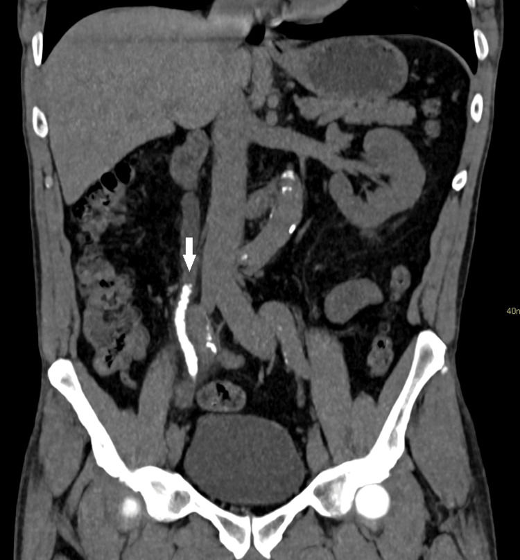
The patient was sent to the emergency room. Computed tomography revealed renal lithiasis: calculus measuring 9 mm in the right lower calyx and multiple calculi in the right kidney, with the largest of which being 1.2 cm in the lower calyx; multiple small stones situated along the right ureter (some overlapping), in greater quantity at the crossing of the iliac vessels; and moderate dilation of the right collector system and density of all calculi ranging from 420 to 505 UH (Figure 1).
Figure 1. Computed tomography (coronal axis) showing right-side steinstrasse and ipsilateral ureteral dilation (arrow).
Laboratory exams revealed kidney function, with serum creatinine of 1.78 mg/dL, urea of 59 mg/dL, and discrete hyperkalemia (5.4 mg/dL), with no associated infection based on the urine exam (Table 1).
Table 1. Results of the laboratory exams.
| Variables | Admission | Metabolic investigation | Normal range values |
| Hemoglobin (g/dL) | 14.4 | - | 12.8-16.5 |
| Hematocrit (%) | 43.3 | - | 40.0-54.0 |
| White blood cells (/mm3) | 9940 | - | 4.000-11.000 |
| Platelets (/mm3) | 324000 | - | 140-450 |
| Creatinine (mg/dL) | 1.78 | - | 0.60-1.20 |
| Urea (mg/dL) | 59.50 | - | <50 |
| Sodium (mmol/L) | 136 | 141 | 135-145 |
| Potassium (mmol/L) | 5.4 | 5.3 | 3.50-5.10 |
| Ionized calcium (mmol/L) | - | 1.3 | 1.10-1.40 |
| Total calcium (mg/dL) | - | 9.7 | 8.6-10.2 |
| Chlorine (mmol/L) | - | 106 | 98-107 |
| Urinary data | |||
| pH | 5.0 | - | 5.0-7.0 |
| Density | 1,008 | - | 1,015-1,025 |
| Nitrite | Negative | - | Negative |
| White blood cells (/mL) | 3,000 | - | Up to 25,000 |
| Red blood cells (/mL) | 1,000 | - | Up to 25,000 |
| Cylinders | Absent | - | Absent |
| Crystals | Absent | - | Absent |
| Uroculture | Negative | - | Negative |
| Venous blood gas | |||
| pH | - | 7.36 | 7.33-7.43 |
| HCO3 (mmoL/L) | - | 23.9 | 23-27 |
| BE (mmoL/L) | - | -1.2 | -3-3 |
| PO2 (mmHg) | - | 23 | 30-50 |
| PCO2 (mmHg) | - | 43.5 | 38-50 |
The patient was submitted to two sessions of ureteroscopy by rigid ureteroscope and laser lithotripter, with a six-week interval because of the stone burden, without complications, resulting in the complete resolution of the ureteral calculi. The patient is currently undergoing follow-up at a nephrology clinic for metabolic investigation of the calculi.
Discussion
Cases of spontaneous steinstrasse are rare, and different factors contribute to the indication of the best therapeutic option to adopt. In the present case, the patient had type 3 steinstrasse and altered kidney function.
Treatment for this condition can be conservative or surgical, and the decision is directly related to the clinical presentation. In the present case, surgical intervention was performed because of the altered kidney function.
The literature describes the association between spontaneous steinstrasse and nephrocalcinosis with renal tubular acidosis [8]. In the present case, the patient had bilateral nephrolithiasis but no indication of tubular acidosis or nephrocalcinosis. Currently, the patient remains in metabolic investigation and urological follow-up because of the nephrolithiasis.
Analyzing 958 patients with renal stones who underwent ESWL, Kim et al. verified that 63.6% of cases have spontaneous resolution [9]. However, the therapeutic approach to patients with spontaneous steinstrass requires more clinical studies, as the rarity of cases makes the standardization of conduct difficult.
Although conservative conduct is a therapeutic option, patients with persistent symptoms and ureteral obstruction are preferably treated surgically, as in the present case. Thus, when conservative treatment (spontaneous elimination of calculi) is not satisfactory, the conduct should include temporary urinary deviation for the monitoring of infection. With the resolution of this condition, definitive treatment is instituted: surgical removal of the steinstrasse.
Conclusions
Spontaneous steinstrasse is an uncommon event, for which the therapeutic approach lacks scientific evidence. The most adequate therapeutic option depends on the patient’s clinical condition and the size of the calculi. Patients with persistent symptoms and ureteral obstruction are preferably treated surgically.
Acknowledgments
The authors would like to thank the staff of the Radiology Unit of Hospital de Base/FUNFARME for the radiological analysis.
The authors have declared that no competing interests exist.
Author Contributions
Concept and design: Luís Cesar Fava Spessoto, Rafael S. Aguiar, Guilherme C. Gonzales, Ana Clara N. Spessoto, Fernando Nestor Facio Jr.
Acquisition, analysis, or interpretation of data: Luís Cesar Fava Spessoto, Rafael S. Aguiar, Fernando Nestor Facio Jr.
Drafting of the manuscript: Luís Cesar Fava Spessoto, Rafael S. Aguiar, Guilherme C. Gonzales, Ana Clara N. Spessoto, Fernando Nestor Facio Jr.
Critical review of the manuscript for important intellectual content: Luís Cesar Fava Spessoto, Fernando Nestor Facio Jr.
Supervision: Luís Cesar Fava Spessoto, Guilherme C. Gonzales, Ana Clara N. Spessoto
Human Ethics
Consent was obtained or waived by all participants in this study
References
- 1.The steinstrasse: a legacy of extracorporeal lithotripsy? Coptcoat MJ, Webb DR, Kellet MJ, Whitfield HN, Wickham JE. Eur Urol. 1988;14:93–95. doi: 10.1159/000472910. [DOI] [PubMed] [Google Scholar]
- 2.Large spontaneous steinstrasse: our experience and management issues in tertiary care centre. Parmar K, Manoharan V, Kumar S, Ranjan KR, Chandna A, Chaudhary K. Urologia. 2022;89:226–230. doi: 10.1177/03915603211001174. [DOI] [PubMed] [Google Scholar]
- 3.Risk factors for formation of steinstrasse after extracorporeal shock wave lithotripsy for pediatric renal calculi: a multivariate analysis model. El-Assmy A, El-Nahas AR, Elsaadany MM, El-Halwagy S, Sheir KZ. Int Urol Nephrol. 2015;47:573–577. doi: 10.1007/s11255-015-0938-8. [DOI] [PubMed] [Google Scholar]
- 4.Risk factors for the formation of a steinstrasse after extracorporeal shock wave lithotripsy: a statistical model. Madbouly K, Sheir KZ, Elsobky E, Eraky I, Kenawy M. J Urol. 2002;167:1239–1242. [PubMed] [Google Scholar]
- 5.EAU guidelines. [ Oct; 2023 ];https://uroweb.org/guidelines Milan. 2023 2023 [Google Scholar]
- 6.The complications of extracorporeal shockwave lithotripsy: management and prevention. Coptcoat MJ, Webb DR, Kellett MJ, et al. Br J Urol. 1986;58:578–580. doi: 10.1111/j.1464-410x.1986.tb05888.x. [DOI] [PubMed] [Google Scholar]
- 7.Massive steinstrasse without lithotripsy: a rare case report. Abdulmajed MI, Anandaram PS, Wyn Jones V, Shergill IS. Turk J Urol. 2013;39:61–63. doi: 10.5152/tud.2013.013. [DOI] [PMC free article] [PubMed] [Google Scholar]
- 8.Bilateral spontaneous steinstrasse and nephrocalcinosi associated with distal renal tubular acidosis. Van Savage JG, Fried FA. J Urol. 1993;150:467–468. doi: 10.1016/s0022-5347(17)35516-7. [DOI] [PubMed] [Google Scholar]
- 9.Treatment of steinstrasse with repeat extracorporeal shock wave lithotripsy: experience with piezoelectric lithotriptor. Kim SC, Oh CH, Moon YT, Kim KD. J Urol. 1991;145:489–491. doi: 10.1016/s0022-5347(17)38376-3. [DOI] [PubMed] [Google Scholar]


