Transcatheter edge‐to‐edge repair (TEER) of the mitral valve (MV) is an increasingly common technique to treat patients with severe degenerative or functional mitral regurgitation (MR). Although this procedure is well studied in the United States and Europe, fewer data are available for Asian populations.
In this issue of the Journal of the American Heart Association (JAHA), Kubo and colleagues present data from the OCEAN (Optimized Catheter Valvular Intervention)‐Mitral registry. 1 This registry is a large cohort of 2150 high surgical risk patients from 21 Japanese institutions who underwent mitral TEER between April 2019 and June 2021. Degenerative MR (or primary MR [PMR] was noted in 34.6% of patients, 55.7% had ventricular functional MR (or secondary MR [SMR]), and 19.5% had atrial functional MR/SMR. The cause of MR is commensurate with other large mitral TEER registries.
Similar to other large‐scale studies in the United States 2 , 3 and Europe, 4 the authors found that mitral TEER was safe and effective, with a 1‐year all‐cause mortality rate of 12.3% and heart failure (HF) hospitalization rate of 15.0%. The combined end point of death or HF hospitalization was 22.9%. MR reduction was found to be durable with 1‐month follow‐up data showing MR 2+ or less in 96.7% of patients, which was sustained at 1 year with MR 2+ or less in 94.1% of patients. New York Heart Association functional class was improved at 1 month with 96.7% of patients reporting New York Heart Association class I or II symptoms, and this was also sustained at 95% at 1 year. Overall, these findings are consistent with the aforementioned studies and extends the published experience to Asia.
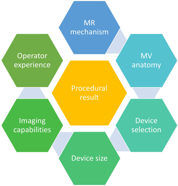
The more critical question at this point is less so the safety and efficacy of mitral TEER, but more so the definition of an ideal procedural result, and how that result can be achieved to deliver optimal outcomes (Figure 1). To this point, Kubo and colleagues also evaluated the impact of residual MR on patient outcomes. In patients with PMR, 0/1+ residual MR was associated with a lower incidence of death or HF hospitalization compared with those with 2+ or more MR. In patients with secondary MR, those with 0/1+ residual MR had a lower trend of death or HF hospitalization compared with those with 2+ MR and had significantly lower events compared with those with 3+/4+ residual MR. These data highlight the importance of MR reduction and the relationship of residual MR on outcomes.
Figure 1. Factors that contribute to an optimal mitral transcatheter edge‐to‐edge repair (TEER).

MR indicates mitral regurgitation; and MV, mitral valve.
Although historically a successful result has been classified as achieving 2+ residual MR or less, there is a growing body of literature that suggests that we should strive to achieve even further MR reduction. Among almost 20 000 patient undergoing TEER for PMR in the US Transcatheter Valve Therapy (TVT) registry, Makkar and colleagues demonstrated the lowest mortality among those who achieved mild or less MR (as well as mean gradient [MG] <5 mm Hg) based on site‐reported echocardiograms. 3 Similarly, Kar and colleagues demonstrated that patients with SMR in the COAPT (Cardiovascular Outcomes Assessment of the Mitra Clip Percutaneous Therapy for Heart Failure Patients with Functional Mitral Regurgitation) trial whose MR was reduced to at most 2+ demonstrated greater freedom from death or HF hospitalization compared with patients with 3+ or more residual MR based on core‐lab adjudicated echocardiogram review. 5
Although it is easy to suggest more aggressive MR reduction as a goal, the procedural result of mitral TEER is a combination of MR reduction and residual MV area (MVA). Thus, intraprocedural decision making is a balancing act between the desire to reduce MR and the concern for the development of mitral stenosis. As MG is typically used to monitor for the development of mitral stenosis, several prior studies have evaluated the impact of post‐TEER gradients on outcomes, albeit with variable results.
In a study of 713 patients who underwent TEER, Koell and colleagues found that an elevated MG >5 was associated with an increased risk of the primary end point (death or HF hospitalization at 5 years), in patients with PMR but not in SMR. 6 Yoon and colleagues studied 419 patients with PMR and divided patients into 4 groups based on postprocedure mitral gradient: <1.9 mm Hg, 3.0 mm Hg, 4.0 mm Hg, and 6.0 mm Hg. 7 No significant difference was found for all‐cause mortality at 2 years across the 4 quartiles. The discordance between these 2 studies, as well as between patients with PMR versus SMR, highlight the limitations of using mean mitral gradient as the primary determinant for the development of mitral stenosis.
As many factors contribute to mitral gradients including cardiac output, heart rate, and degree of mitral regurgitation, it has been suggested that this method is inaccurate for the assessment of mitral stenosis, and other techniques, such as direct measurement of MVA by 3‐dimensional (3D) planimetry (3D MVA) should be considered. Utsunomiya et al evaluated the residual 3D MVA and relationship to pulmonary hypertension and outcomes. They not only found a 40% discordance between mitral stenosis defined by MVA versus MG but also found that a post TEER 3D MVA <1.94 cm2 was associated with an increased risk of all‐cause mortality and HF hospitalization. Although MG is a fast and easy metric to follow, this study points out the limitation of this technique while highlighting the utility of measuring 3D MVA.
Ultimately, the unifying theme of these studies is the emphasis on optimizing the procedural result, which includes consideration of MR and mitral stenosis, to achieve the best outcomes for the patient. Given the many interrelated factors that contribute to procedural success (Figure 1), this is a challenging, yet critical, topic to study. In addition to understanding MV anatomic criteria and device capabilities, one must also recognize the importance of the procedural team.
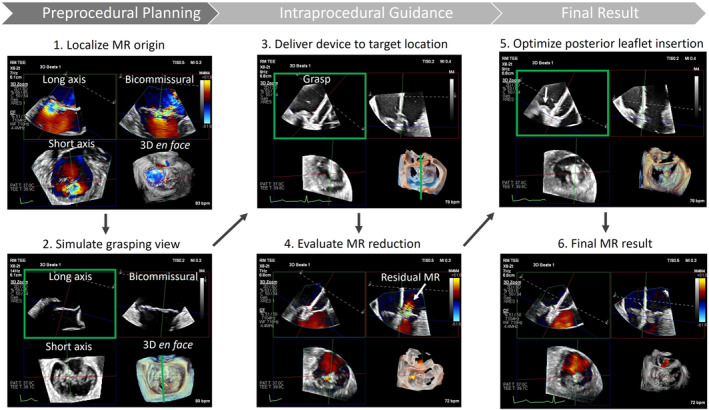
Operator experience has previously been shown to be an important contributor to procedural success. A large analysis of more than 12 000 patients from the US TVT registry demonstrated that increasing institutional experience was associated with higher rates of procedural success and optimal MR reduction, with inflections at 50 cases and continued improvement up to 200 cases. 8 It should also be noted that only 29% of patients in the OCEAN registry were treated using the contemporary G4 MitraClip system that consists of a larger spectrum of device sizes (length and width of the clip) and allows independent leaflet grasping. In our and others' experience, and as highlighted in Figure 2, this has been an important device iteration to improve procedural results. 9
Figure 2. Pre‐ and intraprocedural transesophageal echocardiographic imaging using 3‐dimensional multiplanar reconstruction (3D MPR).

The detail provided by MPR imaging and versatility of the current generation device allows a granular analysis of the result and further optimization (for example, the residual MR seen lateral to the clip at the posterior leaflet in panel 4 is treated by lifting the posterior gripper and slight clockwise rotation of the clip with resolution of MR seen in panel 6). MR indicates mitral regurgitation.
A similarly critical (but often underrecognized) component of procedural success is pre‐ and intraprocedural imaging. Echocardiography plays a role in every step of the procedural pathway, starting with the definition of MR mechanism and anatomy, which determines procedural candidacy and device selection. Intraprocedural imaging guidance is then necessary for device manipulation and evaluation of procedural results. Imaging strengths or weaknesses at any stage in this process can affect procedural success. Most centers still use 2‐dimensional X‐plane and gastric short‐axis imaging supplemented by episodic single‐view 3D left atrial views to guide TEER.
Unfortunately, this ignores the significant advances in 3D echocardiographic technology that allow for more detailed evaluation of MV anatomy and procedural guidance. Our center relies heavily on 3D multiplanar reconstruction (3D MPR) for preprocedural evaluation of patients with MR to determine candidacy for TEER, and we have found this technique to be invaluable for understanding MR mechanism, origin, and graspability of the leaflets at the MR origin. 10 Furthermore, the use of 3D MPR allows for direct translation of the preprocedural plan into intraprocedural guidance using the same imaging platform (Figure 2). Preprocedurally, the exact location and orientation of the device is planned using 3D MPR. This allows simulation of the potential grasping plane to understand leaflet anatomy in that area. Potential challenges can be identified such as short leaflet length, chords, or calcification, which then assist in device selection. Intraprocedurally, the same 3D MPR images are created to carry out the previously determined plan. This technique is especially helpful with challenging anatomy, such as commissural MR, or in patient with difficult imaging windows. 11
An additional benefit of real time 3D MPR guidance is continuous monitoring of device position and orientation while crossing the valve and grasping. If any changes occur, they can be recognized and corrected without delay. Once a device is placed, it is easy to immediately understand the procedural result as all key 2‐dimensional images are available for review in addition to the 3D en face. If residual MR is present, the exact location and mechanism of the MR jet can be assessed and a decision can be made to either adjust the current device or release and plan for an additional device. Monitoring for the development of mitral stenosis is also easily performed by measuring MVA using 3D planimetry.
As powerful as 3D MPR echocardiography is for preprocedural evaluation and intraprocedural guidance, experience and expertise is necessary, and these advanced techniques are not widely used at the majority of centers that offer TEER. Conscious and consistent change (perhaps better described as evolution) requires both an acknowledgment of the additional value of 3D imaging as well as an understanding by imagers and program administrators of the time commitment necessary to allow for learning and application of contemporary imaging to create the expertise necessary for guiding structural cardiac interventions. Although our group was also initially reticent to change our practice from traditional imaging, we have clearly been able to treat more complex pathology with better granular detail and optimize our results since making this shift.
With the knowledge that significant residual MR is detrimental (both to subjective and objective outcomes), it remains to be seen how the evolving landscape of transcatheter MV replacement devices will affect patient selection for these treatments rather than TEER. We should also acknowledge that the versatility of TEER, as well as the well‐established safety of the procedure, often leads us to bring patients for this treatment who we know are anatomically not well suited for an optimal result but we feel would still benefit functionally from even a modest reduction in their MR. Although it is difficult to find an appropriate comparator group to systematically study the outcomes of such patients, anecdotal clinical practice would suggest that this is an important group needing percutaneous therapy and caregivers should keep this in mind.
Overall, the OCEAN registry is an important step forward in demonstrating the safety and efficacy of MitraClip TEER to the broader worldwide patient population and the operators should be congratulated on their procedural results. We applaud the detailed analysis by the investigators that highlights the importance of optimal MR reduction on outcomes, as the pursuit of perfection often requires this type of self‐reflection and critical appraisal. Further studies are necessary to better understand the ideal balance of MR reduction and residual MVA to optimize procedural result and long‐term outcomes. Recognition of the evolution in echocardiographic imaging with 3D MPR and its application to preprocedural patient selection and intraprocedural guidance is an important step forward in this regard and should be supported by imagers as well as program administrators. Future study focusing on prediction models based on MV anatomy could also assist in determining ideal device selection and placement, yet even then it is up to the skill of the operator and imager to deliver the optimal result.
Disclosures
Rhonda Miyasaka is a consultant for Abbott. Amar Krishnaswamy has no disclosures to report.
Acknowledgments
“The relentless pursuit of perfection” is an original slogan of the Lexus Automobile Company, 1989.
This article was sent to Amgad Mentias, MD, Associate Editor, for editorial decision and final disposition.
See Article by Kubo et al.
For Disclosures, see page 4.
References
- 1. Kubo S, Yamamoto M, Saji M, Asami M, Enta Y, Nakashima M, Shirai S, Izumo M, Mizuno S, Watanabe Y, et al. One‐year outcomes and their relationship to residual mitral regurgitation after transcatheter edge‐to‐edge repair with MitraClip device: insights from the OCEAN‐Mitral registry. J Am Heart Assoc. 2023;12:e030747. doi: 10.1161/JAHA.123.030747 [DOI] [PMC free article] [PubMed] [Google Scholar]
- 2. Sorajja P, Mack M, Vemulapalli S, Holmes DR Jr, Stebbins A, Kar S, Lim DS, Thourani V, McCarthy P, Kapadia S, et al. Initial experience with commercial transcatheter mitral valve repair in the United States. J Am Coll Cardiol. 2016;67:1129–1140. doi: 10.1016/j.jacc.2015.12.054 [DOI] [PubMed] [Google Scholar]
- 3. Makkar RR, Chikwe J, Chakravarty T, Chen Q, O'Gara PT, Gillinov M, Mack MJ, Vekstein A, Patel D, Stebbins AL, et al. Transcatheter mitral valve repair for degenerative mitral regurgitation. JAMA. 2023;329:1778–1788. doi: 10.1001/jama.2023.7089 [DOI] [PMC free article] [PubMed] [Google Scholar]
- 4. von Bardeleben RS, Rogers JH, Mahoney P, Price MJ, Denti P, Maisano F, Rinaldi M, Rollefson WA, De Marco F, Chehab B, et al. Real‐world outcomes of fourth‐generation mitral transcatheter repair: 30‐day results from EXPAND G4. JACC Cardiovasc Interv. 2023;16:1463–1473. doi: 10.1016/j.jcin.2023.05.013 [DOI] [PubMed] [Google Scholar]
- 5. Kar S, Mack MJ, Lindenfeld J, Abraham WT, Asch FM, Weissman NJ, Enriquez‐Sarano M, Lim DS, Mishell JM, Whisenant BK, et al. Relationship between residual mitral regurgitation and clinical and quality‐of‐life outcomes after transcatheter and medical treatments in heart failure: COAPT trial. Circulation. 2021;144:426–437. doi: 10.1161/CIRCULATIONAHA.120.053061 [DOI] [PubMed] [Google Scholar]
- 6. Koell B, Ludwig S, Weimann J, Waldschmidt L, Hildebrandt A, Schofer N, Schirmer J, Westermann D, Reichenspurner H, Blankenberg S, et al. Long‐term outcomes of patients with elevated mitral valve pressure gradient after mitral valve edge‐to‐edge repair. JACC Cardiovasc Interv. 2022;15:922–934. doi: 10.1016/j.jcin.2021.12.007 [DOI] [PubMed] [Google Scholar]
- 7. Yoon SH, Makar M, Kar S, Chakravarty T, Oakley L, Sekhon N, Koseki K, Enta Y, Nakamura M, Hamilton M, et al. Prognostic value of increased mitral valve gradient after transcatheter edge‐to‐edge repair for primary mitral regurgitation. JACC Cardiovasc Interv. 2022;15:935–945. doi: 10.1016/j.jcin.2022.01.281 [DOI] [PubMed] [Google Scholar]
- 8. Chhatriwalla AK, Vemulapalli S, Holmes DR Jr, Dai D, Li Z, Ailawadi G, Glower D, Kar S, Mack MJ, Rymer J, et al. Institutional experience with transcatheter mitral valve repair and clinical outcomes: insights from the TVT registry. JACC Cardiovasc Interv. 2019;12:1342–1352. doi: 10.1016/j.jcin.2019.02.039 [DOI] [PubMed] [Google Scholar]
- 9. Rogers JH, Asch F, Sorajja P, Mahoney P, Price MJ, Maisano F, Denti P, Morse MA, Rinaldi M, Bedogni F, et al. Expanding the spectrum of TEER suitability: evidence from the EXPAND G4 post approval study. JACC Cardiovasc Interv. 2023;16:1474–1485. doi: 10.1016/j.jcin.2023.05.014 [DOI] [PubMed] [Google Scholar]
- 10. Ramchand J, Miyasaka R. Periprocedural echocardiographic guidance of transcatheter mitral valve edge‐to‐edge repair using the MitraClip. Cardiol Clin. 2021;39:267–280. doi: 10.1016/j.ccl.2021.01.009 [DOI] [PubMed] [Google Scholar]
- 11. Harb SC, Krishnaswamy A, Kapadia SR, Miyasaka RL. The added value of 3D real‐time multiplanar reconstruction for Intraprocedural guidance of challenging MitraClip cases. JACC Cardiovasc Imaging. 2020;13:1809–1814. doi: 10.1016/j.jcmg.2019.11.014 [DOI] [PubMed] [Google Scholar]


