Abstract
AAA+ proteolytic machines unfold proteins prior to degradation. Cryo-EM of a ClpXP-substrate complex reveals a postulated but heretofore unseen intermediate in substrate unfolding/degradation. The natively folded substrate is drawn tightly against the ClpX channel by interactions between axial pore loops and the substrate degron tail, and by contacts with the native substrate that are, in part, enabled by movement of one ClpX subunit out of the typically observed hexameric spiral.
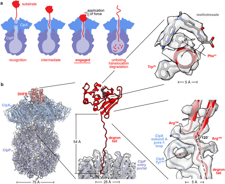
From bacteria to mammals, ATP-fueled AAA+ proteases degrade regulatory, unneeded, or damaged intracellular proteins (Sauer and Baker 2011). For target proteins with stable three-dimensional structures, ClpXP and other AAA+ proteases harness the energy of ATP hydrolysis to unfold this structure before translocating the denatured polypeptide through a narrow axial channel and into a self-compartmentalized peptidase chamber for degradation. Within the ClpXP complex, the ClpX unfoldase mediates substrate specificity by recognizing short ‘degron’ peptides within target substrates. Unfolding is thought to occur when ClpX translocates a disordered substrate segment, containing a recognition degron, until the attached native structure is pulled against the axial channel. Subsequent power strokes then attempt to further translocate the substrate polypeptide through the channel, resulting in repeated application of a force that increases the cumulative probability of unfolding and eventually succeeds in denaturing the substrate to allow sequence-independent translocation of the denatured polypeptide into the ClpP peptidase chamber for proteolysis (Fig. 1a). This model accounts for many studies (Sauer, Fei et al. 2022). However, visualization of a native substrate being pulled against the axial channel of any AAA+ protease has not, until now, been achieved.
Figure 1: ClpXP bound to degron-tagged DHFR.
(a) Cartoon of steps in ClpXP degradation of a protein substrate. (b) Overlay of cryo-EM map and model of ClpXP bound to the degron-tagged DHFR substrate (left). Density for the degron tail could be modeled extending from the folded domain of DHFR to the ClpP entry portal (center). The insets (right) display density for methotrexate and the DHFR Trp30 and Phe31 side chains (top), and the last two residues of native DHFR, Arg158, and Arg159, contacted by the pore-1 loop of ClpX subunit A, which includes Tyr153.
To visualize this postulated ‘engaged’ complex, we prepared cryo-EM grids using Escherichia coli ClpXΔN, a variant lacking the N-terminal domain, E. coli ClpP, and a protein substrate consisting of E. coli dihydrofolate reductase (DHFR) with a C-terminal tail containing a ssrA degron (see Methods). Along with ATP, the reaction mix included methotrexate (MTX), a small-molecule that binds to and stabilizes DHFR (Ainavarapu, Li et al. 2005), and prevents its degradation by ClpXP (Lee, Schwartz et al. 2001). After grid screening, data collection, and image processing (see Methods; Figs. S1-S4), we determined a structure at ~2.8 Å resolution (Table 1; Fig. 1b), which showed natively folded DHFR in close contact with the ClpX ring and 19 residues of the DHFR degron tail in a β-ribbon conformation that extended ~50 Å through the axial channel of ClpX and into the entry portal of ClpP.
Table 1:
Cryo-EM data collection, processing, model building, and validation statistics.
| Sample and data deposition information | |
|---|---|
| Sample name | ClpXΔN•ClpP•DHFR•MTX |
| Nucleotide added | ATP |
| PDB ID | 8V9R |
| EMDB ID | 43081 |
| EMPIAR ID | to be deposited |
| Data collection and image processing | |
| Microscope | Titan Krios G3i |
| Camera | Gatan K3 (counting mode) |
| Magnification (nominal) | 130,000 X |
| Accelerating voltage (keV) | 300 |
| Total electron dose (e−/Å2) | 51.94 |
| Defocus range (μm) | −0.5 to −1.8 |
| Micrographs collected | 15,920 |
| Pixel size (Å) initial/calibrated | 0.679 / 0.654 |
| Map reconstruction | |
| Image processing package | cryoSPARC |
| Total extracted ClpXP particles | 1,005,797 |
| Final particle count | 449,424 |
| Symmetry imposed | C1 |
| Resolution (Å) | |
| 0.143 GSFSC unmasked | 3.3 |
| 0.143 GSFSC spherical mask | 3.1 |
| 0.143 GSFSC tight/local mask | 2.8 |
| 3DFSC sphericity (out of 1.0) | 0.986 |
| Model composition | |
| Number of atoms | 72,633 |
| Protein residues | 4,955 |
| Ligands | 11 (ATP:3; ADP:3; MTX:1; Mg:4) |
| Model refinement | |
| Refinement package | Phenix and Coot |
| Map-to-model cross correlation | |
| masked | 0.81 |
| unmasked | 0.83 |
| RMSD bond lengths (Å) [#>4σ] | 0.003 [0] |
| RMSD bond angles (°) [#>4σ] | 0.666 [0] |
| Model validation | |
| MolProbity score | 0.94 |
| Clash score | 1.80 |
| C-beta outliers (%) | 0.02 |
| Rotamer outliers (%) | 0.0 |
| Ramachandran favored (%) | 99.5 |
| Q-Score [mean] | |
| Global | 0.62 |
| ClpX | 0.61 |
| ClpPcis | 0.71 |
| DHFR | 0.60 |
Density for DHFR and MTX was well resolved in the map (Fig. 1b), and the refined model aligned to a DHFR•MTX crystal structure (Sawaya and Kraut 1997) with a Cα RMSD of 1.25 Å. Thus, ClpX binding per se does not induce partial denaturation of DHFR. Indeed, ClpX supports ClpP degradation of a very large number of structurally diverse proteins (Roche and Sauer 2001, Flynn, Neher et al. 2003, Neher, Villen et al. 2006), making it unlikely that an ability to selectively bind partially denatured conformations of any particular protein would have evolved.
As newly highlighted in our structure, packing between the native DHFR domain and ClpX determines the angle between the degron tail and terminal DHFR structural element (Figs. 1b, 2a). The C-terminal β-strand of native DHFR, which is embedded in a β-sheet, connected to the degron tail at an angle of ~120° and without slack (Fig. 1b). Arg158, the penultimate residue of untagged DHFR, had the same conformation as in an MTX-bound crystal structure (Sawaya and Kraut 1997), whereas the side chain of Arg159, the last native residue, assumed a new conformation that allowed the top axial pore-1 loop of ClpX to contact the extreme C-terminus of the native DHFR domain (Fig. 1b). We posit that the absence of slack would ensure that force from a power stroke is directly applied to the native substrate. We expect that the observed contact angle between substrate and unfoldase also likely influences unfolding efficiency, as some angles would optimize peeling of secondary-structure elements, whereas others would require shearing of multiple hydrogen bonds (Rohs, Etchebest et al. 1999).
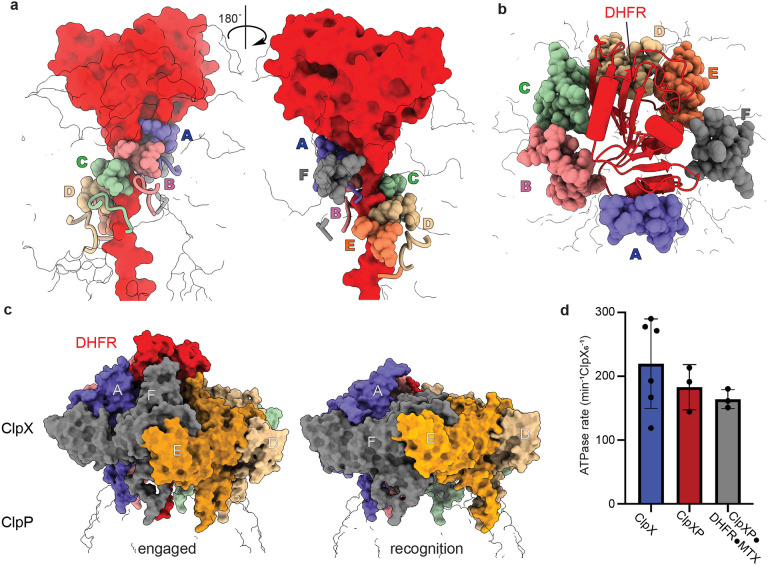
Figure 2: ClpX•DHFR interactions and the consequences of binding.
(a) Front and back views of contacts between pore-1 loops of ClpX (residues 150-155; sphere representation), pore-2 loops of ClpX (residues 198-205; cartoon representation), and DHFR and its degron tail (red surface representation). The six ClpX subunits are annotated A-F and colored purple, salmon, green, wheat, orange, and grey, respectively. The ClpX subunit F pore-1 loop is disengaged from the degron tail but adjacent to the folded domain of DHFR. (b) Top view showing packing of the ClpX RKH loops (residues 220-238; sphere representation colored by subunit as above) around native DHFR (red cartoon representation). (c) Side views of ClpX complexes with DHFR (left) and an ssrA degron (right; pdb 6WRF) illustrating major upward movement of subunit F. (d) Bars show mean rates of ATP hydrolysis for ClpX, ClpXP, or ClpXP in the presence of degron tagged DHFR•MTX ± 1 SD, with symbols denoting replicate assays.
Grip between the axial-channel loops of ClpX and a degron tail also affects unfolding efficiency, with larger non-polar side chains in the tail providing superior grip and faster unfolding (Sauer and Baker 2011, Bell, Baker et al. 2019, Sauer, Fei et al. 2022). In our structure, five pore-1 loops (subunits A,B,C,D,E) and four pore-2 loops (subunits A,B,C,D) of ClpX6 contacted 10 DHFR-degron residues in the channel (Fig. 2a), with many interactions involving large side chains of the degron tail. The pore-1 loop of subunit F was disengaged from the degron tail but close to native DHFR. Hundreds of unsuccessful ClpX power strokes can be needed to unfold and degrade a stable substrate (Kenniston, Baker et al. 2003, Cordova, Olivares et al. 2014). In this regard, the extensive contacts between ClpX and the degron tail are likely to minimize substrate dissociation after an unsuccessful power stroke, thereby increasing the cumulative probability of unfolding for MTX-free DHFR. Our structure also resolved a peripheral collar of contacts between the six RKH loops of ClpX6 and native DHFR (Fig. 2b), which would also minimize substrate dissociation during denaturation attempts. Moreover, we expect that the marked flexibility of individual RKH loops observed in this and previous structures (Fig. S5) allows ClpX to accommodate the highly divergent structures of the very large number of substrates it degrades. We compared Cα positions of Arg228 in the RKH loops of our DHFR complex with the corresponding positions in an ssrAdegron complex (Fei, Bell et al. 2020) (pdb 6WRF) and found movements ranging from 12 to 30 Å (Fig. S5).
In most ClpX hexamers, the axial pore-1 loops form a spiral with the A loop at or near the top and the F loop near the bottom (Fei, Bell et al. 2020, Fei, Bell et al. 2020, Ripstein, Vahidi et al. 2020, Ghanbarpour, Cohen et al. 2023, Ghanbarpour, Fei et al. 2023). By contrast, subunit F moved ‘up’ in our DHFR-bound structure (Fig. 2c), resulting in its pore-1 loop being ~20 Å higher in the channel than the pore-1 loop of subunit E, disengaged from the DHFR degron tail, and at roughly the same level as the pore-1 loop of subunit B (Fig. 2a). Only one other ClpX6 structure, which was engaged with the SspB adaptor and a GFP substrate (Ghanbarpour, Fei et al. 2023), has a similar ‘up’ conformation of subunit F. These engaged structures may be specialized for substrate denaturation. In both the DHFR- and SspB/GFP-bound structures, the ClpP-binding IGF loop of subunit E also hopped into a cleft in the ClpP7 ring that is unoccupied in subunit-F ‘down’ structures (Fig. S6).
DHFR-bound ClpX had ATP bound in subunits A/B/C and ADP in subunits D/E/F (Table S1; Fig. S7), whereas other ClpX6 structures (Fei, Bell et al. 2020, Fei, Bell et al. 2020, Ripstein, Vahidi et al. 2020, Ghanbarpour, Cohen et al. 2023, Ghanbarpour, Fei et al. 2023) contain four or five ATP or ATPγS molecules and one or two ADPs. We considered the possibility that DHFR•MTX binding might trap ClpXP in an ATPase inactive conformation by keeping subunit F in an ‘up’ conformation, but the complex hydrolyzed ATP at a rate only slightly slower than ClpX or ClpXP alone (Fig. 2d). We also used cryoDRGN (Zhong, Bepler et al. 2021, Kinman, Powell et al. 2023, Sun, Kinman et al. 2023) to test for minor populations of ClpX or DHFR structures in the complex, specifically looking for structures in which subunit F was ‘down’ or the IGF-loop of subunit E ‘hopped’, as such conformers were expected had ClpX successfully unfolded DHFR and begun translocating the denatured polypeptide. No such state was observed and, instead, we observed DHFR moving subtly or becoming unresolved above the pore, possibly as a result of ATP-hydrolysis-dependent power strokes within ClpX (Movie S1). Combined with our ATPase assay, this dynamic view of the structure suggests that ClpX readily hydrolyzes ATP as it attempts to denature DHFR•MTX, even when this outcome is highly improbable.
MATERIALS AND METHODS
Protein purification.
E. coli ClpP-His6 and a C-terminally His6-tagged ClpXΔN variant consisting of three ClpXΔN subunits connected by genetically encoded peptide linkers were expressed separately in E. coli and purified by Ni++-NTA, ion-exchange, and gel-filtration chromatography (Martin, Baker et al. 2005). The E. coli DHFR gene was fused to the GSHLGLIEVEKPLYCVEPFVGETAHFEIELSEPDVHGQWKLTSH6 peptide tail at its C-terminus and cloned into a pETDuet expression vector. This tagged DHFR protein was expressed in BL21 (DE3) cells at 18 °C overnight and purified using Ni++-NTA affinity chromatography by loading and washing in buffer N1 [20 mM HEPES pH 7.8, 400 mM NaCl, 100 mM KCl, 10% glycerol, 1 mM DTT, 15 mM imidazole] and eluting in buffer N1 supplemented with imidazole to a concentration of 250 mM. The eluate was concentrated and then subjected to gel-filtration chromatography in buffer GF [20 mM HEPES pH 7.5, 300 mM KCl, 10% glycerol, and 1 mM TCEP]. A synthetic degron-tag peptide consisting of maleimide-GSGSWSHPQFEKAANDENYALAA (21st Century Biochemicals, Inc.), where the underlined sequence is the ssrA tag, was crosslinked to the tagged DHFR protein by reacting ~20 equivalents of the peptide with one equivalent of tagged DHFR for 2 h at room temperature in buffer CL [100 mM HEPES, pH 7.5, 500 mM NaCl, 10% glycerol, 1 mM TCEP] that had been degassed using argon. Unreacted peptide was quenched using 20 mM DTT, removed using a PD- 10 desalting column, and the crosslinked DHFR-C15-ssrA protein was concentrated and flash-frozen in liquid nitrogen for storage. We used this branched-tail substrate hoping to visualize how ClpX accommodates multiple polypeptides in its axial channel, but the branch was not well ordered and could not be modeled in the structure.
Cryo-EM single particle analysis.
The ClpXΔN variant (5.7 μM pseudohexamer) and ClpP (1.5 μM tetradecamer) were incubated with ATP (5 mM) and DHFR-C15-ssrA (20 μM) in buffer R1 [20 mM HEPES pH 7.5, 100 mM KCl, and 25 mM MgCl2] at room temperature for 5 min. Prior to vitrification, 2.5 μL sample aliquots were placed on 200-mesh Quantifoil 2/1 copper grids, which had been glow-discharged at −15 mA for 60 s using an easiGlow discharger (Pelco), and samples were blotted using a FEI Vitrobot Mk IV with a 0 blot force at 6 °C and 95% relative humidity for 4 s.
Movies were collected with EPU (Thermo Fisher Scientific) using aberration-free image shift (AFIS) and hole-clustering methods on a Titan Krios G3i (Thermo Fisher Scientific) with an acceleration voltage of 300 keV and magnification of 130,000 X and detected in super-resolution mode on a K3 detector (Gatan) with a pixel size of 0.679 Å (binned by 2). Movies were collected as 40 frames with a total exposure per specimen of 51.94 e−/Å2 and a defocus range from −0.5 to −1.8 μm. Data processing was performed in cryoSPARC (v.3.3.1) (Punjani, Rubinstein et al. 2017) as depicted in Figure S1. Raw movies (15,290) were pre-processed using ‘patch motion correction’, and ‘patch CTF estimation’. Particles (~47,000) picked using the blob-picker tool from 1,000 random micrographs were extracted (box size 440 px, Fourier cropped to 256 px) and subjected to ‘2D classification’. A set of 4 well-resolved 2D classes composed of side and shoulder views were provided to the ‘template picker’ tool and applied to the full dataset. The particles and micrographs were subjected to ‘manually curate exposures’, filtering out low resolution and low particle count exposures. Particles were extracted (box size 440 px, Fourier cropped to 256 px) and after two rounds of 2D classification, the preliminary stack included 1,005,797 particles.
Multiclass ‘ab-initio’ reconstruction was performed using three classes. One class consisted of 629,143 ClpXP particles (group I) and another of 251,567 ClpXP particles (group II). Inspection of the remaining class via 2D classification revealed a mixture of free ClpP, a low-resolution ClpXP complex, and ‘junk’ particles likely corresponding to errantly picked particles, and these particles were not further considered. After separate homogeneous refinements of groups I and II, heterogeneous refinement was performed using six sub-classes of group I and four sub-classes of groups II. The resulting maps were visually inspected, and four classes from group I and three classes from group II were selected for another round of homogeneous refinement (801,567 particles) that used an ab initio map from group I as an initial model. These aligned particles were then recentered on ClpX using ‘volume tools’ followed by homogeneous refinement with per-particle defocus estimation enabled and subjected to another round of heterogeneous refinement (four classes) that identified the 449,424 particles used for final reconstructions. The final map was obtained through homogeneous refinement, followed by local refinement employing a mask focused on ClpX and the cis ClpP ring. The final map was rescaled using a calibrated pixel size of 0.654 Å (Ghanbarpour, Cohen et al. 2023) in cryoSPARC and had a GSFSC of ~2.8 Å after FSC-mask auto-tightening. After centering particles on the ClpP equatorial ring and extracting the centered particles with a larger box size, we also visualized a second ClpX•DHFR complex bound to the second heptameric ring of ClpP14, albeit at lower resolution (Fig. S8).
Local resolution was estimated using MonoRes (Vilas, Gomez-Blanco et al. 2018) within cryoSPARC; angular FSCs were calculated using the 3DFSC server (Tan, Baldwin et al. 2017); and Q-scores were calculated using a ChimeraX Q-score plugin (Pintilie, Zhang et al. 2020). Model building was performed using a combination of ChimeraX-1.3 (Pettersen, Goddard et al. 2021), Coot-0.9.4 (Casanal, Lohkamp et al. 2020), and Phenix −1.14 (Liebschner, Afonine et al. 2019). The final map was sharpened using CryoSPARC with a b-factor of −50.
ATPase assays.
ATP hydrolysis was measured using a coupled enzymatic reaction (Norby 1988) in which NADH oxidation to NAD+ reduces absorbance at 340 nm (Δε = 6.22 mM−1 cm−1) using a SpectraMax M5 plate reader and a 384-well assay plate (Corning, 3575). A stock ATPase reaction mix (20X) contained 20 μL of a mixture of pyruvate kinase and lactic dehydrogenase from rabbit muscle (P0294, Sigma Aldrich), 10 μL of 200 mM NADH (CAS# 606688), 15 μL of 1 M phosphoenolpyruvate (Sigma Aldrich, 10108294001) in 25 mM HEPES-KOH (pH 7.6), and 25 μL of 200 mM ATP (pH 6.5). For assays, ClpX6 (1 μM) with or without ClpP14 (3 μM) and DHFR-GSYLAALAA (Bell 2020) plus MTX (16 μM each) was present in 10 μL of buffer AB [25 mM HEPES pH 7.8, 100 mM KCl, 20 mM MgCl2, 10% glycerol]. After 5 min of incubation at 30 °C with 2 mM ATP, the ATPase assay was initiated by addition of an equal volume of 2X ATPase reaction mix in buffer AB. Final reaction concentrations were: 0.5 μM ClpX6, 1.5 μM ClpP14 (if present), 8 μM DHFR-GSYLAALAA with MTX (if present), ATPase reaction mix (1X) in a total reaction volume of 20 μL.
CryoDRGN analysis.
CryoDRGN was used to analyze the full set of 449,424 particles where the signal for the trans ring of ClpP had been subtracted in cryoSPARC by: 1) aligning particles on ClpP14 through a local refinement with a mask surrounding ClpP14; 2) subtracting signal of the trans ClpP7 ring; and 3) performing a final local refinement using the signal subtracted particle stack and a mask encompassing DHFR, ClpX6, and the ClpP7 cis ring. These particles were down-sampled to a box size of 254 px (1.13 Å/pixel) and used to train an eight-dimensional latent-variable model in cryoDRGN v2.3.0 using 1024x3 encoder and decoder architectures. The poses and ctf parameters for cryoDRGN training were supplied from the aforementioned local refinement. Following 20 epochs of training, 100 volumes were sampled from the k-means cluster centers of latent embeddings. After extensive visual inspection of these volumes using our atomic model as a reference, we observed neither the ‘down’ conformation in F subunit nor any instances of IGF ‘hopping’. However, the sampled volumes did reveal subtle movements of DHFR or instances where it was unresolved (Supplemental Movie 1).
Supplementary Material
ACKNOWLEDGMENTS
We thank Ed Brignole, Laurel Kinman, and Barrett Powell for helpful discussion and feedback, and the MIT-IBM Satori team and the MIT SuperCloud Supercomputing Center for high performance computing resources and support. This work was supported by NIH grants R01-GM144542 and R35-GM141517, and NSF-CAREER grant 2046778. Samples were prepared at the Automated Cryogenic Electron Microscopy Facility in MIT.nano and screened on a Talos Arctica microscope, which was a gift from the Arnold and Mabel Beckman Foundation.
Footnotes
CONFLICTS OF INTEREST
The authors declare no conflicts of interest.
REFERENCES
- Ainavarapu S. R., Li L., Badilla C. L. and Fernandez J. M. (2005). “Ligand binding modulates the mechanical stability of dihydrofolate reductase.” Biophys J 89(5): 3337–3344. [DOI] [PMC free article] [PubMed] [Google Scholar]
- Bell T. A. (2020). “Intersubunit communication and coordinated mechanical activity in the AAA+ protease ClpXP.” PhD. thesis. Massachusetts Institute of Technology. [Google Scholar]
- Bell T. A., Baker T. A. and Sauer R. T. (2019). “Interactions between a subset of substrate side chains and AAA+ motor pore loops determine grip during protein unfolding.” Elife 8. [DOI] [PMC free article] [PubMed] [Google Scholar]
- Casanal A., Lohkamp B. and Emsley P. (2020). “Current developments in Coot for macromolecular model building of Electron Cryo-microscopy and Crystallographic Data.” Protein Sci 29(4): 1069–1078. [DOI] [PMC free article] [PubMed] [Google Scholar]
- Cordova J. C., Olivares A. O., Shin Y., Stinson B. M., Calmat S., Schmitz K. R., Aubin-Tam M. E., Baker T. A., Lang M. J. and Sauer R. T. (2014). “Stochastic but highly coordinated protein unfolding and translocation by the ClpXP proteolytic machine.” Cell 158(3): 647–658. [DOI] [PMC free article] [PubMed] [Google Scholar]
- Fei X., Bell T. A., Barkow S. R., Baker T. A. and Sauer R. T. (2020). “Structural basis of ClpXP recognition and unfolding of ssrA-tagged substrates.” Elife 9. [DOI] [PMC free article] [PubMed] [Google Scholar]
- Fei X., Bell T. A., Jenni S., Stinson B. M., Baker T. A., Harrison S. C. and Sauer R. T. (2020). “Structures of the ATP-fueled ClpXP proteolytic machine bound to protein substrate.” Elife 9. [DOI] [PMC free article] [PubMed] [Google Scholar]
- Flynn J. M., Neher S. B., Kim Y. I., Sauer R. T. and Baker T. A. (2003). “Proteomic discovery of cellular substrates of the ClpXP protease reveals five classes of ClpX-recognition signals.” Mol Cell 11(3): 671–683. [DOI] [PubMed] [Google Scholar]
- Ghanbarpour A., Cohen S. E., Fei X., Kinman L. F., Bell T. A., Zhang J. J., Baker T. A., Davis J. H. and Sauer R. T. (2023). “A closed translocation channel in the substrate-free AAA+ ClpXP protease diminishes rogue degradation.” Nat Commun 14(1): 7281. [DOI] [PMC free article] [PubMed] [Google Scholar]
- Ghanbarpour A., Fei X., Baker T. A., Davis J. H. and Sauer R. T. (2023). “The SspB adaptor drives structural changes in the AAA+ ClpXP protease during ssrA-tagged substrate delivery.” Proc Natl Acad Sci U S A 120(6): e2219044120. [DOI] [PMC free article] [PubMed] [Google Scholar]
- Kenniston J. A., Baker T. A., Fernandez J. M. and Sauer R. T. (2003). “Linkage between ATP consumption and mechanical unfolding during the protein processing reactions of an AAA+ degradation machine.” Cell 114(4): 511–520. [DOI] [PubMed] [Google Scholar]
- Kinman L. F., Powell B. M., Zhong E. D., Berger B. and Davis J. H. (2023). “Uncovering structural ensembles from single-particle cryo-EM data using cryoDRGN.” Nat Protoc 18(2): 319–339. [DOI] [PMC free article] [PubMed] [Google Scholar]
- Lee C., Schwartz M. P., Prakash S., Iwakura M. and Matouschek A. (2001). “ATP-dependent proteases degrade their substrates by processively unraveling them from the degradation signal.” Mol Cell 7(3): 627–637. [DOI] [PubMed] [Google Scholar]
- Liebschner D., Afonine P. V., Baker M. L., Bunkoczi G., Chen V. B., Croll T. I., Hintze B., Hung L. W., Jain S., McCoy A. J., Moriarty N. W., Oeffner R. D., Poon B. K., Prisant M. G., Read R. J., Richardson J. S., Richardson D. C., Sammito M. D., Sobolev O. V., Stockwell D. H., Terwilliger T. C., Urzhumtsev A. G., Videau L. L., Williams C. J. and Adams P. D. (2019). “Macromolecular structure determination using X-rays, neutrons and electrons: recent developments in Phenix.” Acta Crystallogr D Struct Biol 75(Pt 10): 861–877. [DOI] [PMC free article] [PubMed] [Google Scholar]
- Martin A., Baker T. A. and Sauer R. T. (2005). “Rebuilt AAA + motors reveal operating principles for ATP-fuelled machines.” Nature 437(7062): 1115–1120. [DOI] [PubMed] [Google Scholar]
- Neher S. B., Villen J., Oakes E. C., Bakalarski C. E., Sauer R. T., Gygi S. P. and Baker T. A. (2006).”Proteomic profiling of ClpXP substrates after DNA damage reveals extensive instability within SOS regulon.” Mol Cell 22(2): 193–204. [DOI] [PubMed] [Google Scholar]
- Norby J. G. (1988). “Coupled assay of Na+,K+-ATPase activity.” Methods Enzymol 156: 116–119. [DOI] [PubMed] [Google Scholar]
- Pettersen E. F., Goddard T. D., Huang C. C., Meng E. C., Couch G. S., Croll T. I., Morris J. H. and Ferrin T. E. (2021). “UCSF ChimeraX: Structure visualization for researchers, educators, and developers.” Protein Sci 30(1): 70–82. [DOI] [PMC free article] [PubMed] [Google Scholar]
- Pintilie G., Zhang K., Su Z., Li S., Schmid M. F. and Chiu W. (2020). “Measurement of atom resolvability in cryo-EM maps with Q-scores.” Nat Methods 17(3): 328–334. [DOI] [PMC free article] [PubMed] [Google Scholar]
- Punjani A., Rubinstein J. L., Fleet D. J. and Brubaker M. A. (2017). “cryoSPARC: algorithms for rapid unsupervised cryo-EM structure determination.” Nat Methods 14(3): 290–296. [DOI] [PubMed] [Google Scholar]
- Ripstein Z. A., Vahidi S., Houry W. A., Rubinstein J. L. and Kay L. E. (2020). “A processive rotary mechanism couples substrate unfolding and proteolysis in the ClpXP degradation machinery.” Elife 9. [DOI] [PMC free article] [PubMed] [Google Scholar]
- Roche E. D. and Sauer R. T. (2001). “Identification of endogenous SsrA-tagged proteins reveals tagging at positions corresponding to stop codons.” J Biol Chem 276(30): 28509–28515. [DOI] [PubMed] [Google Scholar]
- Rohs R., Etchebest C. and Lavery R. (1999). “Unraveling proteins: a molecular mechanics study.” Biophys J 76(5): 2760–2768. [DOI] [PMC free article] [PubMed] [Google Scholar]
- Sauer R. T. and Baker T. A. (2011). “AAA+ proteases: ATP-fueled machines of protein destruction.” Annu Rev Biochem 80: 587–612. [DOI] [PubMed] [Google Scholar]
- Sauer R. T., Fei X., Bell T. A. and Baker T. A. (2022). “Structure and function of ClpXP, a AAA+ proteolytic machine powered by probabilistic ATP hydrolysis.” Crit Rev Biochem Mol Biol 57(2): 188–204. [DOI] [PMC free article] [PubMed] [Google Scholar]
- Sawaya M. R. and Kraut J. (1997).”Loop and subdomain movements in the mechanism of Escherichia coli dihydrofolate reductase: crystallographic evidence.” Biochemistry 36(3): 586–603. [DOI] [PubMed] [Google Scholar]
- Sun J., Kinman L. F., Jahagirdar D., Ortega J. and Davis J. H. (2023). “KsgA facilitates ribosomal small subunit maturation by proofreading a key structural lesion.” Nat Struct Mol Biol. [DOI] [PMC free article] [PubMed] [Google Scholar]
- Tan Y. Z., Baldwin P. R., Davis J. H., Williamson J. R., Potter C. S., Carragher B. and Lyumkis D. (2017). “Addressing preferred specimen orientation in single-particle cryo-EM through tilting.” Nat Methods 14(8): 793–796. [DOI] [PMC free article] [PubMed] [Google Scholar]
- Vilas J. L., Gomez-Blanco J., Conesa P., Melero R., Miguel de la Rosa-Trevin J., Oton J., Cuenca J., Marabini R., Carazo J. M., Vargas J. and Sorzano C. O. S. (2018). “MonoRes: Automatic and Accurate Estimation of Local Resolution for Electron Microscopy Maps.” Structure 26(2): 337–344 e334. [DOI] [PubMed] [Google Scholar]
- Zhong E. D., Bepler T., Berger B. and Davis J. H. (2021). “CryoDRGN: reconstruction of heterogeneous cryo-EM structures using neural networks.” Nat Methods 18(2): 176–185. [DOI] [PMC free article] [PubMed] [Google Scholar]
Associated Data
This section collects any data citations, data availability statements, or supplementary materials included in this article.


