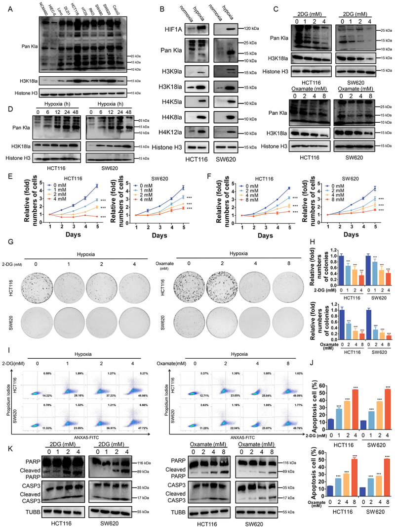
Figure 2.

Effects of histone lactylation inhibition on colorectal cancer cells proliferation and survival in hypoxia.
Note: (A) Western blotting analysis of lactylation or H3K18la levels in normal colon epithelial cells and CRC cells. (B) Western blotting analysis of Pan- and site-specific histone lactylation in HCT116 and SW620 cells in normoxia compared with in hypoxia (1% oxygen, 24 h). (C) Lactylation and H3K18la levels were detected in HCT116 and SW620 cells cultured in different concentrations of 2-DG or oxamate for 24 h in hypoxia (1% oxygen) by western blot. (D) Lactylation and H3K18la levels were detected in HCT116 and SW620 cells cultured in hypoxia (1% oxygen) at the indicated time. (E and F) Proliferation of HCT116 and SW620 cells cultured in different concentrations of 2-DG (E) or oxamate (F) in hypoxia (1% oxygen) was analyzed using CCK8 assay. ***P ≤0.001. (G) Tumor growth of HCT116 and SW620 cells treated with different concentrations of 2-DG or oxamate in hypoxia (1% oxygen) was evaluated by colony formation assay. (H) Statistical analysis of the colony formation assay performed using HCT116 and SW620 cells treated with different concentrations of 2-DG or oxamate in hypoxia (1% oxygen). All of the experiments were performed in triplicate, and relative colony numbers are shown as means ± SD. ***P ≤0.001. (I) ANXA5/annexin V-FITC and PI staining showing apoptosis in HCT116 and SW620 cells cultured in different concentrations of 2-DG or oxamate in hypoxia (1% oxygen). (J) Quantification of apoptotic cells. ***P ≤0.001. (K) Western blotting analysis of cleaved CASP3 and cleaved PARP in in HCT116 and SW620 cells cultured in different concentrations of 2-DG or oxamate in hypoxia (1% oxygen).
