Abstract
Histamine receptors are a group of G protein-coupled receptors (GPCRs) that play important roles in various physiological and pathophysiological conditions. Antihistamines that target the histamine H1 receptor (H1R) have been widely used to relieve the symptoms of allergy and inflammation. Here, to uncover the details of the regulation of H1R by the known second-generation antihistamines, thereby providing clues for the rational design of newer antihistamines, we determine the cryo-EM structure of H1R in the apo form and bound to different antihistamines. In addition to the deep hydrophobic cavity, we identify a secondary ligand-binding site in H1R, which potentially may support the introduction of new derivative groups to generate newer antihistamines. Furthermore, these structures show that antihistamines exert inverse regulation by utilizing a shared phenyl group that inserts into the deep cavity and block the movement of the toggle switch residue W4286.48. Together, these results enrich our understanding of GPCR modulation and facilitate the structure-based design of novel antihistamines.
Subject terms: Cryoelectron microscopy, Cryoelectron microscopy, G protein-coupled receptors, Drug discovery
Histamine receptor H1R has been extensively targeted in the development of antihistamines. Here, Wang et al. determine structures of H1R alone and bound to different antihistamines, providing insights into the structure-based design of next-generation drugs.
Introduction
Histamine is a biogenic amine that plays an important role in various physiological and pathophysiological conditions by activating four different G-protein coupled receptors (GPCRs), classified as H1R, H2R, H3R, and H4R1–3. These four receptors are either well-established drug targets (H1R, H2R, and H3R) or are being evaluated for related diseases, for example, by targeting H4R for anti-inflammatory diseases4–6. Among them, H1R is widely distributed throughout the body in epithelial cells and smooth vascular, neuronal, glial, and immune cells7,8. When activated by histamine released by mast cells or basophils, H1R can cause allergic and inflammatory symptoms and has therefore been extensively targeted in the development of antihistamines9,10. To date, more than 45 H1R-antihistamines are available worldwide, and moreover, new antihistamines are continuously being investigated11,12. Since the first antihistamine came out in 1937, antihistamines have gone through the development process of first-generation, second-generation and now the new second-generation antihistamines5,13,14. Although first-generation antihistamines such as chlorpheniramine and diphenhydramine were once widely used clinically, they cause adverse central nervous system (CNS) responses due to their poor receptor selectivity and ability to cross the blood-brain barrier (BBB) and bind to H1R in the CNS11,15–17. By introducing new chemical groups, such as carboxyl groups combined with protonated amines, second-generation antihistamines have been successively developed. Second-generation antihistamines have high H1R selectivity, rarely cross the BBB, and preferentially bind peripheral H1R11,18. Therefore, second-generation antihistamines cause few or no CNS side effects19,20. However, second-generation H1R antihistamines may still have adverse effects on the heart21. For example, the second-generation drug astemizole was reported to cross-interact with several other targets and thus cause cardiotoxicity in vivo22.
Recently, new second-generation antihistamines, mainly derived from the active metabolites or optical isomers of second-generation antihistamines, have been introduced23. For example, as a metabolite of the second-generation antihistamine loratadine, desloratadine has been developed and defined as a new-generation antihistamine24,25; however, it remains doubtful whether these newer antihistamines are actually superior to their predecessors in terms of efficacy and safety. In other words, great challenges still exist in the development of new antihistamines, especially the lack of interaction details between H1R and second-generation antihistamines, which hinders the rational design of new drugs. In addition to guiding drug development, elucidating the structural states of H1R will also help to understand the mechanism of inverse modulation of ligands on GPCRs. Since H1R has a high basal signaling capacity, a prominent feature of many GPCRs, and all antihistamines have been shown to be inverse agonists rather than antagonists26–28, how H1R obtains its constitutive activity and how it is blocked by antihistamines remain poorly elucidated. Here, we determine the structure of H1R in the apo form and bound to different antihistamines. The structural information provides insights into the interaction and modulation of H1R with antihistamines and will facilitate the structure-based design of next-generation drugs.
Results
Structure determination of H1R in apo form and bound to inverse agonists
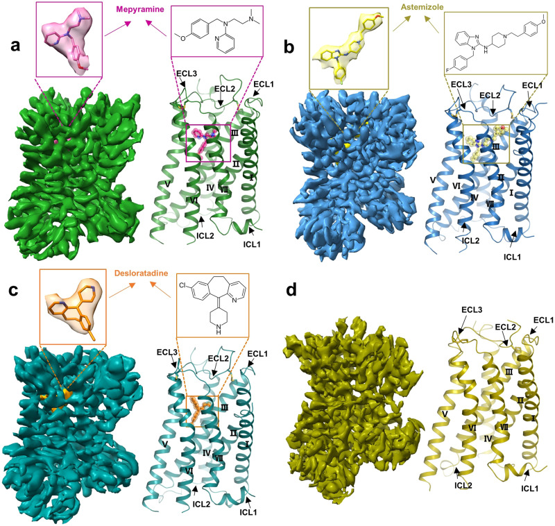
To solve the structure of H1R in complex with inverse agonists, we employed the mBril fusion and gluing strategy recently developed by our group29. Briefly, mBril was fused between TM5 and TM6 in a way that would form two continuous helices with the receptor (Supplementary Fig. 1a). Meanwhile, the helical tag K3-ALFA was added to the C-terminus of H8 in the desired configuration (Supplementary Fig. 1a). We next used Alphafold2 to predict the structure of the fusion protein. By docking the known 1B3-Bril structure onto the predicted receptor, we obtained a complex model, and based on this model, the “4-9” glue molecule was chosen for the first attempt to solve the structure of H1R bound to the first-generation antihistamine mepyramine. However, this sample did not show many particles with the desired shape in the 2D analysis (Supplementary Fig. 1b). The reason seems to be that this “4-9” glue molecule is too harsh since the mBril domain is bent in most of the particles (Supplementary Fig. 1b). We then reprepared the grid by using the “6-13” glue molecule. This new sample indeed displayed much better particles in the preanalysis (Supplementary Fig. 1c), and we thus conducted large-scale data collection. After data processing, a cryo-EM map with a global resolution of 3.2 Å was obtained (Supplementary Fig. 2 and Supplementary Table 1). A further refinement focused on the TM region produced an improved map in which the side chains of most H1R residues were traceable (Fig. 1a, Supplementary Fig. 2). Guided by the map, mepyramine and H1R residues consisting of S24-Q171, V174-L221 and N408-R481 were built.
Fig. 1. Cryo-EM map and overall structure of H1R bound to inverse agonist and H1R in apo form.
a Cryo-EM map and overall structure of the H1R-mepyramine complex. H1R and mepyramine are colored green and hot pink, respectively. b Cryo-EM map and overall structure of the H1R-astemizole complex. H1R and astemizole are colored sky blue and pale yellow, respectively. c Cryo-EM map and overall structure of the H1R-desloratadine complex. H1R and desloratadine are colored in deep teal and orange, respectively. d Cryo-EM map and overall structure of H1R in apo form, H1R color as olive.
The same structure determination strategy was then applied to H1R in apo form and complexed with the second-generation antihistamines astemizole and desloratadine, and their structures were finally determined at 3.5, 3.0 and 3.4 Å resolution, respectively (Fig. 1b–d, Supplementary Figs. 3–5 and Supplementary Table 1). Interestingly, although following exactly the same conditions, the apo form of H1R had lower quality density maps compared to those complexed with inverse agonists (Fig. 1 and Supplementary Figs. 2–5), suggesting that without inverse agonist stabilization, H1R may have a dynamic structure (Supplementary Fig. 6). Guided by the density map, models of H1R in apo form and bound to astemizole and desloratadine were built (Fig. 1b–d). Finally, the model of H1R in apo form contains residues P29-E55, L58-R134, R139-Q170, R176-L221 and N408-R481. The astemizole-H1R model contains residues T20-Q171, V174-L221, and N408-R481. The desloratadine-H1R model contains residues P25-Q171, D178-L221, and N408-R481. As expected, all the inverse agonists were bound at the orthosteric pocket with the core pharmacophore buried at roughly similar positions (Fig. 1a–c). H1R shares an almost identical overall conformation in the different inverse agonist bound states, including that in complex with doxepin30, as reflected by the root-mean-square deviation (RMSD) values of 0.5-0.7 Å for the Cα within the receptor. Although all these H1R structures reside in a typical inactive conformation, featuring the close arrangement of TM6 with the receptor core on the intracellular side (Fig. 1), noticeable structural differences are also observed around the ligand binding pocket, as discussed below.
Recognition mechanism of mepyramine with H1R
Mepyramine is a first-generation antihistamine that targets H1R31,32 and has been approved for the treatment of allergic reactions and urticaria. Like many other first-generation antihistamines, mepyramine can cross the blood‒brain barrier and often causes serious side effects, such as drowsiness. Recently, mepyramine has been shown to bind to a variety of voltage-gated sodium channels and directly inhibit their activity, so mepyramine has the potential to be developed as a topical analgesic agent33. Deciphering the recognition details of mepyramine by each individual target receptor will provide valuable clues for further optimization of the drug for improved specificity and efficacy.
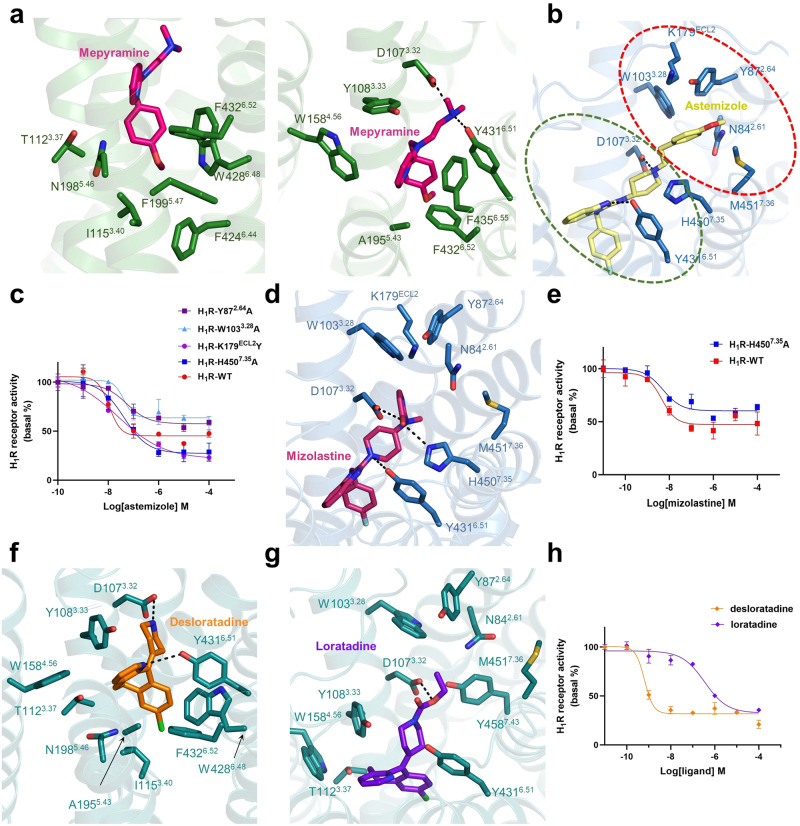
The determined structure of mepyramine-H1R shows that mepyramine is captured in the orthosteric pocket mainly constituted by residues from TM3, TM5, TM6, and TM7 (Fig. 2a). The three functionality groups of mepyramine establish an interaction network with H1R. Among them, the methoxyphenyl group of mepyramine, especially the methoxy moiety, is deeply inserted into a hydrophobic cavity formed by H1R residues T1123.37, I1153.40, N1985.46, F1995.47, F4246.44, W4286.48 and F4326.52 (Fig. 2a). Consistent with the structural observation, all the single mutations of F1995.47, W4286.48 and F4326.52 abolished the H1R affinity with mepyramine34. In contrast, the N198A mutation did not cause obvious affinity loss35, suggesting that hydrophobic contacts dominate the H1R interaction with the methoxyphenyl group. Notably, W4286.48 is the toggle switch residue that normally initiates GPCR activation through ligand-induced conformational changes36. In addition, F4246.44 and I1153.40 are derived from the P5.50-I3.40-F6.44 triadmotif, which also plays a key role in GPCR activation36. In general, GPCR ligands bind receptors at sites above these motifs, as is the case for H1R recognition of the natural ligand histamine (Supplementary Fig. 7b); however, when coordinating an inverse agonist, H1R adopts a completely different binding mode, and to the best of our knowledge, the site holding mepyramine is the deepest location ever observed. The pyridine moiety of mepyramine is again mainly coordinated by hydrophobic interactions with H1R residues Y1083.33, W1584.56, A1955.43, F4326.52 and F4356.55 (Fig. 2a). Among them, as anticipated, the single mutations of Y1083.33 and W1584.56 completely disrupted the mepyramine binding activity34. The F435A mutation (F436A in guinea pig H1R) also resulted in a 4-fold and 20-fold decrease in the affinity of mepyramine for human and guinea pig H1R35,37, respectively. The third functionality group, the dimethylamino moiety, adopts a pose similar to that in the doxepin-H1R complex30 and forms a salt bridge with the H1R residue D1073.32 (Fig. 2a and Supplementary Fig. 7b). D1073.32 is a highly conserved residue in aminergic receptors and has been repeatedly confirmed to be indispensable for ligand binding30,38; therefore, D1073.32-mediated polar contact may represent a universal mode in H1R ligand interactions. Moreover, Y4316.51 forms a hydrogen bond with the N atom in the dimethylamino group (Fig. 2a). The single mutant of Y4316.51 also completely disrupted the binding activity of mepyramine34. In summary, the structural and functional data reveal that H1R engages the first-generation drug mepyramine through a highly hydrophobic pocket and a conserved salt bridge.
Fig. 2. Detailed interactions of inverse agonists in the H1R ligand-binding pocket.
a The ligand-binding pocket of mepyramine. H1R is shown as green ribbons, with critical residues for ligand binding shown as green sticks and mepyramine shown as hot pink sticks. b The ligand-binding pocket of astemizole. H1R and astemizole are sky blue and pale yellow, respectively. The main and secondary pockets are indicated with green and red circles, respectively. c Dose-dependent responses of astemizole measured by cellular IP1 accumulation assays in wild-type and mutant H1R. d Mizolastine docked in H1R. Mizolastine is shown as warm pink sticks, and the critical residues of H1R are shown as blue sticks. The H4507.35-mediated hydrogen bond is indicated with a dashed line. e Dose-dependent responses of mizolastine measured by cellular IP1 accumulation assays in wild-type and mutant H1R. f The ligand-binding pocket of desloratadine. H1R and desloratadine color as deep teal and orange, respectively. g Loratadine docked in H1R. Loratadine and H1R are colored purple‒blue and deep teal, respectively. Hydrogen bonds and salt bridges are highlighted as black dashed lines. h Dose-dependent responses of desloratadine (orange line) and loratadine (purple line) and loratadine at wild-type H1R measured by cellular IP1 accumulation assays. The data from the cellular IP1 accumulation assays are represented as the mean ± SEM, n = 3 independent samples.
Recognition mechanism of astemizole with H1R
Astemizole is a second-generation antihistamine drug, but it was withdrawn from the market due to its potential to cause arrhythmias at high doses. The reason is that astemizole not only targets H1R but also blocks the hERG potassium channel39,40. In addition, astemizole has also been shown to disrupt the protein‒protein interaction within polycomb repressive complex 2 (PRC2) and thus arrest the proliferation of cancer cells41. Repurposing astemizole for new clinical uses has been constantly explored42–44. Delineating the molecular recognition mechanism with astemizole is a prerequisite for precise modification of astemizole to eliminate its pleiotropic effect.
The H1R structure bound to astemizole shows that the fluorophenyl and benzimidazole groups of astemizole occupy positions similar to those accommodating the methoxyphenyl and pyridine groups of mepyramine, respectively (Fig. 2b). A hydrogen bond, also mediated by Y4316.51, and the remaining extensive hydrophobic interactions are responsible for the coordination of astemizole (Fig. 2b). The piperidine group extends as the dimethylamino moiety of mepyramine and establishes an electrostatic interaction with the H1R residue D1073.32 (Fig. 2b). The shared pose of mepyramine and astemizole in the orthosteric pocket suggests that H1R adopts a conserved binding mode for ligands with similar chemotypes. Interestingly, the additional methoxyphenyl moiety of astemizole is extended from the main ligand binding pocket toward the site defined by residues from ECL2, TM2, TM3 and TM7 (Fig. 2b). We refer to this site as a secondary binding pocket. Surrounding H1R residues Y872.64, W1033.28, and M4517.36 make hydrophobic contacts with the methoxyphenyl moiety (Fig. 2b). Mutation of Y872.64 and W1033.28 markedly impaired the ability of astemizole to inhibit H1R signaling (Fig. 2c), confirming the contribution of this secondary pocket to astemizole binding. Apart from the hydrophobic residues, three polar residues, N842.61, K179ECL2 and H4507.35, also decorate this secondary pocket (Fig. 2b); however, they do not form any polar interactions with the methoxyphenyl moiety and even induce forces inconsistent with the hydrophobicity of the methoxyphenyl moiety. Mutating K179ECL2 and H4507.35 to hydrophobic residues improves the efficacy of astemizole in inhibiting H1R (Fig. 2c and Supplementary Table 2). Therefore, modifying the methoxyphenyl moiety with alternative derivative groups, for example, introducing hydrogen bond donors/acceptors, may further improve the specificity of astemizole. In other words, making the most use of this secondary pocket could lead to new selective or effective antihistamines. Consistent with this notion, another second-generation nonsedating antihistamine, mizolastine, which differs from astemizole only by the methoxyphenyl moiety, does not cross-interact with any non-H1R targets, including the potassium channel45. To probe how mizolastine engages H1R, especially how the polar group dihydropyrimidine of mizolastine fits into the secondary pocket, we docked mizolastine onto H1R (Supplementary Table 3). In the resulting docked structure, mizolastine occupies the expected position in the pocket, and its fluorophenyl and benzimidazole groups adopt the same poses as the corresponding groups in astemizole, indicating that the docking of mizolastine is plausible (Fig. 2d). Interestingly, instead of inducing an unfavorable environment for the methoxyphenyl moiety of astemizole, H4507.35 mediates a hydrogen bond with the dihydropyrimidine of mizolastine (Fig. 2d), suggesting the compatibility of this derivative group with the secondary pocket. Accordingly, different from the boosting effect of astemizole, the H450A mutation obviously crippled the ability of mizolastine to inhibit H1R signaling (Fig. 2e and Supplementary Table 2). Therefore, while the incorporation of dihydropyrimidine abolishes the mizolastine affinity for other non-H1R targets, the dihydropyrimidine group can be well matched to the secondary pocket of H1R, thereby still retaining a high H1R affinity. Together, the astemizole-H1R structure reveals that in addition to the main ligand binding pocket, H1R possesses a secondary pocket that can be explored for the development of new antihistamines.
Recognition mechanism of desloratadine with H1R
Desloratadine, an antihistamine for the treatment of seasonal allergic rhinitis, is a metabolic derivative of another second-generation drug, loratadine46. For this reason, desloratadine has been called a new-generation antihistamine; however, except for displaying a higher affinity and slower disassociation rate with the target receptor H1R47, desloratadine does not appear to confer improved efficacy or additional clinical benefit compared to loratadine. Nevertheless, it is still of great significance to uncover the molecular mechanism leading to the difference in affinity to provide clues for the design of next-generation antihistamines.
The solved structure of desloratadine-H1R shows that desloratadine is buried in the main orthosteric pocket, and its tricyclic group is enclosed by a series of hydrophobic residues such as Y1083.33, Y4316.51 and F4326.52 in a manner similar to the other two ligands (Fig. 2f). In addition to the hydrophobic interactions, Y4316.51 also forms a hydrogen bond with the N atom in the pyridine moiety, as in the mepyramine-H1R complex (Fig. 2f). Mutation of Y4316.51 and F4326.52, two residues in which mutation does not significantly affect the basal signaling ability of H1R34,48, dramatically impairs the ability of mepyramine and desloratadine to inhibit H1R signaling (Supplementary Fig. 8 and Supplementary Table 2), demonstrating the indispensability of main pocket residues in the coordination of ligands. Finally, the piperidine ring also forms an electrostatic interaction with D1073.32 (Fig. 2f). Interestingly, the only difference between desloratadine and loratadine is that the ethoxycarbonyl group attached to the piperidine ring in loratadine is replaced with hydrogen in desloratadine. This substitution causes desloratadine to have approximately two orders of magnitude higher affinity for H1R than loratadine. To uncover the underlying mechanism, we again removed the desloratadine molecule and docked loratadine onto this structure (Supplementary Table 3). As anticipated, the tricyclic group of loratadine is located in the main pocket, similar to desloratadine, in the docked model. The ethoxycarbonyl group extends into the secondary binding pocket (Fig. 2g). However, unlike the methoxyphenyl moiety in astemizole, the ethoxycarbonyl group is much smaller in size and cannot form effective hydrophobic contacts with the surrounding residues Y872.64, W1033.28, and M4517.36. Instead, the carbonyl tail even faces two hydrophilic atoms from H1R residues N842.61 and Y4587.43 (Fig. 2g). Obviously, when the tricyclic group of loratadine is positioned in the main ligand binding pocket, very reasonable accommodation of the ethoxycarbonyl group in the secondary binding pocket will not be achieved. Consistently, the measured potency of loratadine in inhibiting H1R was much lower than that of desloratadine (Fig. 2h and Supplementary Table 2). Structure-guided introduction of hydrophilic derivatives into loratadine at the corresponding position of the ethoxycarbonyl group may result in antihistamines with better clinical performance.
Comparison of the ligand pocket in different states
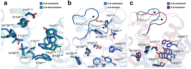
With the availability of H1R structures bound to different ligands, we next performed a detailed comparison of the ligand pocket shaped by different agents. Superposition of the H1R structures bound to second-generation antihistamines from this study reveals that the residues lining the entire ligand pocket adopt roughly similar configurations (Fig. 3a), which suggests that H1R may employ a general recognition mechanism to engage antihistamines, especially when the antihistamines share a similar chemotype. However, when comparing the astemizole-bound ligand pocket to that bound with the first-generation antihistamine doxepin, remarkable conformational differences were observed in the region surrounding the secondary pocket (Fig. 3b). In the doxepin-bound structure, H1R ECL2 occupies the vestibule above the secondary pocket, but in the astemizole-bound structure, ECL2 adopts a lifted conformation, releasing more space for accommodation of the methoxyphenyl moiety of astemizole (Fig. 3b). Meanwhile, the side chain of Y872.64 also rotates to participate in the construction of the secondary pocket (Fig. 3b). Further comparison of the astemizole-bound H1R with that bound to the endogenous agonist histamine revealed a more extensive conformational change in the ligand pocket (Fig. 3c). Due to the smaller size of the agonist histamine, the active pocket becomes more constricted to make full contact with the ligand. For example, residues Y4316.51, F4326.52, H4507.35 and Y4587.43 in the active structure all move closer toward the receptor core (Fig. 3c). Again, ECL2 sits on top of the secondary pocket, although it is not inserted as deep into the pocket as that in the doxepin-bound structure (Fig. 3c). Together, these results demonstrate that there is structural plasticity in the ligand-binding pocket of H1R, and it is worth making full use of these different states of the pocket for the development of next-generation drugs.
Fig. 3. The ligand-binding pocket of different H1R complexes.
a Superposition of H1R-astemizole (sky blue) and H1R-desloratadine (deep teal). b Comparison of the selective ligand-binding pockets of H1R-astemizole (sky blue) and H1R-doxepin (light blue) (PDB ID: 3RZE). c Comparison of the crucial residues and ECL2 from H1R-astemizole (sky blue) and H1R-histamine (raspberry). Key different residues are shown as sticks.
Mechanism of H1R activation
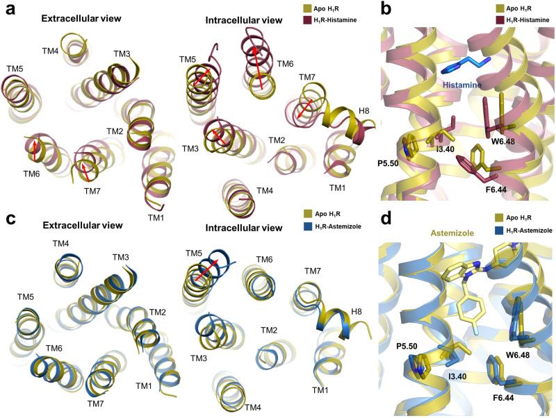
An important property of H1R is its high basal signaling capacity, a distinctive feature observed in many GPCRs. Due to the lack of efficient methods to solve the structure of GPCRs in the apo state, whether these GPCRs maintain an activity-like structure or what conformation they have in the apo state remains unknown. Enabled by our recently developed method, we successfully solved the structure of H1R in the apo state (Fig. 1d). The determined structure shows that it adopts a typical inactive conformation in the apo state (Fig. 1d). Therefore, the high basal signal of H1R may not be attributed to the preexisting stable open conformation of TM6. Interaction with a G protein appears to still be required to stabilize H1R in a fully active state. Although a ‘squash to activate and expand to deactivate’ model has been proposed for the activation of H1R through structural comparison of inverse agonist-bound and agonist-bound H1R48, the existence of an additional apo structure provides a more comprehensive understanding of H1R activation and regulation (Fig. 4). In the apo state, TM6 and TM7 appear to adopt an intermediate conformation, and this conformation is induced to move in opposite directions by inverse agonists and agonists (Supplementary Fig. 9). Specifically, histamine binding induces a further contraction of TM6 and TM7 on the extracellular side (Fig. 4a). The resulting closed packing on the extracellular region drives the downward movement of key activation motifs, the toggle residue W4286.48 and the P5.50-I3.40-F6.44 triadmotif (Fig. 4b). These conformational rearrangements then propagate to the intracellular side and result in the outward movement of TM6 (Fig. 4a). In contrast, engagement of an inverse agonist at the orthosteric site seems to insulate the contact between TM3 and TM6 and results in a wider opening of the extracellular core of the agonist-stabilized structures (Fig. 4c). Therefore, the ready accessibility between TM3 and TM6 in the apo state of H1R is probably one of the reasons for its basal signaling ability. Next, we probed the mechanism of inverse modulation of histamines. Taking the H1R structure bound to astemizole as an example, the fluorophenyl group of astemizole inserted deeply into the vicinity of W4286.48 and completely prevented its downward movement (Fig. 4d). As a result, the W4286.48 and P5.50-I3.40-F6.44 triadmotif are constantly fixed in inactive positions, leading to the closed packing of TM6 against the intracellular core and ultimately preventing basal activation of H1R (Fig. 4d). Therefore, all these compounds are classified as inverse agonists. In summary, agonist and inverse agonist binding to H1R induces an opposite conformational change in the extracellular region, which ultimately causes different signaling outputs on the intracellular side.
Fig. 4. Conformational changes during H1R activation.
a Structural comparisons of H1R bound to histamine (PDB ID: 7DFL) and in apo H1R. Significant conformational changes are indicated with red arrows. b Comparison of the P5.50-I3.40-F6.44 triadmotif and W6.28 in the H1R apo form (olive) and histamine-bound active form (raspberry). Histamine is shown as marine sticks. c Structural comparisons of H1R bound to astemziole (sky blue) and in apo form (olive). Significant conformational changes are indicated with red arrows. d Comparison of the P5.50-I3.40-F6.44 triadmotif and W6.28 in the H1R apo form (olive) and the astemizole-bound H1R inactive form (sky blue). Astemizole color as pale yellow.
Discussion
Antihistamines that block H1R activity are commonly used to prevent and treat symptoms of allergic rhinitis, allergic conjunctivitis, and urticaria12. Although second-generation antihistamines have offered many advantages over first-generation antihistamines, such as minimal blood‒brain barrier penetration, efforts are still being made to develop newer generations of antihistamines with higher efficacy and safety14. In recent years, structure-guided drug screening and design has become an effective strategy in the field of GPCRs49–52; however, access to different structural states of target receptors is often a prerequisite. In this regard, a detailed analysis of the recognition mechanism between H1R and known antihistamines will be of great significance and can provide valuable clues for the development of a new generation of drugs. Therefore, in this study, we performed structural studies on H1R in complex with three representative antihistamines, mepyramine, astemizole and desloratadine. In these structures, a secondary pocket composed of ECL2, TM2, TM3 and TM7 was identified in addition to the conventional orthosteric site (Fig. 2b). Unlike the hydrophobic feature of the orthosteric site, this secondary pocket is decorated with several polar residues. Therefore, the introduction of optimal derivative groups into existing antihistamines can be explored in the future.
All four histamine receptors display constitutive activity11; however, the ligands for each receptor subtype have distinct pharmacological profiles53. Of these, all validated H1R blockers act as inverse agonists, in contrast to the existence of neutral antagonists for the other three histamine receptors54–56. Examination of all reported H1R blockers shows that almost all of them contain a phenyl group at the hydrophobic core (Supplementary Fig. 10), and according to the structural information in this study, this phenyl group is inserted into the deep cavity and will block the activation switch of the toggle switch residue W4286.48. As a result, all these H1R blockers most likely function as inverse agonists as well. Comparison of the ligand pocket of H1R with those of the other three histamine receptors reveals clear differences in the main pocket, which should result in the specificity profile of various antihistamines. For example, residues 3.33, 5.46 and 6.52 make close contact with the three antihistamines used in this study; however, they exhibit sequence divergence in the other three histamine receptors (Supplementary Fig. 11). This difference may account for the exclusive selectivity for H1R of mepyramine, astemizole, and desloratadine. Detailed comparison of ligand pockets, including those from aminergic receptors, further reveals that the secondary ligand pockets are even less conserved (Supplementary Fig. 12). Therefore, taking full advantage of the features of secondary pockets may lead to the development of new antihistamine drugs that are more selective and effective. Consistent with the different configurations of the ligand pocket, the common ligands for each receptor subtype also have accordingly different chemical structures (Supplementary Fig. 13). For example, the ligands for H2R and H3R have a more linear structure57. Furthermore, their binding sites to H2R and H3R are not as deep as those involved in H1R (Supplementary Fig. 14); therefore, these ligands cannot directly impose conformational constraints on the toggle switch residue. As a result, they cannot employ the H1R-ligand-like mechanism to modulate the target receptors, and thus, different modulation mechanisms, such as neutral antagonism, emerge56,58. Taken together, the results here provide insights into the molecular regulation of H1R inverse agonists and a framework for optimizing a new generation of antihistamines.
Methods
Expression and purification of Fab and glue molecules
The coding sequences of the Fab or glue molecule were cloned and inserted into the pET-22b (+) vector with an N-terminal pelB signal peptide and a C-terminal 6xHis tag. Plasmids were transformed into E. coli BL21 (DE3) cells. Cells were grown to OD600 = 0.8 at 37 °C in LB medium containing 1 ‰ ampicillin, and a final concentration of 0.5 mM isopropyl-β-D-thiogalactopyranoside (IPTG) was added to the medium and then cultured for 18 ~ 24 hours at 16 °C. Cells were harvested and disrupted by sonication. Both Fab and glue molecules were purified by Ni-NTA chromatography. The unwanted proteins were removed with wash buffer (20 mM HEPES pH 7.5, 200 mM NaCl, 20 mM imidazole), and then the target protein was eluted with wash buffer supplemented with 300 mM imidazole. The elution was concentrated using a 10-kDa molecular weight cutoff concentrator (Millipore). The concentrated proteins were aliquoted, flash frozen in liquid nitrogen and stored at −80 °C before use.
Construct design of H1R-mBRIL
The codon-optimized human H1R gene was cloned and inserted into the pFastbac A vector (Thermo Fisher Cat# 10360014) with a hemagglutinin (HA) signal peptide followed by a FLAG tag at the N-terminus and a 10× His tag at the C-terminus. ICL3 residues 217-408 and C-terminal residues 484-487 of H1R were deleted and replaced by mBRIL and K3-ALFA tags, respectively.
Complex formation and purification
Recombinant baculovirus for insect cell expression was made using the Bac-to-Bac baculovirus expression system (Thermo Fisher Cat# 10360014). Spodoptera frugiperda Sf9 cells (Invitrogen Cat# A35243) were grown in SIM SF Medium (Sino Biological Inc.) at 27 °C and were infected with recombinant baculovirus containing the H1R-mBRIL gene at a density of 4 × 10⁶ cells per mL. After 48 hours of infection, the cells were spun down, and cell pellets were stored at −80 °C until use.
Thawed cell pellets were resuspended in lysis buffer composed of 10 mM HEPES pH 7.5, 1 mM PMSF, and 0.5 mM EDTA. Cell membranes were then spun down and solubilized with a buffer of 20 mM HEPES pH 7.5, 500 mM NaCl, 5 μM mepyramine maleate (inverse agonist, TOPSCIENCE Cat# T1232), 1% (w/v) n-dodecyl-B-D-maltoside (DDM, Anatrace Cat# D310), and 0.1% (w/v) cholesteryl hemisuccinate (CHS, Sigma Cat# C6512) at 4 °C for 2 hours. The solubilized receptor was isolated by centrifugation and incubated with Ni-NTA chromatography at 4 °C for 2 hours. The resin was collected in a column, washed with a buffer composed of 20 mM HEPES pH 7.5, 150 mM NaCl, 5 μM mepyramine maleate, 0.03% (w/v) DDM, and 0.02% (w/v) CHS, and eluted by wash buffer supplemented with 250 mM imidazole. Then, 2 mM CaCl2, excess purified Fab and glue molecules were added to the elution and incubated with the anti-FLAG M1 affinity resin (M1 resin, Sigma–Aldrich Cat# A4596) at 4 °C for 1 hour. M1 resin was then collected and washed with a buffer containing 20 mM HEPES pH 7.5, 150 mM NaCl, 5 μM mepyramine maleate, 0.03% DDM, 0.01% CHS, and 2 mM CaCl2. The complex was then gradually exchanged into a buffer containing 20 mM HEPES pH 7.5, 150 mM NaCl, 5 μM mepyramine maleate, 0.1% (w/v) LMNG, 0.01% (w/v) CHS, and 2 mM CaCl2 and then eluted with a buffer containing 20 mM HEPES pH 7.5, 150 mM NaCl, 5 μM mepyramine maleate, 0.00075% (w/v) LMNG, 0.00025% (w/v) glycol-diosgenin (GDN, Anatrace Cat#GDN101), 0.0001% CHS, 5 mM EDTA and 200 µg/ml synthesized Flag peptide. The complex was further purified by size-exclusion chromatography using a Superdex 200 Increase 10/300 column (GE Healthcare) preequilibrated with buffer containing 20 mM HEPES pH 7.5, 150 mM NaCl, 5 μM mepyramine maleate, 0.00075% (w/v) LMNG, 0.00025% (w/v) GDN, and 0.0001% (w/v) CHS. The monodisperse peak fractions were collected and concentrated to ∼5 mg/ml for cryo-EM analysis. For the H1R-mBRIL complex bound to astemizole and desloratadine, 10 μM astemizole (inverse agonist, TOPSCIENCE Cat# T1278) or 10 μM desloratadine (inverse agonist, TOPSCIENCE Cat# T2520) was added at each step during purification.
Cryo-EM sample preparation and data acquisition
To prepare the cryo-EM grids of the H1R-mBril complexes, 3.5 μL of sample was applied onto a glow-charged amorphous alloy film grid (CryoMatrix nickel titanium alloy film, R1.2/1.3, Zhenjiang Lehua Electronic Technology Co., Ltd.)59. The grids were vitrified in liquid ethane using a Vitrobot Mark IV (Thermo Fisher Scientific) instrument with a blot force of 4, blot time of 4 s, humidity of 100%, and temperature of 8 °C. Grids were first screened on an FEI 200 kV Arctica transmission electron microscope (TEM), and grids with evenly distributed thin ice and promising were transferred to an FEI 300 kV Titan Krios TEM (Thermo Fisher Scientific FEI, the Center for Integrative Imaging, Hefei National Laboratory for Physical Sciences at the Microscale, University of Science and Technology of China) equipped with a Gatan Quantum energy filter and a spherical corrector for data collection. Images were taken by a Gatan K3 direct electron detector at a magnitude of 81,000, superresolution counting model at a pixel size of 0.535 Å. Each image was dose-fractionated in 32 frames using a total exposure time of 4.1 s at a dose rate of 14.174 e/pixel/second. All image stacks were collected by the EPU program of FEI, and the nominal defocus value varied from −1.2 to 2.0 µm.
Cryo-EM data processing
For the apo H1R-mBRIL complex, mepyramine-H1R-mBRIL complex, astemizole-H1R-mBRIL complex and desloratadine-H1R-mBRIL complex, 4536, 3644, 3572 and 4066 movies were collected, respectively, and then binned 2-fold in cryoSPARC v.3.2.060 using patch motion correction, yielding a pixel size of 1.07 Å. Contrast transfer function (CTF) parameters for each micrograph were estimated by patch CTF estimation in cryoSPARC. A total of 4739217, 4085241, 4034639, and 4562548 particle projections were produced by autopicking the apo H1R-mBRIL complex, mepyramine-H1R-mBRIL complex, astemizole-H1R-mBRIL complex and desloratadine-H1R-mBRIL, respectively. Then, all particles were used to perform several cycles of 2D classification and 3D classification to discard false-positive particles. The final datasets of 327890, 387910, 357476 and 425634 particle projections from the best class were further applied for final homogenous refinement, particle subtraction and local refinement in cryoSPARC, and density maps were obtained with nominal resolutions of 3.5 Å, 3.2 Å, 3.0 Å and 3.4 Å (determined by FSC using the 0.143 criterion) for the apo H1R-mBRIL complex, mepyramine-H1R-mBRIL complex, astemizole-H1R-mBRIL complex and desloratadine-H1R-mBRIL complex, respectively.
Model building and refinement
The crystal structures of human doxepin-bound H1R (PDB ID: 3RZE) were used as initial models for model rebuilding and refinement against the electron microscopy map. The structure of E3/K3 in the complexes was from the structure of the E3/K3 coiled coil (PDB ID: 1U0I). The ALFA tag and ALFA-Nb were obtained from the crystal structure of ALFA-Nb bound to the ALFA-tag peptide (PDB ID: 6I2G). The structure of Fab-Nb in the complexes was from the crystal structure of pinatuzumab Fab with an anti-Kappa VHH domain (PDB ID: 6AND). All the models were docked into the EM density map using Chimera61, followed by iterative manual adjustment and rebuilding in COOT62 and phenix real_space refine in Phenix63. The final model statistics were validated using MolProbity64. Model refinement statistics are summarized in Supplementary Table 1. The molecular graphic figures were prepared with UCSF ChimeraX65 and PyMOL.
Molecular docking
Molecular docking was performed using Auto Dock Tools (ADT) (version 1.5.7)66 and Auto Dock Vina (version 1_1_2 docking programs)67 to understand the drug molecule interaction with the protein, the potential binding mode, and energy. The structure of H1R reported here was used as the receptor, and the structures of antagonists downloaded from the PubChem database were used as ligands. The receptor and ligands were prepared by AutoDockTools to produce the corresponding low-energy three-dimensional conformation and the correct ionization state (pH 7.0). A 3D docking grid centered in the H1R structure was generated, and residues around the pocket were treated as flexible. Then, the processed inverse agonists were docked into the binding pocket of H1R, outputting the top 10 conformations for each ligand. The most reliable binding poses were selected according to the interaction energy and visual inspection. All results were analyzed and visualized using PyMOL (http://www.pymol.org).
Inositol phosphate accumulation assay
IP1 production was detected by using the IP-One Gq HTRF kit (Cisbio). HEK293 cells were seeded into 24-well culture plates (Corning) at a density of 0.1 million per well and incubated overnight at 37 °C with 5% CO2. Plasmids expressing wild-type H1R or its mutants were transiently transfected using Lipofectamine™ 3000 regent (Invitrogen) when the cells reached 75% confluence. Twenty-four hours after transfection, the culture media was removed, and transfected cells were harvested and washed with Dulbecco’s phosphate buffered saline (DPBS) buffer (Gibco) twice and then resuspended in Hank’s balanced salt solution (HBSS) buffer (Beyotime) at a density of 1.0 × 10⁶ cells per milliliter. The 7 µL cell resuspension was seeded into a 384-well plate (Perkin Elmer) and incubated with 7 µL agonist or inverse agonist with various concentration gradients for 1 hour at 37 °C. Afterward, 3 µL IP1 d2 reagent and IP1 Tb cryptate antibody were added to the 384‐well plate and incubated for another 1 h at room temperature. Then, an EnVision multimode plate reader (Perkin Elmer) was employed to measure the HTRF ratio at 620/665 nm. The accumulation of IP1 was calculated according to a standard dose–response curve in GraphPad Prism 8 (GraphPad Software). Data are represented as the mean ± SEM, n = 3 independent samples.
Reporting summary
Further information on research design is available in the Nature Portfolio Reporting Summary linked to this article.
Supplementary information
Source data
Acknowledgements
The cryo-EM data were collected at the Center of Cryo-Electron Microscopy, University of Science and Technology of China with the help of Dr. Yongxiang Gao. This work was supported by the National Key Research and Development Program of China Grants (2022YFF1203100, 2018YFA0902700 to Y.T. and 2022YFC2303700, 2022YFA1302700 to K.Z.), Anhui Provincial Natural Science Foundation 2308085QC77 (to Q.G.), the Strategic Priority Research Program of the Chinese Academy of Sciences (XDB0490000 to K.Z.) and the Center for Advanced Interdisciplinary Science and Biomedicine of IHM (QYZD20220006 to Y.T. and QYPY20220019 to K.Z.).
Author contributions
D.W. and Q.G. designed, expressed, purified, and prepared the cryo-EM samples. D.W. and B.H. collected the cryo-EM data and helped with data processing. M.L. and K.Z. processed the cryo-EM data. D.W., Z.W. and Y.D. conducted the biochemical experiment. Y.T. conceived and designed the overall project, supervised the research, and wrote the manuscript with input from all co-authors.
Peer review
Peer review information
Nature Communications thanks Bryan Roth, Youwen Zhuang and the other, anonymous, reviewer(s) for their contribution to the peer review of this work. A peer review file is available.
Data availability
The structural data generated in this study have been deposited in the Electron Microscopy Database (EMDB) and the protein data bank (PDB) with the following accession codes: EMD-38078 and 8 X63 [10.2210/pdb8X63/pdb] for the mepyramine-H1R; EMD-38075 and 8X5Y for the astemizole-H1R; EMD-38079 and 8X64 for the desloratadine-H1R; EMD-38074 and 8X5X for the apo H1R. The PDB datasets used for analysis in this study include 3RZE, 7DFL, 7UL3, 7F61, 1U0I, 6I2G and 6AND. All the other data generated in this study are provided in the Supplementary information and source data files. Source data are provided with this paper.
Competing interests
The authors declare no competing interests.
Footnotes
Publisher’s note Springer Nature remains neutral with regard to jurisdictional claims in published maps and institutional affiliations.
These authors contributed equally: Dandan Wang, Qiong Guo, Zhangsong Wu.
Contributor Information
Kaiming Zhang, Email: kmzhang@ustc.edu.cn.
Yuyong Tao, Email: taoyy@ustc.edu.cn.
Supplementary information
The online version contains supplementary material available at 10.1038/s41467-023-44477-4.
References
- 1.Panula P, et al. International union of basic and clinical pharmacology. xcviii. histamine receptors. Pharmacol. Rev. 2015;67:601–655. doi: 10.1124/pr.114.010249. [DOI] [PMC free article] [PubMed] [Google Scholar]
- 2.Monczor F, Fernandez N. Current knowledge and perspectives on histamine h1 and h2 receptor pharmacology: functional selectivity, receptor crosstalk, and repositioning of classic histaminergic ligands. Mol. Pharm. 2016;90:640–648. doi: 10.1124/mol.116.105981. [DOI] [PubMed] [Google Scholar]
- 3.Lieberman P. The basics of histamine biology. Ann. Allergy Asthma Immunol. 2011;106:S2–5. doi: 10.1016/j.anai.2010.08.005. [DOI] [PubMed] [Google Scholar]
- 4.Blaya B, et al. Histamine and histamine receptor antagonists in cancer biology. Inflamm. Allergy Drug Targets. 2010;9:146–157. doi: 10.2174/187152810792231869. [DOI] [PubMed] [Google Scholar]
- 5.Tiligada E, Ennis M. Histamine pharmacology: from Sir Henry Dale to the 21st century. Br. J. Pharmacol. 2020;177:469–489. doi: 10.1111/bph.14524. [DOI] [PMC free article] [PubMed] [Google Scholar]
- 6.Thangam EB, et al. The role of histamine and histamine receptors in mast cell-mediated allergy and inflammation: the hunt for new therapeutic targets. Front Immunol. 2018;9:1873. doi: 10.3389/fimmu.2018.01873. [DOI] [PMC free article] [PubMed] [Google Scholar]
- 7.Hill SJ. Distribution, properties, and functional characteristics of three classes of histamine receptor. Pharm. Rev. 1990;42:45–83. [PubMed] [Google Scholar]
- 8.Jutel M, Akdis M, Akdis CA. Histamine, histamine receptors and their role in immune pathology. Clin. Exp. Allergy. 2009;39:1786–1800. doi: 10.1111/j.1365-2222.2009.03374.x. [DOI] [PubMed] [Google Scholar]
- 9.Huang H, Li YP, Liang JY, Finkelman FD. Molecular regulation of histamine synthesis. Front Immunol. 2018;9:1392. doi: 10.3389/fimmu.2018.01392. [DOI] [PMC free article] [PubMed] [Google Scholar]
- 10.Moriguchi T, Takai J. Histamine and histidine decarboxylase: immunomodulatory functions and regulatory mechanisms. Genes Cells. 2020;25:443–449. doi: 10.1111/gtc.12774. [DOI] [PMC free article] [PubMed] [Google Scholar]
- 11.Simons FE, Simons KJ. Histamine and H1-antihistamines: celebrating a century of progress. J. Allergy Clin. Immunol. 2011;128:1139–1150.e1134. doi: 10.1016/j.jaci.2011.09.005. [DOI] [PubMed] [Google Scholar]
- 12.Simons FE. Advances in H1-antihistamines. N. Engl. J. Med. 2004;351:2203–2217. doi: 10.1056/NEJMra033121. [DOI] [PubMed] [Google Scholar]
- 13.Staub AM, Bovet D. The effect of thymoxy-ethyl-diethylamine (929 F.) and phenol ethers on anaphylactic shock in the guinea pig. Comptes Rendus Seances Soc. Biol. Fil. 1937;125:818–821. [Google Scholar]
- 14.Li LQ, Liu RQ, Peng C, Chen X, Li J. Pharmacogenomics for the efficacy and side effects of antihistamines. Exp. Dermatol. 2022;31:993–1004. doi: 10.1111/exd.14602. [DOI] [PubMed] [Google Scholar]
- 15.Okamura N, et al. Functional neuroimaging of cognition impaired by a classical antihistamine, d-chlorpheniramine. Br. J. Pharmacol. 2000;129:115–123. doi: 10.1038/sj.bjp.0702994. [DOI] [PMC free article] [PubMed] [Google Scholar]
- 16.Serra-Grabulosa JM, Sanchez-Turet M, Grau C. The secondary effects of the antihistamine chlorpheniramine on the central nervous system. Rev. Neurologia. 2002;34:1178–1182. [PubMed] [Google Scholar]
- 17.Wolfson AR, Wong DN, Abrams EM, Waserman S, Sussman GL. Diphenhydramine: Time to Move on? J. Aller Cl. Imm-Pr. 2022;10:3124–3130. doi: 10.1016/j.jaip.2022.07.018. [DOI] [PubMed] [Google Scholar]
- 18.Hu Y, Sieck DE, Hsu WH. Why are second-generation H1-antihistamines minimally sedating? Eur. J. Pharm. 2015;765:100–106. doi: 10.1016/j.ejphar.2015.08.016. [DOI] [PubMed] [Google Scholar]
- 19.Timmerman H. Factors involved in the absence of sedative effects by the second-generation antihistamines. Allergy. 2000;55:5–10. doi: 10.1034/j.1398-9995.2000.055supp60005.x. [DOI] [PubMed] [Google Scholar]
- 20.Yanai K, et al. The clinical pharmacology of non-sedating antihistamines. Pharm. Ther. 2017;178:148–156. doi: 10.1016/j.pharmthera.2017.04.004. [DOI] [PubMed] [Google Scholar]
- 21.Woosley RL. Cardiac actions of antihistamines. Annu Rev. Pharm. Toxicol. 1996;36:233–252. doi: 10.1146/annurev.pa.36.040196.001313. [DOI] [PubMed] [Google Scholar]
- 22.Garcia-Quiroz J, Camacho J. Astemizole: an old anti-histamine as a new promising anti-cancer drug. Anti-Cancer Agent Me. 2011;11:307–314. doi: 10.2174/187152011795347513. [DOI] [PubMed] [Google Scholar]
- 23.Holgate ST, et al. Consensus Group on New-Generation Antihistamines (CONGA): present status and recommendations. Clin. Exp. Allergy. 2003;33:1305–1324. doi: 10.1046/j.1365-2222.2003.01769.x. [DOI] [PubMed] [Google Scholar]
- 24.Devillier P, Roche N, Faisy C. Clinical pharmacokinetics and pharmacodynamics of desloratadine, fexofenadine and levocetirizine: a comparative review. Clin. Pharmacokinet. 2008;47:217–230. doi: 10.2165/00003088-200847040-00001. [DOI] [PubMed] [Google Scholar]
- 25.Geha RS, Meltzer EO. Desloratadine: A new, nonsedating. oral antihistamine. J. Allergy Clin. Immun. 2001;107:752–762. doi: 10.1067/mai.2001.114239. [DOI] [PubMed] [Google Scholar]
- 26.Nijmeijer S, Leurs R, Vischer HF. Constitutive activity of the histamine H(1) receptor. Methods Enzymol. 2010;484:127–147. doi: 10.1016/B978-0-12-381298-8.00007-1. [DOI] [PubMed] [Google Scholar]
- 27.Roumestan C, et al. Histamine H1-receptor antagonists inhibit nuclear factor-kappaB and activator protein-1 activities via H1-receptor-dependent and -independent mechanisms. Clin. Exp. Allergy. 2008;38:947–956. doi: 10.1111/j.1365-2222.2008.02990.x. [DOI] [PubMed] [Google Scholar]
- 28.Leurs R, Church MK, Taglialatela M. H-1-antihistamines: inverse agonism, anti-inflammatory actions and cardiac effects. Clin. Exp. Allergy. 2002;32:489–498. doi: 10.1046/j.0954-7894.2002.01314.x. [DOI] [PubMed] [Google Scholar]
- 29.Guo Q. et al. A method for structure determination of GPCRs in various states. Nat. Chem. Biol.10.1038/s41589-023-01389-0 (2023). [DOI] [PubMed]
- 30.Shimamura T, et al. Structure of the human histamine H1 receptor complex with doxepin. Nature. 2011;475:65–70. doi: 10.1038/nature10236. [DOI] [PMC free article] [PubMed] [Google Scholar]
- 31.Hill SJ, Young JM, Marrian DH. Specific binding of 3H-mepyramine to histamine H1 receptors in intestinal smooth muscle. Nature. 1977;270:361–363. doi: 10.1038/270361a0. [DOI] [PubMed] [Google Scholar]
- 32.Fitzsimons CP, Monczor F, Fernandez N, Shayo C, Davio C. Mepyramine, a histamine H1 receptor inverse agonist, binds preferentially to a G protein-coupled form of the receptor and sequesters G protein. J. Biol. Chem. 2004;279:34431–34439. doi: 10.1074/jbc.M400738200. [DOI] [PubMed] [Google Scholar]
- 33.Hao J, et al. The widely used antihistamine mepyramine causes topical pain relief through direct blockade of nociceptor sodium channels. FASEB J. 2021;35:e22025. doi: 10.1096/fj.202100976RR. [DOI] [PubMed] [Google Scholar]
- 34.Cordova-Sintjago TC, Fang L, Bruysters M, Leurs R, Booth RG. Molecular determinants of ligand binding at the human histamine H(1) receptor: Site-directed mutagenesis results analyzed with ligand docking and molecular dynamics studies at H(1) homology and crystal structure models. J. Chem. Pharm. Res. 2012;4:2937–2951. [PMC free article] [PubMed] [Google Scholar]
- 35.Bruysters M, et al. Mutational analysis of the histamine H1-receptor binding pocket of histaprodifens. Eur. J. Pharm. 2004;487:55–63. doi: 10.1016/j.ejphar.2004.01.028. [DOI] [PubMed] [Google Scholar]
- 36.Weis WI, Kobilka BK. The molecular basis of G protein-coupled receptor activation. Annu Rev. Biochem. 2018;87:897–919. doi: 10.1146/annurev-biochem-060614-033910. [DOI] [PMC free article] [PubMed] [Google Scholar]
- 37.Wieland K, et al. Mutational analysis of the antagonist-binding site of the histamine H(1) receptor. J. Biol. Chem. 1999;274:29994–30000. doi: 10.1074/jbc.274.42.29994. [DOI] [PubMed] [Google Scholar]
- 38.Ohta K, et al. Site-directed mutagenesis of the histamine H1 receptor: roles of aspartic acid107, asparagine198 and threonine194. Biochem Biophys. Res Commun. 1994;203:1096–1101. doi: 10.1006/bbrc.1994.2295. [DOI] [PubMed] [Google Scholar]
- 39.Zhou Z, Vorperian VR, Gong Q, Zhang S, January CT. Block of HERG potassium channels by the antihistamine astemizole and its metabolites desmethylastemizole and norastemizole. J. Cardiovasc Electrophysiol. 1999;10:836–843. doi: 10.1111/j.1540-8167.1999.tb00264.x. [DOI] [PubMed] [Google Scholar]
- 40.Asai T, et al. Cryo-EM structure of K(+)-bound hERG channel complexed with the blocker astemizole. Structure. 2021;29:203–212 e204. doi: 10.1016/j.str.2020.12.007. [DOI] [PubMed] [Google Scholar]
- 41.Kong X, et al. Astemizole arrests the proliferation of cancer cells by disrupting the EZH2-EED interaction of polycomb repressive complex 2. J. Med Chem. 2014;57:9512–9521. doi: 10.1021/jm501230c. [DOI] [PubMed] [Google Scholar]
- 42.Styczynska-Soczka K, Zechini L, Zografos L. Validating the predicted effect of astemizole and ketoconazole using a drosophila model of Parkinson’s Disease. Assay. Drug Dev. Technol. 2017;15:106–112. doi: 10.1089/adt.2017.776. [DOI] [PubMed] [Google Scholar]
- 43.Wang X, et al. Astemizole as a drug to inhibit the effect of SARS-COV-2 in vitro. Micro. Pathog. 2021;156:104929. doi: 10.1016/j.micpath.2021.104929. [DOI] [PMC free article] [PubMed] [Google Scholar]
- 44.Ellegaard AM, et al. Repurposing cationic amphiphilic antihistamines for cancer treatment. EBioMedicine. 2016;9:130–139. doi: 10.1016/j.ebiom.2016.06.013. [DOI] [PMC free article] [PubMed] [Google Scholar]
- 45.Simons FE. Mizolastine: antihistaminic activity from preclinical data to clinical evaluation. Clin. Exp. Allergy. 1999;29:3–8. doi: 10.1046/j.1365-2222.1999.00002.x. [DOI] [PubMed] [Google Scholar]
- 46.Murdoch D, Goa KL, Keam SJ. Deslorataidine - an update of its efficacy in the management of allergic disorders. Drugs. 2003;63:2051–2077. doi: 10.2165/00003495-200363190-00010. [DOI] [PubMed] [Google Scholar]
- 47.Anthes JC, et al. Biochemical characterization of desloratadine, a potent antagonist of the human histamine H(1) receptor. Eur. J. Pharm. 2002;449:229–237. doi: 10.1016/S0014-2999(02)02049-6. [DOI] [PubMed] [Google Scholar]
- 48.Xia R, et al. Cryo-EM structure of the human histamine H(1) receptor/G(q) complex. Nat. Commun. 2021;12:2086. doi: 10.1038/s41467-021-22427-2. [DOI] [PMC free article] [PubMed] [Google Scholar]
- 49.Congreve M, et al. Discovery of 1,2,4-triazine derivatives as adenosine a(2a) antagonists using structure based drug design. J. Medicinal Chem. 2012;55:1898–1903. doi: 10.1021/jm201376w. [DOI] [PMC free article] [PubMed] [Google Scholar]
- 50.Congreve M, Brown GA, Borodovsky A, Lamb ML. Targeting adenosine A(2A) receptor antagonism for treatment of cancer. Expert Opin. Drug Discov. 2018;13:997–1003. doi: 10.1080/17460441.2018.1534825. [DOI] [PubMed] [Google Scholar]
- 51.Stein RM, et al. Virtual discovery of melatonin receptor ligands to modulate circadian rhythms. Nature. 2020;579:609–614. doi: 10.1038/s41586-020-2027-0. [DOI] [PMC free article] [PubMed] [Google Scholar]
- 52.Brown AJH, et al. From structure to clinic: design of a muscarinic M1 receptor agonist with potential to treatment of Alzheimer’s disease. Cell. 2021;184:5886–5901.e5822. doi: 10.1016/j.cell.2021.11.001. [DOI] [PMC free article] [PubMed] [Google Scholar]
- 53.Walter M, Stark H. Histamine receptor subtypes: a century of rational drug design. Front Biosci. (Sch. Ed.) 2012;4:461–488. doi: 10.2741/s279. [DOI] [PubMed] [Google Scholar]
- 54.Hamann J, et al. International Union of Basic and Clinical Pharmacology. XCIV. Adhesion G protein-coupled receptors. Pharm. Rev. 2015;67:338–367. doi: 10.1124/pr.114.009647. [DOI] [PMC free article] [PubMed] [Google Scholar]
- 55.Ghamari N, et al. Histamine H(3) receptor antagonists/inverse agonists: Where do they go? Pharm. Ther. 2019;200:69–84. doi: 10.1016/j.pharmthera.2019.04.007. [DOI] [PubMed] [Google Scholar]
- 56.Echizen H, Ishizaki T. Clinical pharmacokinetics of famotidine. Clin. Pharmacokinet. 1991;21:178–194. doi: 10.2165/00003088-199121030-00003. [DOI] [PubMed] [Google Scholar]
- 57.Falkenstein M, Elek M, Stark H. Chemical Probes for Histamine Receptor Subtypes. Curr. Top. Behav. Neurosci. 2022;59:29–76. doi: 10.1007/7854_2021_254. [DOI] [PubMed] [Google Scholar]
- 58.Wager, T. T. et al. Discovery of two clinical histamine H(3) receptor antagonists: trans-N-ethyl-3-fluoro-3-[3-fluoro-4-(pyrrolidinylmethyl)phenyl]cyclobutanecarboxamide (PF-03654746) and trans-3-fluoro-3-[3-fluoro-4-(pyrrolidin-1-ylmethyl)phenyl]-N-(2-methylpropyl)cyclobutanecarboxamide (PF-03654764). J. Med. Chem.54, 7602–7620 (2011). [DOI] [PubMed]
- 59.Huang X, et al. Amorphous nickel titanium alloy film: A new choice for cryo electron microscopy sample preparation. Prog. Biophys. Mol. Biol. 2020;156:3–13. doi: 10.1016/j.pbiomolbio.2020.07.009. [DOI] [PubMed] [Google Scholar]
- 60.Punjani A, Rubinstein JL, Fleet DJ, Brubaker MA. cryoSPARC: algorithms for rapid unsupervised cryo-EM structure determination. Nat. Methods. 2017;14:290–296. doi: 10.1038/nmeth.4169. [DOI] [PubMed] [Google Scholar]
- 61.Pettersen EF, et al. UCSF Chimera–a visualization system for exploratory research and analysis. J. Comput Chem. 2004;25:1605–1612. doi: 10.1002/jcc.20084. [DOI] [PubMed] [Google Scholar]
- 62.Emsley P, Lohkamp B, Scott WG, Cowtan K. Features and development of Coot. Acta Crystallogr D. Biol. Crystallogr. 2010;66:486–501. doi: 10.1107/S0907444910007493. [DOI] [PMC free article] [PubMed] [Google Scholar]
- 63.Adams PD, et al. PHENIX: a comprehensive Python-based system for macromolecular structure solution. Acta Crystallogr D. Biol. Crystallogr. 2010;66:213–221. doi: 10.1107/S0907444909052925. [DOI] [PMC free article] [PubMed] [Google Scholar]
- 64.Chen VB, et al. MolProbity: all-atom structure validation for macromolecular crystallography. Acta Crystallogr D. Biol. Crystallogr. 2010;66:12–21. doi: 10.1107/S0907444909042073. [DOI] [PMC free article] [PubMed] [Google Scholar]
- 65.Goddard TD, et al. UCSF ChimeraX: meeting modern challenges in visualization and analysis. Protein Sci. 2018;27:14–25. doi: 10.1002/pro.3235. [DOI] [PMC free article] [PubMed] [Google Scholar]
- 66.Morris GM, et al. AutoDock4 and autoDockTools4: automated docking with selective receptor flexibility. J. Comput Chem. 2009;30:2785–2791. doi: 10.1002/jcc.21256. [DOI] [PMC free article] [PubMed] [Google Scholar]
- 67.Trott O, Olson AJ. AutoDock Vina: improving the speed and accuracy of docking with a new scoring function, efficient optimization, and multithreading. J. Comput Chem. 2010;31:455–461. doi: 10.1002/jcc.21334. [DOI] [PMC free article] [PubMed] [Google Scholar]
Associated Data
This section collects any data citations, data availability statements, or supplementary materials included in this article.
Supplementary Materials
Data Availability Statement
The structural data generated in this study have been deposited in the Electron Microscopy Database (EMDB) and the protein data bank (PDB) with the following accession codes: EMD-38078 and 8 X63 [10.2210/pdb8X63/pdb] for the mepyramine-H1R; EMD-38075 and 8X5Y for the astemizole-H1R; EMD-38079 and 8X64 for the desloratadine-H1R; EMD-38074 and 8X5X for the apo H1R. The PDB datasets used for analysis in this study include 3RZE, 7DFL, 7UL3, 7F61, 1U0I, 6I2G and 6AND. All the other data generated in this study are provided in the Supplementary information and source data files. Source data are provided with this paper.


