Abstract
Background
Type 2 diabetes mellitus (T2DM) is an established risk factor for acute ischemic stroke (AIS). Although there are reports on the correlation of diabetes and stroke, data on its pathogenesis is limited. This study aimed to explore the underlying biological mechanisms and promising intervention targets of diabetes-related stroke.
Methods
Diabetes-related datasets (GSE38642 and GSE44035) and stroke-related datasets (GSE16561 and GSE22255) were obtained from the Gene Expression omnibus (GEO) database. The key modules for stroke and diabetes were identified by weight gene co-expression network analysis (WGCNA). Gene Ontology (GO) and Kyoto Encyclopedia of Genes Genomes (KEGG) analyses were employed in the key module. Genes in stroke- and diabetes-related key modules were intersected to obtain common genes for T2DM-related stroke. In order to discover the key genes in T2DM-related stroke, the Cytoscape and protein–protein interaction (PPI) network were constructed. The key genes were functionally annotated in the Reactome database.
Results
By intersecting the diabetes- and stroke-related crucial modules, 24 common genes for T2DM-related stroke were identified. Metascape showed that neutrophil extracellular trap formation was primarily enriched. The hub gene was granulin precursor (GRN), which had the highest connectivity among the common genes. In addition, functional enrichment analysis indicated that GRN was involved in neutrophil degranulation, thus regulating neutrophil extracellular trap formation.
Conclusions
This study firstly revealed that neutrophil extracellular trap formation may represent the common biological processes of diabetes and stroke, and GRN may be potential intervention targets for T2DM-related stroke.
Supplementary Information
The online version contains supplementary material available at 10.1186/s12920-023-01752-z.
Keywords: Stroke, Diabetes, Bioinformatics, Weight gene co-expression network analysis (WGCNA), Neutrophil extracellular trap formation (NETs)
Introduction
Stroke is the second leading cause of death and disability worldwide, accounting for 17% of total deaths [1]. Type 2 diabetes (T2D) and ischemic stroke (IS) are common disorders that often arise together. Patients with diabetes have more than double risk of IS, relative to individuals without diabetes [2]. Despite significantly increased risk, there is a paucity of available treatments that specifically target the risk of stroke in subjects with diabetes [3]. Instead, current strategies for managing diabetes related stroke focus on the control of multiple risk factors, such as lipid profiles, blood pressure, smoking cessation, weight control, and glycemic management using lifestyle or drug interventions [3, 4]. Studying mechanism and downstream signaling of neuronal injury allows development of better stroke treatments. The effects of hyperglycaemia on the risk of cardiovascular disease are largely tissue-specific and pathway-specific. Impaired endothelial function, low-grade inflammation, AGEs, thrombosis, fibrinolysis and modifications of lipoprotein particles increase the risk of cardiovascular events [5]. The pathophysiology of diabetes-related stroke involves abnormalities in the endothelial, vascular smooth muscle cell, and platelet function [6]. Diabetes alters the structure and function of blood vessels, modulates immune function, and increases production of several prothrombotic factors. Over time, capillaries, arterioles, and arteries become increasingly stiff, tortuous, and narrowed [7]. The physiological processes critical to thrombogenesis, such as neuroinflammation, neuroplasticity, cerebral vasoreactivity, and blood–brain barrier (BBB) permeability, are compromised [8]. The molecular biological mechanisms by which diabetes exacerbates brain injury, however, has not been completely elucidated.
Detection of changes in gene expression about the process of diabetes and ischemic stroke using multiple functional genomic approaches can improve our understanding of the molecular mechanisms involved in T2DM-related stroke. But in the vast majority of cases, it focuses more on the effect of individual genes during analysis of gene differential expression, while ignoring the interaction of genes in complex biological gene networks, and fails to establish the relationship between illnesses and genes. Currently, the researches about WGCNA and diabetes were primarily focused on the function of pancreas [9], diabetic kidney [10], diabetic nephropathy [11], diabetic cardiomyopathy [12], and etc. Genetic factors occupy an irreplaceable role both in the pathogenesis of stroke and diabetes mellitus. Hence, exploring interactions at the gene level contributes to understand the correlation between diabetes and stroke. Weight gene co-expression network analysis (WGCNA) is an advanced analytical approach for discovering genetic network-disease relationship and gene–gene relationship, with the advantages of high sensitivity and system-level insight to genes with small fold change or low abundance [13]. The WGCNA approach has provided functional interpretation tools in system biology and has been increasingly used to construct co-expressed gene networks employed in the cardiovascular field [14, 15].
The purpose of this research was to reveal the biological processes of T2DM-related stroke by identifying the shared biological processes in stroke and diabetes co-expression networks. We identified key genes from the common genes of diabetes and stroke to pinpoint promising therapeutic targets for T2DM-related stroke.
Materials and methods
Dataset download
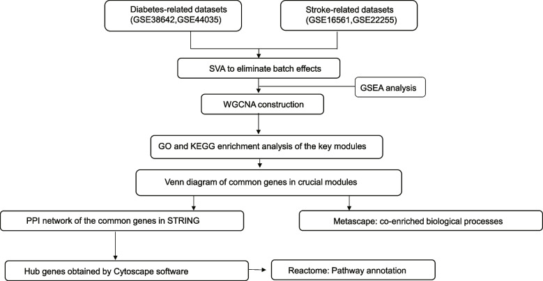
A flow chart for the present research is shown in Fig. 1. We used the term “ischemic stroke” and “diabetes” to search for ischemic stroke and diabetes’ gene expression profiles in GEO database (http://www.ncbi.nlm.nih.gov/geo/). The obtained mRNA microarray datasets were screened by the following criteria. First, these datasets must provide raw data that can be further analyzed. Second, profile information should include both case and control groups. Two stroke-related datasets (GSE38642 and GSE44035) and two diabetes-related datasets (GSE16561 and GSE22255) were chosen for next research (Table 1). The GSE38642 dataset comprised 54 non-diabetic controls and 9 diabetic patients and is based on the GPL6244 platform (Affymetrix Human Gene 1.0 ST Array). The GSE44035 dataset, which is also based on the GPL6244, comprised 9 non-diabetes controls and 1 patient with diabetes. In dataset GSE16561, which was produced on the GPL6883 platform (Illumina HumanRef-8 v3.0 expression beadchip), peripheral blood from 39 ischemic stroke patients and 24 healthy control subjects. Finally, the GSE22255 dataset was based on GPL570 platform (Affymetrix Human Genome U133 Plus 2.0 Array) and comprised 20 patients with ischemic stroke and 20 healthy controls.
Fig. 1.
The research flowchart of data preparation and analysis. SVA: surrogate variable analysis; WGCNA: weighted gene co-expression network analysis; GSEA: Gene set enrichment analysis; GO: Gene Ontology; KEGG: Kyoto Encyclopedia of Genes and Genomes; PPI: protein–protein interaction
Table 1.
Data collection
Data preprocessing
Batch effects were removed with a time specific algorithm within LIMBR, based on Surrogate Variable Analysis (SVA) [16]. The results of batch effect elimination were presented through a PCA graph and box plot. Probe identifications (IDs) in the gene expression matrix were reannotated as gene symbols. Changes in gene expression levels were reported as log2 values. Subsequently, the stroke datasets were combined and a new gene expression profile for all samples was formed. The two stroke datasets were combined together into one new gene expression profile for all samples. Finally, we annotated the gene symbols of gene expression matrix as Entrez IDs by utilizing the org.Hs.eg.db package for subsequent analysis.
Differential gene expression analysis
The limma package was applied to identify differentially expressed genes (DEGs) between case and control groups in diabetes and stroke with the following selection criteria: P-value of < 0.05, thresholds of |log FC|≥ 0.4. The hierarchical clustering analysis and volcano plot were represented by the R packages “pheatmap” and “ggplot2”, respectively.
Gene set enrichment analysis
Gene set enrichment analysis (GSEA), based on functional categories, has been proved to be one of the most powerful and popular tools for analyzing gene enrichment pathway [17]. We used GSEA to compare the biological pathways between case and control groups. KEGG gene sets as Gene Symbols were chosen as the gene set database. The settings for the GSEA run include: (1) number of gene set permutations were set to 1000 and (2) collapse dataset to gene symbols = TRUE.
Weight gene co-expression network analysis
Correlation networks are increasingly being used in bioinformatics applications. WGCNA is known as an algorithm in R-studio software for discovering the co-expressed gene modules, summarizing such clusters using an intramodular hub gene or the modules eigengene, relating modules to external sample traits, and calculating module membership measures. Correlated network based on gene screening methods can be used to identify candidate biomarkers [18]. In this research, we used WGCNA to create the co-expressed gene networks of diabetes and stroke. Firstly, we performed sample clustering to detect outliers. The “pickSoftThreshold” algorithm was used to select an appropriate soft threshold (β) and to obtain a biologically significant scale-free network (scale independence of > 0.8). Then gene–gene correlation matrix was built to describe the degree of association among nodes. The adjacency matrix was converted into a topological overlap matrix (TOM) using the “TOMsimilarity” algorithm. In order to identify co-expression modules, the gene hierarchical clustering dendrogram was obtained. The module eigengene (ME), as well as the correlation between clinical traits and ME were then calculated by spearman correlation analysis and hierarchical clustering to identify clinical-related modules.
GO and KEGG enrichment analyses of genes in disease-related key modules
Gene ontology (GO) describes the overrepresented biological functions. Three independent ontologies accessible are being constructed: molecular function, cellular component, and biological process [19]. Kyoto Encyclopedia of Genes and Genomes (KEGG) enrichment analyses is a reference knowledge base for biological interpretation of large-scale molecular datasets, such as metagenome and genome sequences [20–23]. GO and KEGG analyses for diabetes and stroke related key modules were performed using the R clusterProfiler package.
Detection of shared and key genes in T2DM-related stroke
The common genes of T2DM-related stroke were discovered by intersecting diabetes-related crucial modules with stroke-related crucial modules. Furthermore, the STRING website (https://string-db.org/) was used to build the protein–protein interaction (PPI) relationships. STRING (version 11.5) covers 67,592,464 proteins from 14094 organisms and 20,052,394,042 interactions. We selected the gene symbol as input of website https://cn.string-db.org/, then chose multiple proteins, used gene symbol as list of names, and select homo sapiens as organism. The cytoHubba plug-in in Cytoscape (version 3.9.1) was employed to obtain the hub genes of T2DM-related stroke.
Enriched biological processes of common and crucial genes in T2DM-related stroke
The GO vocabularies, which include biological processes (BPs), molecular functions (MFs), and cellular components (CCs), were performed using Metascape (http://metascape.Org/gp/) to find significantly enriched terms (P value ≤ 0.01) [24]. The biological pathways associated with hub genes were annotated and visualized using Reactome Database (https://reactome.org), which is an open-access, open-source, peer-reviewed, and manually curated database [25]. The R code is available in Supplementary material.
Results
Integrated screening for genes and GSEA analysis in diabetes and stroke
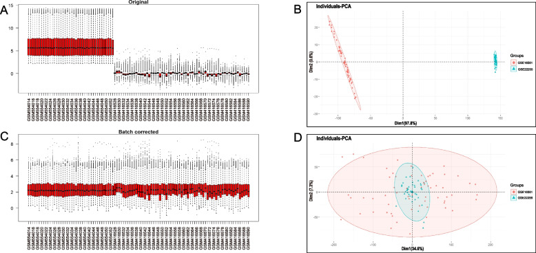
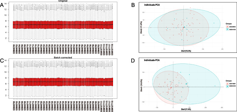
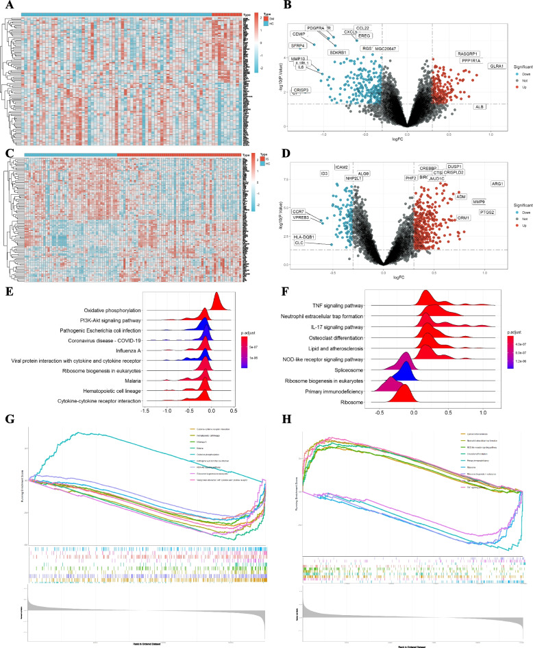
Four GEO datasets were used for the identification of diabetes and stroke-associated genes (Table 1). As shown in Figs. 2 and 3, batch effects have been removed by sva package in all samples from the stroke and diabetes datasets. We then used “limma” R package to identified differentially expressed genes (DEGs) between case and control group in diabetes and stroke. In total, 81 upregulated genes and 153 downregulated genes were included in patients with diabetes (Fig. 4A and B). 158 upregulated genes and 29 downregulated genes were included in IS patients (Fig. 4C and D) based on the criteria p < 0.05 and |logFC|≥ 0.4. GSEA was used to reveal the potential molecular mechanisms of diabetes and stroke based on all gene information in the gene expression matrix. The most highly enriched pathways by enrichment score in diabetes related datasets were related to oxidative phosphorylation, ribosome biogenesis in eukaryotes, hematopoietic cell lineage, and cytokine-cytokine receptor interaction (Fig. 4E and G). The enrichment analysis of gene sets about stroke revealed that compared to control samples, TNF signaling pathway, neutrophil extracellular trap formation, IL-17 signaling pathway, lipid and atherosclerosis, ribosome biogenesis in eukaryotes, and primary immunodeficiency (Fig. 4F and H). According to GSEA analysis, ribosome biogenesis in eukaryotes was the common biological pathway relevant to the pathogenesis of diabetes and stroke.
Fig. 2.
Batch effects considered in analysis. A, B the distribution of stroke samples before elimination of batch effect. C, D the distribution of stroke samples after eliminating the batch effect
Fig. 3.
Batch effects considered in analysis. A, B the distribution of diabetes samples before elimination of batch effect. C, D the distribution of diabetes samples after eliminating the batch effect
Fig. 4.
Identification and pathway analyses of differentially expressed genes (DEGs). A Heatmap of DEGs in diabetes related datasets; B Volcano plots showing the differential genes in diabetes related dataset. C Heatmap of DEGs in stroke related datasets; D Volcano plots showing the differential genes in stroke related dataset. E, F Ridgeline plot showing KEGG pathways enrichment in diabetes (E) and stroke (F). G, H Gene set enrichment analysis (GSEA) plots showing the most enriched gene sets of all detected genes in the diabetes (G) and stroke (H) samples
Stroke-related key module identification
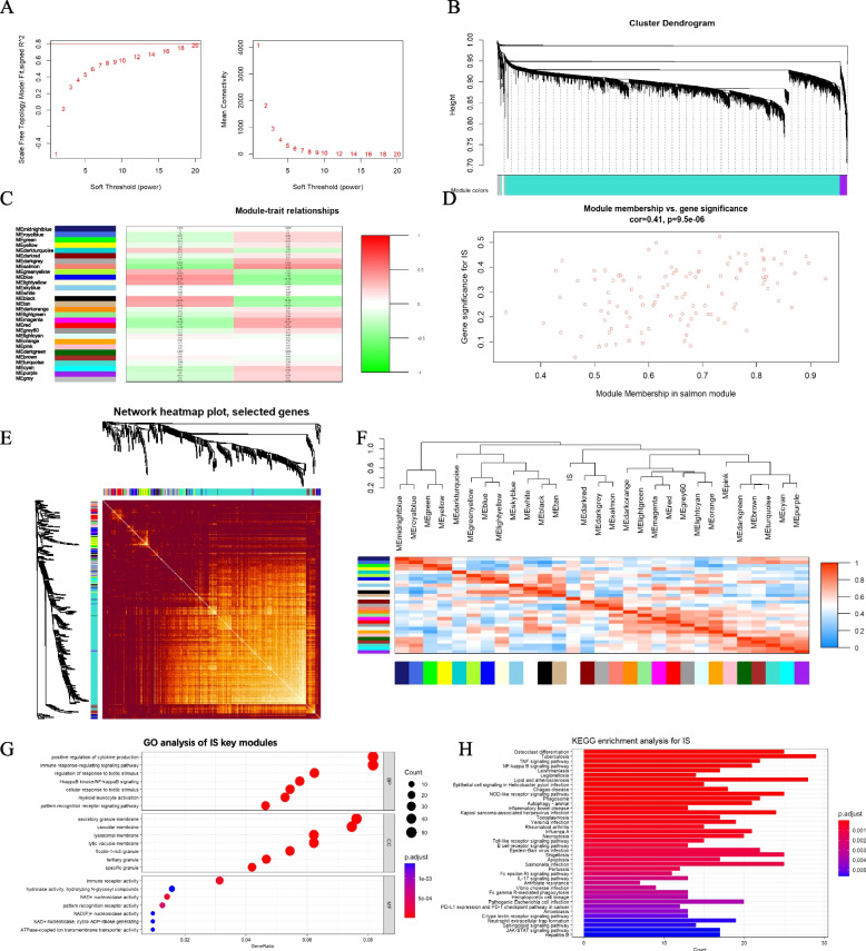
In stroke datasets, the appropriate soft-thresholding power was chosen based on the number of samples to make the resulting networks conservative (if number of samples ≥ 40, unsigned and signed hybrid networks = 6). Network topology analysis of different soft threshold power was shown in Fig. 5A. Each module was represented by a different color. Based on spearman coefficient, the relationships between modules were assessed through a heatmap about module-trait relationships (Fig. 5B and C). The heatmaps of the correlation between values of clinical features and module eigengene showed that the salmon (r = 0.44, p < 0.05), darkgrey (r = 0.3, p = 0.002), red (r = 0.37, p < 0.05), and magenta (r = 0.36, p < 0.05) modules were highly positively related with stroke (Fig. 5D-F). As a result, the four modules were classified as stroke-related crucial modules. GO and KEGG analyses were performed on the genes of key modules (Fig. 5G and H). The GO analyses demonstrated that these genes were primarily related to positive regulation of cytokine production, immune response-regulating signaling pathway, regulation of response to biotic stimulus, NF-kappa B signaling, cellular response to biotic stimulus, and myeloid leukocyte activation in biological processes. In terms of cell components, genes were primarily enriched in secretory granule membrane, vacuolar membrane, lysosomal membrane, lytic vacuole membrane, and ficolin-1-rich granule. Furthermore, molecular function was primarily enriched in the immune receptor activity, hydrolase activity, NAD + nucleosidase activity, pattern recognition receptor activity, and ATPase-coupled ion transmembrane transporter activity. Next, we performed KEGG analysis on the genes in the four modules (Fig. 5H). The findings revealed an association between the TNF signaling pathway, NF-kappa B signaling pathway, lipid and atherosclerosis, toll-like receptor signaling pathway, apoptosis, neutrophil extracellular trap formation.
Fig. 5.
Construction of weighted co-expression network for stroke-related datasets. A Network topology analysis of different soft threshold power. B Dendrograms of genes acquired by mean linkage hierarchical clustering. C The relationship between trait and modules. Correlation coefficients and corresponding P value are listed for each module. D The genes in the salmon module were significantly correlated with stroke. E Heatmap depicts the Topological Overlap Matrix (TOM) of genes selected for weighted co-expression network analysis. F Heatmaps and hierarchical cluster dendrograms of clinical traits and module eigengene. G, H The GO and KEGG pathways in key modules
Diabetes-related key module identification
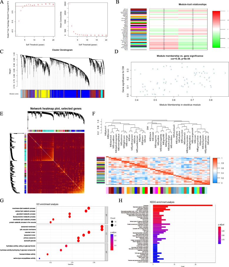
In the stroke and diabetes-related datasets, we found co-expressed gene modules using WGCNA. As illustrated in Fig. 6A, the scale-free topological index was 0.8 when the soft thresholds for diabetes was 6. The derived gene dendrograms and corresponding module colors were showed in Fig. 6B and C. The correlations between the two phenotypes (disease and health states) were calculated by the hierarchical clustering and spearman correlation analyses. Three modules “steelblue”, “dark red”, and “Grey60” had a highly positive association with diabetes, which were chosen as diabetes-related modules (steelblue: r = 0.28, p = 0.02; dark red: r = 0.23, p = 0.05; Grey60: r = 0.24, p = 0.05) (Fig. 6D-F). We conducted GO and KEGG enrichment analysis on the genes included diabetes-related key modules, as well as pathway annotation of key diabetes-related genes, to discover the underlying molecular biological process of diabetes (Fig. 6G and H). The GO terms of biological processes showed that they were primarily enriched in membrane lipid metabolic process, cellular lipid catabolic process, glycolipid metabolic process, and liposaccharide metabolic process (Fig. 6G). With regard to cellular components, these genes were primarily enriched in lysosomal membrane, lytic vacuole membrane, vacuolar lumen, lysosomal lumen, and primary lysosome. In terms of molecular function, these genes were primarily enriched in hydrolase activity, hexosaminidase activity, and serine-type exopeptidase activity. KEGG enrichment analysis suggested that they were mostly involved in pathways of lysosome, glycosaminoglycan degradation, sphingolipid metabolism, phagosome, and autophagy (Fig. 6H).
Fig. 6.
Construction of weighted co-expression network for diabetes-related datasets. A The value of scale independence on the left and the value of mean connectivity on the right. B The relationship between trait and modules. Correlation coefficients and corresponding P value are listed for each module. C The cluster dendrogram of all gene were grouped into different modules. D The genes in the steelblue module were significantly correlated with diabetes. E Network heatmap of all genes (a color change from red to yellow indicates a high degree of overlap between modules). F Heatmaps and hierarchical cluster dendrograms of clinical traits and module eigengene. G, H The GO and KEGG pathways in key modules
The shared genes and functional enrichment analysis in diabetes and stroke
A total of 689 and 476 genes were identified from stroke and diabetes-related modules, respectively. 24 genes were selected by taking the intersection of stroke and diabetes-related genes (Fig. 7A), which were considered to be extremely associated with the pathogenesis T2DM-related stroke. To investigate potential roles of the shared genes in diabetes and stroke, we used metascape to analyze the enriched pathway. As shown in Fig. 7B, the results revealed that these genes were enriched in various biological activities including neutrophil degranulation, lysosomal transport, regulation of NF-kappa B signaling, and inflammatory response. The most enriched ontology clusters belonged to neutrophil degranulation (logP = -53.25, log(q-value) = -48.904).
Fig. 7.
Screening of the common genes between stroke and diabetes. A The common genes screened by intersecting the diabetes and stroke related modules. B The co-enriched biological processes by metascape
Identification of key genes in DM-related stroke
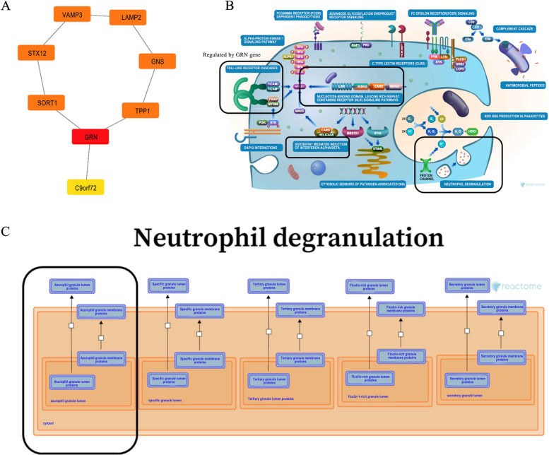
In terms of the 24 shared genes obtained by a disease module-module intersection, we used the STRING database to construct PPI networks (Fig. 8A). Next, cytoHubba in Cytoscape software was used to screen for and visualize the hub genes in the network. As shown in Fig. 8A, GRN (granulin precursor), was identified as the hub gene in T2DM-related stroke. To further understanding how GRN acts in DM-related stroke, the website Reactome was used to analyze its regulatory pathways. We used human reactome database and found that GRN was involved in Toll-like Receptor Cascades, Neutrophil degranulation, DDX58/IFIH1-mediated induction of interferon-alpha/beta, and NLR signaling pathway (Fig. 8B). Notably, GRN was involved in exocytosis of azurophil granule lumen proteins, which contributed to neutrophil degranulation (Fig. 8C). Thus, we believe that GRN might participate in inflammatory response and neutrophil extracellular trap formation by regulating exocytosis of azurophil granule lumen proteins in T2DM-related stroke.
Fig. 8.
Screening the hub genes and the related biological process analysis. A a PPI network of common genes constructed by Cytoscape software. B, C the role of hub gene GRN regulatory pathways in inflammatory response (B) and neutrophil degranulation (C). The schematic art pieces were provided by the Reactome Database (www.reactome.org) under Creative Commons Attribution 4.0 License
Discussion
We used the WGCNA package to investigate key genes and probable biological processes associated with DM-related stroke in this study. Twenty- four common genes were identified to be present in both stroke and diabetes-related crucial modules, indicating that they are the most likely to participate in biological function in T2DM-related stroke. The common genes were enriched highly in biological processes of neutrophil degranulation, regulation of response to biotic stimulus, and inflammatory response, which was associated with the results of GSEA analysis in stroke-related datasets. Most importantly, among these common genes, GRN was identified as the hub gene of DM-related stroke. Functional annotation revealed that GRN acted in T2DM-related stroke by regulating neutrophil degranulation.
Variations in gene expression signatures provide novel insight to the mechanism of DM-related stroke and assists in finding intervention targets. Sentinel variants at PEAR1 and RGS18 were associated with thrombosis risk through a whole genome sequencing, which provide insights regarding the mechanism by genetics may influence cardiovascular disease risk [26]. The mRNA expression of platelet activating factor receptor (PAFR) was significantly higher in patients with diabetes, which might cause macrovascular disease through impaired endothelial function [27]. The study analyzing high-throughput gene expression in blood samples has shown that the circulating gene expression makers including CREM, PELI1, and ZAK were verified to be up-regulated in cardioembolic stroke [28]. In current study, we performed gene co-expression pattern analysis (WGCNA), rather than individual gene/protein-focused analytic strategy, for evaluating expression similarity among genes of T2DM-related stroke. Many of these common genes were involved in immune effector process, apoptosis, and necroptosis. Death-associated protein kinase 1 (DAPK1), a Ca2 + /calmodulin (CaM)-dependent serine/threonine protein kinase, plays important roles in diverse apoptosis pathways in neuronal cell death [29]. A quantitative proteomic analysis revealed DAPK1 as the most prevalent protein recruited to the cytoplasmic tail of glutamate receptor during cerebral ischemia [30]. Targeting DAPK1-related pro-death signals could be considered as a promising therapeutic approach in salvageable brain tissue after ischemic stroke. Sortilin 1 (SORT1) was one of CVD-risk loci identified with genome-wide association studies (GWAS) over the last decade [31]. Sortilin, encoded by SORT1, serving as a key receptor for lipids, cytokines, and enzymes and participating in pathological cargo loading to and trafficking of extracellular vesicles, is also known for its functional role in metabolic disorders and cardiovascular disease [32]. The multiple contributions of sortilin to cardiovascular risk suggest it as a potential drug target. in addition, other common genes identified in our study such as PELI1, LAMP2, and PPM1B, were linked to cell death and inflammation [33–35]. Thus, preventing aberrant cell death and maintaining cellular homeostasis is important to aid in the development of drugs for T2DM-related stroke. Furthermore, we measured correlation between diseases and highly co-expressed gene sets. Compared to conventional differential expression analysis, it is a more effective technique for detecting gene expression with low variation and abundance, as well as being less prone to information loss [36]. Quite a few researches have demonstrated the value of WGCNA in systematically identifying critical genes and relevant mechanisms about stroke or diabetes [36–39].
In our study, we discovered key biological processes in the common genes of diabetes and stroke, including neutrophil degranulation, regulation of response to biotic stimulus, and inflammatory response. Consistently, neutrophil extracellular trap formation (NETs) was highly enriched in both GSEA analysis and function enrichment analysis of stroke-related key modules. This finding indicated that neutrophil extracellular trap formation might be a crucial mechanism for DM-related stroke, explaining the role of neutrophils in pathogenesis for stroke patients with diabetes. Neutrophils, the first line of defense under inflammation and infection, can release their decondensed chromatin and form large extracellular DNA networks [40]. NETs formation mediates the propagation of thrombosis and inflammation and, thereby contributes to stroke. Several studies have demonstrated that NETs contributed to the composition of ischemic stroke thrombi [41, 42] and surrogate markers of NETs in plasma including circulating citrullinated histone H3 (H3Cit) and cell-free DNA (cfDNA) were associated with ischemic stroke outcomes [43, 44]. Hyperglycemia is associated with poor outcome in acute ischemic stroke patients. Recent studies have shown that neutrophil extracellular trap formation in serum of diabetes patients can activate vascular endothelial cells and thrombosis. Cell death and neutrophil activation are considered to be key factors producing endothelial injury under the condition of diabetes, and NETs formation further accelerated the vascular injure [45]. Blocking NET formation has significantly reduced brain infarctions and improved outcomes in diabetic mice [46]. A prominent characteristic of ischemic stroke is platelet activation and immunothrombosis stimulated by NETs [47]. Therefore, NETs formation promotes hypercoagulability and induces ischemic stroke in patients with diabetes, which is a potential biological process of T2DM-related stroke.
GRN (granulin), as a protein coding gene, was expressed in immune cells and epithelia and regulated biological functions including cell proliferation, inflammation, and wound healing [48]. Interactions between inflammation and GRN are complex. Progranulin (PGRN) can bind receptors of TNF-α and block the role of TNF-α to stimulate neutrophile respiratory burst. Therefore, PGRN is considered as anti-inflammatory factor [49]. While this anti-inflammatory activity was degraded by NE-mediated proteolysis of PGNR to GRN peptides [50]. GRN was involved in a pro-inflammatory response at the early stages after cerebral ischemia. Neutrophiles are infiltrated in brain parenchyma after ischemia stroke and the increased activity of neutrophil elastase cleaves PGRN into GRN peptide, exacerbating the inflammation and tissue damage after ischemia [51]. GRN has also been detected in neutrophil-rich peritoneal exudates, induce the release of neutrophil-attracting IL-8 from epithelial cells, and may enhance inflammation [52]. It can also modulate inflammation in neurons by preserving neuronal integrity, axonal outgrowth, and neurons survival [53]. In acute injury and inflammation, GRN is important in the initiation of inflammation by recruiting neutrophils, macrophages, and fibroblasts [54]. GRN, at elevated levels, has been associated with poor prognosis in infectious diseases. Serum GRN levels in sepsis exhibited positively correlation with inflammatory factors including hypersensitive C-reactive protein and procalcitonin [55]. The cell counts of neutrophils and macrophages were increased in transcutaneous puncture wounds under administration of GRN [56]. Elevated PGRN levels may reflect increased pro-inflammatory GRN levels because PGRN and GRN are indistinguishable from each other in ELISA [57]. In addition, several studies have shown that PGRN has proinflammatory effects in diabetes and obesity. PGRN played a role as a novel marker for chronic inflammation associated with diabetes in human [58]. Consistently, PGRN levels were increased in white adipose tissue of the mice fed with a high-fat-diet (HFD) and exerted pro-inflammatory functions [59]. Therefore, PGRN may have two absonant effects, depending on the tissue microenvironment and disease stage. In our study, GRN was shown to be the hub gene for key diabetes and stroke-related modules, suggesting that GRN plays critical roles in DM-related stroke. According to functional annotation, the participation of GRN in the regulation of neutrophil extracellular trap formation and inflammatory response, GRN plays a role in T2DM-related stroke.
Conclusions
A total of 24 common genes may be involved in the mechanisms of T2DM-related stroke through their involvement in neutrophil degranulation, lysosomal transport, regulation of NF-kappa B signaling, and inflammatory response. More crucially, GRN participating in NETs formation may be promising intervention targets for T2DM-related stroke. However, the current research had several limitations. First, the sample size of patients with stroke and diabetes in the GEO database is relatively small. Although we have eliminated batch effects to integrate information from different datasets, studies with larger sample sizes are still needed in subsequent analysis and validation. Second, considering that stroke is a serious complication with type 2 diabetes, our study primarily investigates the molecular mechanism network between type 2 diabetes and stroke, while there is insufficient evidence to support the molecular mechanism network between type 1 diabetes and stroke. Third, the findings in this study are based on bioinformatics analysis. The relationship between GRN and DM-related stroke still needs to be verified by more in vitro and in vivo experiments. Further basic research and clinical research should be focused on a better understanding of the pathophysiological mechanism of GRN in T2DM-related stroke and may contribute to DM-related stroke therapy.
Supplementary Information
Additional file 1: R code 1. Integrated screening for genes and GSEA analysis in diabetes and stroke. R code 2. Stroke-related key module identification. R code 3. Diabetes-related key module identification.
Acknowledgements
We acknowledge GEO (https://www.ncbi.nlm.nih.gov/geo/) database for providing their platforms and contributors for uploading their meaningful datasets.
Authors’ contributions
YH and YB designed the experiments and conducted the study. QH wrote the manuscript. JF and JX revised the manuscript. QH and YH contributed to literature search and data collection. YB performed statistical analysis. All authors read and approved the final manuscript.
Funding
This work was supported by the National Natural Science Foundation of China Youth Project (82001392) and General Project of Hunan Natural Science Foundation (2020JJ4895).
Availability of data and materials
All data from the GSE38642, GSE44035, GSE16561, and GSE22255 datasets are available at the GEO database (https://www.ncbi.nlm.nih.gov/geo/).
Declarations
Ethics approval and consent to participate
Not required.
Consent for publication
Not applicable.
Competing interests
The authors declare no competing interests.
Footnotes
Publisher’s Note
Springer Nature remains neutral with regard to jurisdictional claims in published maps and institutional affiliations.
Yong He and Yang Bai have contributed equally to this work.
References
- 1.Virani SS, Alonso A, Benjamin EJ, Bittencourt MS, Callaway CW, Carson AP, Chamberlain AM, Chang AR, Cheng S, Delling FN, et al. Heart disease and stroke statistics-2020 update: a report from the american heart association. Circulation. 2020;141(9):e139–e596. doi: 10.1161/CIR.0000000000000757. [DOI] [PubMed] [Google Scholar]
- 2.Luitse MJA, Biessels GJ, Rutten GEHM, Kappelle LJ. Diabetes, hyperglycaemia, and acute ischaemic stroke. Lancet Neurol. 2012;11(3):261–271. doi: 10.1016/S1474-4422(12)70005-4. [DOI] [PubMed] [Google Scholar]
- 3.Williams DM, Atkinson M, Evans M. Stroke prevention and treatment in people with type 2 diabetes: is there a role for GLP-1 (Glucagon-Like Peptide-1) Analogues? Stroke. 2023;54(5):1441–1451. doi: 10.1161/STROKEAHA.122.042109. [DOI] [PubMed] [Google Scholar]
- 4.Kernan WN, Forman R, Inzucchi SE. Caring for patients with diabetes in stroke neurology. Stroke. 2023;54(3):894–904. doi: 10.1161/STROKEAHA.122.038163. [DOI] [PubMed] [Google Scholar]
- 5.Laakso M, Kuusisto J. Insulin resistance and hyperglycaemia in cardiovascular disease development. Nat Rev Endocrinol. 2014;10(5):293–302. doi: 10.1038/nrendo.2014.29. [DOI] [PubMed] [Google Scholar]
- 6.Maida CD, Daidone M, Pacinella G, Norrito RL, Pinto A, Tuttolomondo A. Diabetes and ischemic stroke: an old and new relationship an overview of the close interaction between these diseases. Int J Mol Sci. 2022;23(4):2397. doi: 10.3390/ijms23042397. [DOI] [PMC free article] [PubMed] [Google Scholar]
- 7.Yahagi K, Kolodgie FD, Lutter C, Mori H, Romero ME, Finn AV, Virmani R. Pathology of human coronary and carotid artery atherosclerosis and vascular calcification in diabetes mellitus. Arterioscler Thromb Vasc Biol. 2017;37(2):191–204. doi: 10.1161/ATVBAHA.116.306256. [DOI] [PMC free article] [PubMed] [Google Scholar]
- 8.Krinock MJ, Singhal NS. Diabetes, stroke, and neuroresilience: looking beyond hyperglycemia. Ann N Y Acad Sci. 2021;1495(1):78–98. doi: 10.1111/nyas.14583. [DOI] [PubMed] [Google Scholar]
- 9.Li G, Sun J, Zhang J, Lv Y, Liu D, Zhu X, Qi L, Chen Z, Ye Z, Su X, et al. Identification of inflammation-related biomarkers in diabetes of the exocrine pancreas with the use of weighted gene co-expression network analysis. Front Endocrinol. 2022;13:839865. doi: 10.3389/fendo.2022.839865. [DOI] [PMC free article] [PubMed] [Google Scholar]
- 10.Chen J, Luo S-F, Yuan X, Wang M, Yu H-J, Zhang Z, Yang Y-Y. Diabetic kidney disease-predisposing proinflammatory and profibrotic genes identified by weighted gene co-expression network analysis (WGCNA) J Cell Biochem. 2022;123(2):481–492. doi: 10.1002/jcb.30195. [DOI] [PubMed] [Google Scholar]
- 11.Wang Z, Chen X, Li C, Tang W. Application of weighted gene co-expression network analysis to identify novel key genes in diabetic nephropathy. J Diabetes Investigation. 2022;13(1):112–124. doi: 10.1111/jdi.13628. [DOI] [PMC free article] [PubMed] [Google Scholar]
- 12.Dai L, Xie Y, Zhang W, Zhong X, Wang M, Jiang H, He Z, Liu X, Zeng H, Wang H. Weighted gene co-expression network analysis identifies ANGPTL4 as a key regulator in diabetic cardiomyopathy via FAK/SIRT3/ROS pathway in cardiomyocyte. Front Endocrinol. 2021;12:705154. doi: 10.3389/fendo.2021.705154. [DOI] [PMC free article] [PubMed] [Google Scholar]
- 13.Zhang B, Horvath S. A general framework for weighted gene co-expression network analysis. Statistical Appl Genet Mol Biol. 2005;4:1–45. doi: 10.2202/1544-6115.1128. [DOI] [PubMed] [Google Scholar]
- 14.Mo X-G, Liu W, Yang Y, Imani S, Lu S, Dan G, Nie X, Yan J, Zhan R, Li X, et al. NCF2, MYO1F, S1PR4, and FCN1 as potential noninvasive diagnostic biomarkers in patients with obstructive coronary artery: A weighted gene co-expression network analysis. J Cell Biochem. 2019;120(10):18219–18235. doi: 10.1002/jcb.29128. [DOI] [PMC free article] [PubMed] [Google Scholar]
- 15.Wang Y, Liu T, Liu Y, Chen J, Xin B, Wu M, Cui W. Coronary artery disease associated specific modules and feature genes revealed by integrative methods of WGCNA MetaDE and machine learning. Gene. 2019;710:122–130. doi: 10.1016/j.gene.2019.05.010. [DOI] [PubMed] [Google Scholar]
- 16.Crowell AM, Greene CS, Loros JJ, Dunlap JC. Learning and Imputation for Mass-spec Bias Reduction (LIMBR) Bioinformatics. 2019;35(9):1518–1526. doi: 10.1093/bioinformatics/bty828. [DOI] [PMC free article] [PubMed] [Google Scholar]
- 17.Joly JH, Lowry WE, Graham NA. Differential Gene Set Enrichment Analysis: a statistical approach to quantify the relative enrichment of two gene sets. Bioinformatics. 2020;36(21):5247–5254. doi: 10.1093/bioinformatics/btaa658. [DOI] [PMC free article] [PubMed] [Google Scholar]
- 18.Langfelder P, Horvath S. WGCNA: an R package for weighted correlation network analysis. Bmc Bioinformatics. 2008;9:1–3. doi: 10.1186/1471-2105-9-559. [DOI] [PMC free article] [PubMed] [Google Scholar]
- 19.Ashburner M, Ball CA, Blake JA, Botstein D, Butler H, Cherry JM, Davis AP, Dolinski K, Dwight SS, Eppig JT, et al. Gene ontology: tool for the unification of biology. Nat Genet. 2000;25(1):25–29. doi: 10.1038/75556. [DOI] [PMC free article] [PubMed] [Google Scholar]
- 20.Kanehisa M. Toward understanding the origin and evolution of cellular organisms. Protein Sci. 2019;28(11):1947–1951. doi: 10.1002/pro.3715. [DOI] [PMC free article] [PubMed] [Google Scholar]
- 21.Kanehisa M, Furumichi M, Sato Y, Kawashima M, Ishiguro-Watanabe M. KEGG for taxonomy-based analysis of pathways and genomes. Nucleic Acids Res. 2022;51(D1):D587–92. doi: 10.1093/nar/gkac963. [DOI] [PMC free article] [PubMed] [Google Scholar]
- 22.Ogata H, Goto S, Sato K, Fujibuchi W, Bono H, Kanehisa M. KEGG: kyoto encyclopedia of genes and genomes. Nucleic Acids Res. 1999;27(1):29–34. doi: 10.1093/nar/27.1.29. [DOI] [PMC free article] [PubMed] [Google Scholar]
- 23.Kanehisa M, Sato Y. KEGG Mapper for inferring cellular functions from protein sequences. Protein Sci. 2020;29(1):28–35. doi: 10.1002/pro.3711. [DOI] [PMC free article] [PubMed] [Google Scholar]
- 24.Zhou Y, Zhou B, Pache L, Chang M, Khodabakhshi AH, Tanaseichuk O, Benner C, Chanda SK. Metascape provides a biologist-oriented resource for the analysis of systems-level datasets. Nat Commun. 2019;10:1523. doi: 10.1038/s41467-019-09234-6. [DOI] [PMC free article] [PubMed] [Google Scholar]
- 25.Croft D, Mundo AF, Haw R, Milacic M, Weiser J, Wu G, Caudy M, Garapati P, Gillespie M, Kamdar MR, et al. The reactome pathway knowledgebase. Nucleic Acids Res. 2014;42(D1):D472–D477. doi: 10.1093/nar/gkt1102. [DOI] [PMC free article] [PubMed] [Google Scholar]
- 26.Keramati AR, Chen M-H, Rodriguez BAT, Yanek LR, Bhan A, Gaynor BJ, Ryan K, Brody JA, Zhong X, Wei Q, et al. Genome sequencing unveils a regulatory landscape of platelet reactivity. Nat Commun. 2021;12(1):3626. doi: 10.1038/s41467-021-23470-9. [DOI] [PMC free article] [PubMed] [Google Scholar]
- 27.Kurano M, Darestani SG, Shinnakasu A, Yamamoto K, Dochi Y, Uemura K, Ikeda Y, Kikuchi A, Hashiguchi H, Deguchi T, et al. mRNA expression of platelet activating factor receptor (PAFR) in peripheral blood mononuclear cells is associated with albuminuria and vascular dysfunction in patients with type 2 diabetes. Diabetes Res Clin Pract. 2018;136:124–133. doi: 10.1016/j.diabres.2017.11.028. [DOI] [PubMed] [Google Scholar]
- 28.Garcia-Berrocoso T, Pala E, Consegal M, Piccardi B, Negro A, Gill N, Penalba A, Huerga Encabo H, Fernandez-Cadenas I, Meisel A, et al. Cardioembolic ischemic stroke gene expression fingerprint in blood: a systematic review and verification analysis. Transl Stroke Res. 2020;11(3):326–336. doi: 10.1007/s12975-019-00730-x. [DOI] [PubMed] [Google Scholar]
- 29.Lai TW, Zhang S, Wang YT. Excitotoxicity and stroke: Identifying novel targets for neuroprotection. Prog Neurobiol. 2014;115:157–188. doi: 10.1016/j.pneurobio.2013.11.006. [DOI] [PubMed] [Google Scholar]
- 30.Tu W, Xu X, Peng L, Zhong X, Zhang W, Soundarapandian MM, Balel C, Wang M, Jia N, Zhang W, et al. DAPK1 interaction with NMDA receptor NR2B subunits mediates brain damage in stroke. Cell. 2010;140(2):222–234. doi: 10.1016/j.cell.2009.12.055. [DOI] [PMC free article] [PubMed] [Google Scholar]
- 31.Zeller T, Blankenberg S, Diemert P. Genomewide association studies in cardiovascular disease-an update 2011. Clin Chem. 2012;58(1):92–103. doi: 10.1373/clinchem.2011.170431. [DOI] [PubMed] [Google Scholar]
- 32.Goettsch C, Kjolby M, Aikawa E. Sortilin and its multiple roles in cardiovascular and metabolic diseases. Arterioscler Thromb Vasc Biol. 2018;38(1):19–25. doi: 10.1161/ATVBAHA.117.310292. [DOI] [PMC free article] [PubMed] [Google Scholar]
- 33.Choi S-W, Park H-H, Kim S, Chung JM, Noh H-J, Kim SK, Song HK, Lee C-W, Morgan MJ, Kang HC, et al. PELI1 Selectively targets kinase-active rip3 for ubiquitylation-dependent proteasomal degradation. Molecular Cell. 2018;70(5):920. doi: 10.1016/j.molcel.2018.05.016. [DOI] [PubMed] [Google Scholar]
- 34.Kenari AN, Cheng L, Hill AF. Methods for loading therapeutics into extracellular vesicles and generating extracellular vesicles mimetic-nanovesicles. Methods. 2020;177:103–113. doi: 10.1016/j.ymeth.2020.01.001. [DOI] [PubMed] [Google Scholar]
- 35.Steinberg GR. Inflammation in obesity is the common link between defects in fatty acid metabolism and insulin resistance. Cell Cycle. 2007;6(8):888–894. doi: 10.4161/cc.6.8.4135. [DOI] [PubMed] [Google Scholar]
- 36.Pei G, Chen L, Zhang W: WGCNA Application to Proteomic and Metabolomic Data Analysis. In Proteomics in Biology, Pt A. Edited by Shukla AK, vol. 585; 2017: 135-158. Methods in Enzymology. [DOI] [PubMed]
- 37.Fan X, Chen H, Xu C, Wang Y, Yin P, Li M, Tang Z, Jiang F, Wei W, Song J, et al. S1PR3, as a core protein related to ischemic stroke, is involved in the regulation of blood-brain barrier damage. Front Pharmacol. 2022;13:834948. doi: 10.3389/fphar.2022.834948. [DOI] [PMC free article] [PubMed] [Google Scholar]
- 38.Wei R, Qiao J, Cui D, Pan Q, Guo L. Screening and identification of hub genes in the development of early diabetic kidney disease based on weighted gene co-expression network analysis. Front Endocrinol. 2022;13:883658–883658. doi: 10.3389/fendo.2022.883658. [DOI] [PMC free article] [PubMed] [Google Scholar]
- 39.Yan S, Sun M, Gao L, Yao N, Feng T, Yang Y, Li X, Hu W, Cui W, Li B. Identification of key LncRNAs and pathways in prediabetes and type 2 diabetes mellitus for hypertriglyceridemia patients based on weighted gene co-expression network analysis. Front Endocrinol. 2022;12:800123. doi: 10.3389/fendo.2021.800123. [DOI] [PMC free article] [PubMed] [Google Scholar]
- 40.Sorvillo N, Cherpokova D, Martinod K, Wagner DD. Extracellular DNA net-works with dire consequences for health. Circ Res. 2019;125(4):470–488. doi: 10.1161/CIRCRESAHA.119.314581. [DOI] [PMC free article] [PubMed] [Google Scholar]
- 41.Ducroux C, Di Meglio L, Loyau S, Delbosc S, Boisseau W, Deschildre C, Ben Maacha M, Blanc R, Redjem H, Ciccio G, et al. Thrombus Neutrophil extracellular traps content impair tPA-induced thrombolysis in acute ischemic stroke. Stroke. 2018;49(3):754–757. doi: 10.1161/STROKEAHA.117.019896. [DOI] [PubMed] [Google Scholar]
- 42.Laridan E, Denorme F, Desender L, Francois O, Andersson T, Deckmyn H, Vanhoorelbeke K, De Meyer SF. Neutrophil extracellular traps in ischemic stroke thrombi. Ann Neurol. 2017;82(2):223–232. doi: 10.1002/ana.24993. [DOI] [PubMed] [Google Scholar]
- 43.Demyanets S, Stojkovic S, Mauracher L-M, Kopp CW, Wojta J, Thaler J, Panzer S, Gremmel T. Surrogate markers of neutrophil extracellular trap formation are associated with ischemic outcomes and platelet activation after peripheral angioplasty and stenting. J Clin Med. 2020;9(2):304. doi: 10.3390/jcm9020304. [DOI] [PMC free article] [PubMed] [Google Scholar]
- 44.Valles J, Lago A, Teresa Santos M, Maria Latorre A, Tembl JI, Salom JB, Nieves C, Moscardo A. Neutrophil extracellular traps are increased in patients with acute ischemic stroke: prognostic significance. Thromb Haemost. 2017;117(10):1919–1929. doi: 10.1160/TH17-02-0130. [DOI] [PubMed] [Google Scholar]
- 45.Hirota T, Levy JH, Iba T. The influence of hyperglycemia on neutrophil extracellular trap formation and endothelial glycocalyx damage in a mouse model of type 2 diabetes. Microcirculation. 2020;27(5):e12617. doi: 10.1111/micc.12617. [DOI] [PubMed] [Google Scholar]
- 46.Denorme F, Portier I, Rustad JL, Cody MJ, de Araujo CV, Hoki C, Alexander MD, Grandhi R, Dyer MR, Neal MD, et al. Neutrophil extracellular traps regulate ischemic stroke brain injury. J Clin Invest. 2022;132(10):e154225. doi: 10.1172/JCI154225. [DOI] [PMC free article] [PubMed] [Google Scholar]
- 47.Engelmann B, Massberg S. Thrombosis as an intravascular effector of innate immunity. Nat Rev Immunol. 2013;13(1):34–45. doi: 10.1038/nri3345. [DOI] [PubMed] [Google Scholar]
- 48.Diaz-Cueto L, Stein P, Jacobs A, Schultz RM, Gerton GL. Modulation of mouse preimplantation embryo development by acrogranin (epithelin/granulin precursor) Dev Biol. 2000;217(2):406–418. doi: 10.1006/dbio.1999.9564. [DOI] [PubMed] [Google Scholar]
- 49.Tang W, Lu Y, Tian Q-Y, Zhang Y, Guo F-J, Liu G-Y, Syed NM, Lai Y, Lin EA, Kong L, et al. The growth factor progranulin binds to TNF receptors and is therapeutic against inflammatory arthritis in mice. Science. 2011;332(6028):478–484. doi: 10.1126/science.1199214. [DOI] [PMC free article] [PubMed] [Google Scholar]
- 50.Kessenbrock K, Froehlich L, Sixt M, Laemmermann T, Pfister H, Bateman A, Belaaouaj A, Ring J, Ollert M, Faessler R, et al. Proteinase 3 and neutrophil elastase enhance inflammation in mice by inactivating antiinflammatory progranulin. J Clin Investig. 2008;118(7):2438–2447. doi: 10.1172/JCI34694. [DOI] [PMC free article] [PubMed] [Google Scholar]
- 51.Horinokita I, Hayashi H, Oteki R, Mizumura R, Yamaguchi T, Usui A, Yuan B, Takagi N. Involvement of progranulin and granulin expression in inflammatory responses after cerebral ischemia. Intern J Mol Sci. 2019;20(20):5210. doi: 10.3390/ijms20205210. [DOI] [PMC free article] [PubMed] [Google Scholar]
- 52.Zhu J, Nathan C, Jin WW, Sim D, Ashcroft GS, Wahl SM, Lacomis L, Erdjument-Bromage H, Tempst P, Wright CD, et al. Conversion of proepithelin to epithelins: roles of SLPI and elastase in host defense and wound repair. Cell. 2002;111(6):867–878. doi: 10.1016/S0092-8674(02)01141-8. [DOI] [PubMed] [Google Scholar]
- 53.Van Damme P, Van Hoecke A, Lambrechts D, Vanacker P, Bogaert E, Van Swieten J, Carmeliet P, Van den Bosch L, Robberecht W. Progranulin functions as a neurotrophic factor to regulate neurite outgrowth and enhance neuronal survival. J Cell Biol. 2008;181(1):37–41. doi: 10.1083/jcb.200712039. [DOI] [PMC free article] [PubMed] [Google Scholar]
- 54.Jian J, Konopka J, Liu C. Insights into the role of progranulin in immunity, infection, and inflammation. J Leukoc Biol. 2013;93(2):199–208. doi: 10.1189/jlb.0812429. [DOI] [PMC free article] [PubMed] [Google Scholar]
- 55.Shan Y, Zhang X, Zhou G, Ji X, Gu Y. Increased progranulin as an independent predictive biomarker for poor prognosis in sepsis. Cytokine. 2022;155:155911–155911. doi: 10.1016/j.cyto.2022.155911. [DOI] [PubMed] [Google Scholar]
- 56.He ZH, Ong CHP, Halper J, Bateman A. Progranulin is a mediator of the wound response. Nat Med. 2003;9(2):225–229. doi: 10.1038/nm816. [DOI] [PubMed] [Google Scholar]
- 57.Murakoshi M, Gohda T, Sakuma H, Shibata T, Adachi E, Kishida C, Ichikawa S, Koshida T, Kamei N, Suzuki Y. Progranulin and its receptor predict kidney function decline in patients with type 2 diabetes. Front Endocrinol. 2022;13:849457. doi: 10.3389/fendo.2022.849457. [DOI] [PMC free article] [PubMed] [Google Scholar]
- 58.Youn B-S, Bang S-I, Kloeting N, Park JW, Lee N, Oh J-E, Pi K-B, Lee TH, Ruschke K, Fasshauer M, et al. Serum progranulin concentrations may be associated with macrophage infiltration into omental adipose tissue. Diabetes. 2009;58(3):627–636. doi: 10.2337/db08-1147. [DOI] [PMC free article] [PubMed] [Google Scholar]
- 59.Matsubara T, Mita A, Minami K, Hosooka T, Kitazawa S, Takahashi K, Tamori Y, Yokoi N, Watanabe M, Matsuo EI, et al. PGRN is a key adipokine mediating high fat diet-induced insulin resistance and obesity through IL-6 in adipose tissue. Cell Metab. 2012;15(1):38–50. doi: 10.1016/j.cmet.2011.12.002. [DOI] [PubMed] [Google Scholar]
Associated Data
This section collects any data citations, data availability statements, or supplementary materials included in this article.
Supplementary Materials
Additional file 1: R code 1. Integrated screening for genes and GSEA analysis in diabetes and stroke. R code 2. Stroke-related key module identification. R code 3. Diabetes-related key module identification.
Data Availability Statement
All data from the GSE38642, GSE44035, GSE16561, and GSE22255 datasets are available at the GEO database (https://www.ncbi.nlm.nih.gov/geo/).


