Abstract
The physiological role of dihydroxyacetone synthase (DHAS) in Candida boidinii was evaluated at the molecular level. The DAS1 gene, encoding DHAS, was cloned from the host genome, and regulation of its expression by various carbon and nitrogen sources was analyzed. Western and Northern analyses revealed that DAS1 expression was regulated mainly at the mRNA level. The regulatory pattern of DHAS was similar to that of alcohol oxidase but distinct from that of two other enzymes in the formaldehyde dissimilation pathway, glutathione-dependent formaldehyde dehydrogenase and formate dehydrogenase. The DAS1 gene was disrupted in one step in the host genome (das1Δ strain), and the growth of the das1Δ strain in various carbon and nitrogen sources was compared with that of the wild-type strain. The das1Δ strain had completely lost the ability to grow on methanol, while the strain with a disruption of the formate dehydrogenase gene could survive (Y. Sakai et al., J. Bacteriol. 179:4480–4485, 1997). These and other experiments (e.g., those to determine the expression of the gene and the growth ability of the das1Δ strain on media containing methylamine or choline as a nitrogen source) suggested that DAS1 is involved in assimilation rather than dissimilation or detoxification of formaldehyde in the cells.
In methylotrophic yeasts, formaldehyde is the key intermediate in methanol metabolism since it stands at the branchpoint of pathways for methanol assimilation and dissimilation. Since formaldehyde is an extremely toxic compound, its intracellular level should be strictly regulated.
Dihydroxyacetone synthase (DHAS) (EC 2.2.1.3) catalyzes the first reaction in the assimilation pathway by fixing formaldehyde to d-xylulose 5-phosphate, after formaldehyde is generated from methanol via alcohol oxidase (AOD) (EC 1.1.3.13) (3, 11). Otherwise, in a formaldehyde oxidation pathway, formaldehyde is dissimilated to CO2 through enzymes, e.g., glutathione-dependent formaldehyde dehydrogenase (FLD) (EC 1.2.1.1) and formate dehydrogenase (FDH) (EC 1.2.1.2).
So far, the physiological significance of these methanol-metabolizing enzymes has been estimated mainly from the enzymatic properties of purified enzyme or from phenotypes of mutants deficient in the specific enzyme obtained via random mutagenesis. Through such analyses, for example, FDH had been considered to be essential to the energy supply for methylotrophic growth (2, 34). However, through gene disruption analysis with Candida boidinii (23), the physiological role of FDH was revealed to be mainly detoxification of formate rather than stimulated energy generation. Such unexpected results with FDH led us to reevaluate the physiological roles of a key enzyme in formaldehyde assimilation, DHAS, using a gene-disrupted strain.
Similar to other cases, the physiological function of DHAS as a methanol assimilation enzyme has been estimated from its enzymatic properties and by analysis of a mutant obtained by random mutagenesis (13, 14). However, our previous study showed that loss of a peroxisome membrane protein, Pmp47, not only inhibits transport of DHAS into peroxisomes but also leads to loss of DHAS activity (26), raising the possibility that a previously derived DHAS-deficient mutant strain (13, 14) did not represent the specific mutation in the DHAS structural gene. In addition, we needed to clone and disrupt the gene for DHAS in C. boidinii to further investigate the molecular mechanism of peroxisomal transport of DHAS in relation to the function of Pmp47. Furthermore, although the strong and methanol-inducible promoter of the DHAS-encoding gene is expected to be applicable to expression of various heterologous genes in methylotrophic yeasts (37), the regulation of DHAS has not been studied in detail.
This study was conducted (i) to see whether DHAS is involved in detoxification of formaldehyde and (ii) to reveal how DHAS synthesis is regulated by various carbon and nitrogen sources. First, C. boidinii DAS1, encoding DHAS, was cloned from the C. boidinii genome and its primary structure was determined. Next, the regulation of DAS1 expression by various carbon and nitrogen sources was investigated and compared with that of other methanol-metabolizing enzymes. Lastly, the das1Δ strain, a mutant of C. boidinii harboring disrupted DAS1, was constructed and used to study the physiological importance of the enzyme in growth on various carbon and nitrogen sources. Our results suggest that DHAS is involved mainly in the assimilation of formaldehyde and that the physiological significance of DHAS for formaldehyde detoxification is minor.
MATERIALS AND METHODS
Yeast and bacterial strains, media, and cultivation.
C. boidinii TK62 (ura3) (22), which was derived from C. boidinii S2, was used as the host for mutagenesis. C. boidinii GC, which is a URA3 gene convertant from strain TK62 (25), was used as the wild-type control strain. Escherichia coli JM109 (29) was used for plasmid preparation and for the construction of a C. boidinii genomic library.
Complex YPD and synthetic MI media were used for cultivation of C. boidinii strains (24). YPD medium consisted of 1% (wt/vol) Bacto-Yeast Extract and 2% (wt/vol) Bacto-Peptone (Difco Laboratories, Detroit, Mich.) and 2% glucose. Synthetic MI medium consisted of 0.28% (wt/vol) KH2PO4, 0.06% (wt/vol) MgSO4 · 7H2O, 0.045% (wt/vol) EDTA · 2Na, 0.0055% (wt/vol) CaCl2 · 2H2O, 0.004% (wt/vol) FeCl2 · 6H2O, 0.00085% (wt/vol) MnSO4 · 3H2O, 0.0011% (wt/vol) ZnSO4 · 7H2O, 0.0002% (wt/vol) CuSO4 · 5H2O, 0.00014% (wt/vol) CoCl2 · 2H2O, 0.00013% (wt/vol) Na2MoO4 · 2H2O, 0.0002% (wt/vol) H3PO3, 0.00003% (wt/vol) KI, and carbon and nitrogen sources. The carbon source was one of the following, unless stated otherwise: 2% (wt/vol) glucose, 2% (vol/vol) glycerol, 1% (vol/vol) methanol, 0.5% (vol/vol) oleic acid, or 0.6% (wt/vol) d-alanine. Tween 80 (Sigma Chemical, St. Louis, Mo.) was added to oleic acid medium at a concentration of 0.05% (vol/vol). The nitrogen source used was one of the following: 0.76% (wt/vol) ammonium chloride, 0.5% (wt/vol) methylamine hydrochloride, or 0.3% (wt/vol) choline chloride. The initial pH of the medium was adjusted to 6.0. Cultivation was performed aerobically at 28°C with reciprocal shaking, and growth was monitored by measuring the optical density at 610 nm. Methanol induction of the das1Δ strain was performed by transferring YPD-grown cells to methanol-MI medium at an optical density at 610 nm of 0.5 and subsequently incubating them for 16 h.
Preparation of crude extracts and enzyme assays.
Yeast cells were broken on a 3110BX mini-beadbeater (Biospec Products, Bartlesville, Okla.) in buffer A containing glass beads (diameter, 0.5 mm). Buffer A consisted of 50 mM potassium phosphate (pH 7.0), 5 mM MgCl2, 0.5 mM thiamine pyrophosphate, 1 mM dithiothreitol, 1 mM EDTA, and 0.024% phenylmethylsulfonylfluoride. Glass beads and cell debris were removed by centrifugation at 12,000 × g for 10 min at 4°C. DHAS activity was determined as described previously by using β-hydroxypyruvate as the substrate (38). Formaldehyde was determined by the method of Nash (19). One unit of DHAS activity was defined as the amount of protein required to consume 1 μmol of formaldehyde in 1 min.
AOD (35), FLD (33), FDH (33), and catalase (CTA) (1) activities were determined as described previously, and the enzyme activity was defined according to the literature in each case. Protein was determined by the method of Bradford with a protein assay kit (Bio-Rad Laboratories, Hercules, Calif.) by using bovine serum albumin as the standard.
Protein methods.
Standard 10% Laemmli gels (15), with the separating gel at pH 9.2, were employed. Immunoblotting was performed by the method of Towbin et al. (36) with the ECL detection kit (Amersham, Arlington Heights, Ill.). Anti-DHAS polyclonal antibody was as described previously (26).
Determination of the N-terminal amino acid sequence.
DHAS was purified by the method of Kato et al. (12). The N-terminal amino acid sequence of the purified enzyme was determined by automated Edman degradation by using a Shimadzu PSQ-2 protein sequencer (Shimadzu, Kyoto, Japan).
DNA and RNA methods.
Yeast DNA was purified by the method of Cryer et al. (4) or Davis et al. (5). Total RNAs were extracted from C. boidinii cells by using ISOGEN (Nippon Gene, Toyama, Japan). Southern and Northern analyses were performed as described previously (22, 27). The gel-purified DNA fragment was 32P labeled by the random primer method (6). The 1.8-kb EcoRV-BglII fragment harboring the C. boidinii DAS1 coding region and the 0.9-kb ClaI-HindIII fragment harboring the C. boidinii ACT1 (actin) coding region (26) were used for Northern analyses.
Cloning and sequencing of the C. boidinii DAS1 gene.
In order to construct a pBluescript II KS+ gene library (Stratagene, La Jolla, Calif.), C. boidinii S2 genomic DNA was digested with EcoRI. The DNA fragments corresponding to approximately 8.3 kb were inserted into the EcoRI site of pBluescript II KS+ and transformed into E. coli JM109. Transformants were transferred onto a Biodyne nylon membrane (Pall Bio Support, East Hills, N.Y.). After lysis of the transformants and binding of the liberated DNA to nylon membrane, these blots were colony hybridized by using a 32P-labeled partial DHAS cDNA (the 0.5-kb fragment harboring the 3′ half from the second XbaI site in the coding region to the poly(A) tail) as the probe. This DHAS cDNA clone, obtained during screening for methanol-inducible peroxisomal genes, was a generous gift from J. M. Goodman, University of Texas, Southwestern Medical Center at Dallas (8). Positive clones were found to harbor a reactive 8.3-kb EcoRI fragment, and the recovered plasmid was named pDAS1. pDAS1 was sequenced by using a PRISM DyeDeoxy Terminator Cycle sequencing kit and a model 373A DNA sequencer (Applied BioSystems, Foster City, Calif.) or by the dideoxy termination method of Sanger et al. (30).
Disruption and expression of the DAS1 gene.
Transformation of C. boidinii TK62 and the das1Δ ura3 strain was performed by the modified lithium acetate method (21). pDAS1 DNA was digested with EcoRV to liberate the 3.8-kb fragment, including most of the DHAS-encoding region. The remaining linear 7.5-kb fragment and the 4.3-kb SacI-XhoI fragment of C. boidinii URA3 DNA from pSPR (28) were made blunt ended with T4 polymerase (Takara Co. Ltd., Kyoto, Japan) and then ligated, yielding the DAS1 disruption vector, pDASΔ. pDASΔ had the C. boidinii URA3 DNA as a selectable marker and the truncated DAS1-flanking sequences. After digestion of pDASΔ with PstI and SalI, the liberated 8.8-kb fragment was used to transform C. boidinii TK62 to uracil prototrophy. The gene disruption was confirmed by genomic Southern analysis using EcoRI-digested genomic DNA from each transformant and the 32P-labeled 1.8-kb EcoRI-EcoRV fragment harboring the 5′-flanking region of DAS1 as the probe. The das1Δ strain was reverted to uracil auxotrophy after 5-fluoroorotidic acid selection, yielding the das1Δ ura3 strain, by our previously described procedure (28). The das1Δ aod1Δ strain was derived by replacing a 1,579-bp StyI fragment within the AOD1 coding region (27) of the das1Δ ura3 strain with the 4.3-kb SacI-XhoI fragment of C. boidinii URA3 DNA from pSPR (28).
The DAS1 expression plasmid was constructed by introducing the PCR-amplified DAS1 coding region, having two flanking NotI sites, into pNoteI (20). The primers used for PCR amplification were as follows: forward primer, 5′GCGGCCGCAAATGGCTCTCGCAAAAGCTGC3′; reverse primer, 5′GCGGCCGCTTATAAATGATTTTGATCATGTTTTG3′. The identity of the PCR product obtained was confirmed by DNA sequence analysis. The constructed plasmid had the DAS1 coding gene under control of the C. boidinii AOD1 promoter and the C. boidinii URA3 gene. The plasmid was linearized with BamHI and introduced into the C. boidinii das1Δ ura3 strain.
Nucleotide sequence accession number.
The nucleotide sequence of DAS1 has been submitted to GenBank and assigned accession no. AF086822.
RESULTS AND DISCUSSION
Primary structure of C. boidinii DAS1.
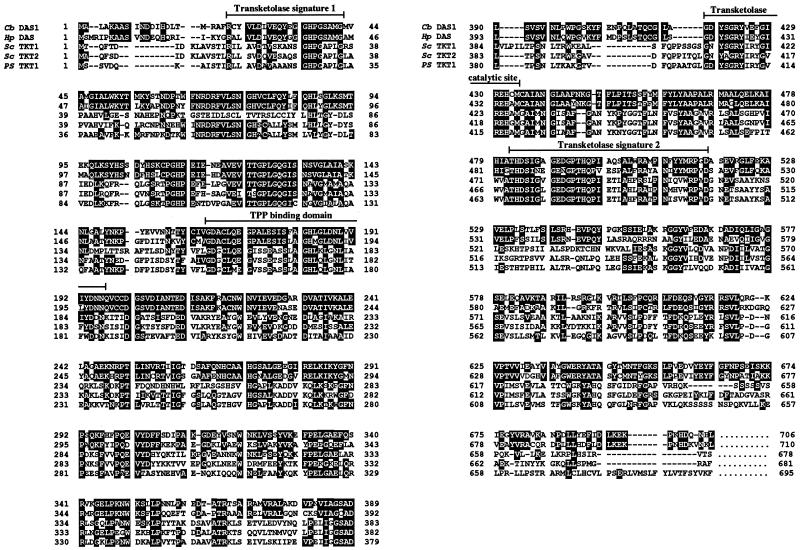
During sequencing of the cDNA library clones obtained from methanol-grown C. boidinii ATCC 32195 (8), Goodman et al. had found a partial clone that coded for an open reading frame (ORF) similar to the deduced amino acid sequence of Hansenula polymorpha DHAS (8a, 10) (unpublished data). Using this putative DAS1 fragment as the probe, we obtained the complete DAS1 clone from the genomic library of C. boidinii S2. DAS1 consists of a 2,118-bp ORF corresponding to a protein of 706 amino acid residues (Fig. 1). This ORF was identified as the gene encoding DHAS based on (i) the identity of the N-terminal amino acid sequence, NH2-ALAKAASINDDIHDLTMRAFR-, derived with that of the purified DHAS protein; (ii) agreement of the calculated molecular mass of this protein (78,132 Da) with that of the purified DHAS as determined by sodium dodecyl sulfate-polyacrylamide gel electrophoresis (SDS-PAGE) (78 kDa); and (iii) the loss of DHAS activity in the das1Δ strain (described below). The sequence encoded by the DAS1 coding region showed greater similarity to the deduced amino acid sequence of the H. polymorpha DAS product (69% identity) (10) than to the sequences of other transketolases from Saccharomyces cerevisiae (7, 31) and Pichia stipitis (17) (39 to 41% identity) (Fig. 1). All of these transketolases contained transketolase signature 1 (amino acid residues 22 to 42) (16), transketolase signature 2 (residues 482 to 517) (16, 32), the catalytic domain of transketolase (residues 418 to 434) (7), and a possible thiamine pyrophosphate binding domain (residues 165 to 196) (9, 32) (Fig. 1).
FIG. 1.
Alignment of the deduced amino acid sequence of the C. boidinii DAS1 (Cb DAS1) product with sequences of H. polymorpha DHAS (Hp DAS) (GenBank accession no. X02424 [10]), S. cerevisiae transketolase 1 (Sc TKT1) (GenBank accession no. X73224 [31]) and transketolase 2 (Sc TKT2) (GenBank accession no. X73532 [31]), and P. stipitis transketolase (Ps TKT1) (GenBank accession no. Z26486 [17]). White letters indicate amino acid residues identical to those of the C. boidinii DAS1 product.
Regulation of DAS1 expression by various carbon and nitrogen sources.
The activities of methanol-metabolizing enzymes in C. boidinii cells grown on different carbon and nitrogen sources were studied. As shown in Table 1, when NH4Cl was used as the sole nitrogen source for growth, DHAS activity was induced by methanol but was not induced by glucose, glycerol, or other peroxisome-inducing carbon sources, i.e., oleate and d-alanine. DHAS induction by methanol was repressed by glucose (Table 1; glucose + methanol) but not by glycerol (Table 1; glycerol + methanol). Methylamine and choline, when used as nitrogen sources, are known to be metabolized to formaldehyde in yeast cells (18). When either of these substrates was used as a nitrogen source together with glycerol as the carbon source, we observed induction of DHAS activity (Table 1; MA/glycerol or Chl/glycerol). In contrast, when glucose was used as the carbon source, DHAS activity was not induced (Table 1; MA/glucose or Chl/glucose). These results indicate that DHAS activity was induced by methanol or formaldehyde and that induction of DHAS activity suffers from repression by glucose but not by glycerol.
TABLE 1.
Relative activities of enzymes related to methanol metabolism during growth on various carbon and nitrogen sourcesa
| N source/C source | Relative activity (%)
|
|||
|---|---|---|---|---|
| DHAS | AOD | FDH | FLD | |
| NH4Cl/methanol | 100 | 100 | 100 | 92 |
| NH4Cl/glucose | NDb | ND | ND | ND |
| NH4Cl/glycerol | ND | 20 | 3.3 | 22 |
| NH4Cl/oleate | ND | ND | 4.4 | 4.5 |
| NH4Cl/d-alanine | ND | ND | 3.3 | 25 |
| NH4Cl/glucose + methanol | ND | ND | ND | ND |
| NH4Cl/glycerol + methanol | 67 | 90 | 32 | 67 |
| MAc/glycerol | 40 | 32 | 36 | 80 |
| Chld/glycerol | 42 | 63 | 49 | 100 |
| MA/glucose | ND | ND | 31 | 89 |
| Chl/glucose | ND | ND | 78 | 75 |
Cells were grown and disrupted as described in Materials and Methods. Specific activities of DHAS, AOD, FLD, and FDH for methanol medium are 0.22 ± 0.06, 2.3 ± 0.18, 0.82 ± 0.10, and 0.55 ± 0.08 U/mg of protein, respectively.
ND, not detected.
MA, methylamine.
Chl, choline.
As shown in Table 1, regulation of DHAS was more like that of AOD than the other two enzymes, FDH and FLD, both involved in the formaldehyde oxidation pathway; i.e., AOD and DHAS showed complete repression by glucose, while FLD and FDH did not, when formaldehyde was generated in the cells via methylamine or choline metabolism.
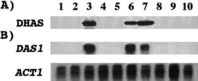
Next, DAS1 expression was monitored at the protein and mRNA levels. We conducted Western and Northern analyses by using crude extracts and total RNAs extracted from C. boidinii cells grown on each carbon and nitrogen source (Fig. 2). These regulatory patterns of DAS1 expression and the regulatory pattern of DHAS enzyme activity (Table 1) coincided each other. Therefore, the regulation of DHAS activity was confirmed to be controlled mainly at the mRNA level.
FIG. 2.
Regulation of DAS1 expression in C. boidinii. (A) Western analysis. Protein (20 μg) was separated by SDS-PAGE and detected with anti-DHAS polyclonal antibodies. (B) Northern analysis. Total RNA (20 μg) was loaded in each lane and probed with either the 32P-labeled DAS1 or the 32P-labeled ACT1 (actin) DNA as described in Materials and Methods. Carbon sources in each lane are as follows: lanes 1, 8, and 9, glucose; lanes 2, 6, and 7, glycerol; lane 3, methanol; lane 4, oleate; lane 5, d-alanine; and lane 10, glucose plus methanol. Nitrogen sources in each lane are as follows: lanes 1, 2, 3, 4, 5, and 10, NH4Cl; lanes 6 and 8, methylamine; and lanes 7 and 9, choline. The concentrations of carbon and nitrogen sources are described in Materials and Methods.
Disruption of the DAS1 gene causes defects in growth on methanol and glycerol-plus-methanol media.
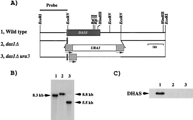
Disruption of the DAS1 gene was confirmed by Southern analysis with EcoRI-digested DNA from each transformant (Fig. 3A). The DNA from the wild-type strain gave a signal of 8.3 kb; this signal shifted to 8.8 and 5.5 kb in the das1Δ and das1Δ ura3 strains, respectively, as expected for disruption and deletion of the URA3 sequence, caused by a homologous recombination (Fig. 3B). Methanol-induced cells of the das1Δ strain did not show any DHAS activity but exhibited AOD (1.8 U/mg of protein), CTA (2,611 U/mg of protein), FLD (0.62 U/mg of protein), and FDH (0.40 U/mg of protein) activities comparable to the levels for the wild-type strain (Table 1). Western analysis using anti-DHAS antibody showed no signal in the cell extracts of the das1Δ strain and the das1Δ ura3 strain (Fig. 3C).
FIG. 3.
One-step disruption of the DAS1 gene in C. boidinii. (A) Restriction map of the cloned fragment and disruption strategy. The lightly shaded boxes and arrows at both ends of URA3 show repeated sequences for homologous recombination to remove the URA3 gene after the gene disruption. (B) Genomic Southern analysis of EcoRI-digested total DNAs (3 μg of each) from the host strain TK62 (lane 1), the das1Δ strain (lane 2), and the das1Δ ura3 strain (lane 3) probed with the 32P-labeled 1.8-kb EcoRI-EcoRV fragment, including the 5′-flanking region of DAS1. (C) Immunoblot analysis of strain TK62 (lane 1), the das1Δ strain (lane 2), and the das1Δ ura3 strain (lane 3) with extracts of methanol-induced cells and anti-DHAS polyclonal antibody.
The das1Δ strain had lost the ability to grow on methanol both in a batch culture (Fig. 4A) and in a methanol-limited chemostat culture (D = 0.05 h−1). Again, these results differed from the growth of the fdh1Δ strain, which was retarded in a methanol batch culture and one-fourth the maximum yield in a methanol-limited chemostat culture (23). The growth of the das1Δ strain on methanol was restored by the expression of the coding region of the DAS1 gene under control of the C. boidinii AOD1 promoter (Fig. 4A).
FIG. 4.
Growth of the wild-type and das1Δ strains on various carbon and nitrogen sources. MA, methylamine. Symbols: •, wild-type strain; ○, das1Δ strain; □, aod1Δ · das1Δ strain; ▵, das1Δ strain expressing DAS1 under control of the AOD1 promoter. O.D. 610, optical density at 610 nm.
Growth of the das1Δ strain had a prolonged lag period on medium containing glycerol plus methanol (Fig. 4C) relative to the rate for the wild-type strain. This growth inhibition may be due mainly to the toxicity of formaldehyde produced by AOD from methanol in the medium, since this growth retardation was not observed in the das1Δ aod1Δ strain, the double disruptant of AOD1 and DAS1 (Fig. 4C). In contrast to its growth on glycerol-plus-methanol medium, the das1Δ strain showed the same growth as the wild-type strain in media containing glycerol and methylamine (Fig. 4D) and glycerol and choline (data not shown).
Physiological role of DHAS as an assimilatory enzyme.
The syntheses of methanol-assimilatory and -dissimilatory enzymes have been considered to be regulated under the same control system through methanol induction and glucose repression (14). In C. boidinii, the regulatory pattern of DHAS was similar to that of AOD. However, the regulation of AOD and DHAS was clearly distinct from the regulation of enzymes in the dissimilation pathway, i.e., FDH and FLD.
Our previous study of FDH1 regulation and gene disruption revealed that the main physiological role of the glutathione-dependent formaldehyde oxidation pathway is detoxification of formaldehyde and formate (23). Comparison of the present results with those obtained in the previous study has revealed several differences in both regulation and knockout effect between the DAS1 and FDH1 genes. (i) The fdh1Δ strain retained the ability to grow on methanol, but the das1Δ strain did not. (ii) The FDH1 expression was observed under all conditions where formaldehyde is generated in the cells, i.e., with media containing glucose and methylamine, glucose and choline, glycerol and methylamine, glycerol and choline, and glycerol plus methanol. However, DAS1 expression was not detected in glucose-methylamine or glucose-choline medium. (iii) The defect in growth of the fdh1Δ strain was observed in all media where FDH1 expression occurred. In contrast, even though DAS1 was expressed in glycerol-methylamine and glycerol-choline media, we could not observe any defect in growth of the das1Δ strain on these media.
These results represent differences in the physiological roles of DAS1 and FDH1: the main role of DHAS may be fixation of formaldehyde into cell constituents, which is different from that of FDH, which is involved in the detoxification of formate. This was supported by the observation that the wild-type strain had a growth yield ca. four times higher than those of the das1Δ strain or the das1Δ aod1Δ strain on medium containing glycerol plus methanol (Fig. 4C). Furthermore, the growth yields of these two das1Δ strains on medium containing methanol plus glycerol were the same as those on glycerol medium (Fig. 4B). These results indicate that methanol was not assimilated in these das1Δ strains during growth on medium containing glycerol plus methanol and strongly suggest that DHAS is involved mainly in the assimilation of formaldehyde.
In a previous study, we showed that Pmp47 is necessary for the translocation and folding of DHAS but not of AOD or CTA (26). Further analysis with the DAS1 gene and the das1Δ strain obtained in this study will enable us to reveal the relationship between DHAS import into peroxisomes and the function of Pmp47 at the molecular level.
ACKNOWLEDGMENTS
We thank Joel M. Goodman for generously donating the partial cDNA clone for DHAS.
This work was partly supported by a grant-in-aid for Scientific Research from the Ministry of Education, Science, Sports and Culture, Japan, to Y.S.
REFERENCES
- 1.Bergmeyer H U. Zur Messung von Katalase Aktivitäten. Biochem Z. 1955;327:255–258. [PubMed] [Google Scholar]
- 2.Bystrykh L V, Aminova L R, Trotsenko Y A. Methanol metabolism in mutants of the methylotrophic yeast, Hansenula polymorpha. FEMS Microbiol Lett. 1988;51:89–94. [Google Scholar]
- 3.Bystrykh L V, Sokolov A P, Trotsenko Y A. Purification and properties of dihydroxyacetone synthase from the methylotrophic yeast, Candida boidinii. FEBS Lett. 1981;132:324–328. [Google Scholar]
- 4.Cryer D R, Eccleshal R, Murmer J. Isolation of yeast DNA. Methods Cell Biol. 1975;12:39–44. doi: 10.1016/s0091-679x(08)60950-4. [DOI] [PubMed] [Google Scholar]
- 5.Davis R W, Thomas M, Cameron J, St. John T P, Scherer S, Padgett R A. Rapid DNA isolations for enzymatic and hybridization analysis. Methods Enzymol. 1980;65:404–411. doi: 10.1016/s0076-6879(80)65051-4. [DOI] [PubMed] [Google Scholar]
- 6.Feinberg A P, Vogelstein B. A technique for radiolabeling DNA restriction endonuclease fragments to high specific activity. Anal Biochem. 1983;132:6–13. doi: 10.1016/0003-2697(83)90418-9. [DOI] [PubMed] [Google Scholar]
- 7.Fletcher T S, Kwee I L, Nakada T, Largman C, Martin B M. DNA sequence of the yeast transketolase gene. Biochemistry. 1992;31:1892–1896. doi: 10.1021/bi00121a044. [DOI] [PubMed] [Google Scholar]
- 8.Garrard L J, Goodman J M. Two genes encode the major membrane-associated protein of methanol-induced peroxisomes from Candida boidinii. J Biol Chem. 1989;264:13929–13937. [PubMed] [Google Scholar]
- 8a.Goodman, J. M. Unpublished data.
- 9.Hawkins C F, Borges A, Perham P N. A common structural motif in thiamin pyrophosphate-binding enzymes. FEBS Lett. 1989;255:77–82. doi: 10.1016/0014-5793(89)81064-6. [DOI] [PubMed] [Google Scholar]
- 10.Janowicz Z, Eckart M, Drewke C, Roggenkamp R, Hollenberg C P, Maat J, Ledeboer A M, Visser C, Verrips C T. Cloning and characterization of the DAS gene encoding the major methanol assimilatory enzyme from the methylotrophic yeast Hansenula polymorpha. Nucleic Acids Res. 1985;13:3043–3062. doi: 10.1093/nar/13.9.3043. [DOI] [PMC free article] [PubMed] [Google Scholar]
- 11.Kato N, Higuchi T, Sakazawa C, Nishizawa T, Tani Y, Yamada H. Purification and properties of a transketolase responsible for formaldehyde fixation in a methanol-utilizing yeast, Candida boidinii (Kloeckera sp.) no. 2201. Biochim Biophys Acta. 1982;715:143–150. [PubMed] [Google Scholar]
- 12.Kato N, Nishizawa T, Sakazawa C, Tani Y, Yamada H. Xylulose 5-phosphate-dependent fixation of formaldehyde in a methanol-utilizing yeast, Kloeckera sp. no. 2201. Agric Biol Chem. 1979;43:2013–2015. [Google Scholar]
- 13.Koning W D, Bonting K, Harder W, Dijkhuizen L. Classical transketolase functions as the formaldehyde-assimilating enzyme during growth of a dihydroxyacetone synthase-negative mutant of the methylotrophic yeast Hansenula polymorpha on mixtures of xylose and methanol in continuous culture. Yeast. 1990;6:117–125. [Google Scholar]
- 14.Koning W D, Gleeson M A G, Harder W, Dijkhuizen L. Regulation of methanol metabolism in the yeast Hansenula polymorpha. Isolation and characterization of mutants blocked in methanol assimilatory enzymes. Arch Microbiol. 1987;147:375–382. [Google Scholar]
- 15.Laemmli U K. Cleavage of structural proteins during the assembly of the head of bacteriophage T4. Nature. 1970;227:680–685. doi: 10.1038/227680a0. [DOI] [PubMed] [Google Scholar]
- 16.Lindqvist Y, Schneider G, Ermler U, Sundstrom M. Three-dimensional structure of transketolase, a thiamine diphosphate dependent enzyme, at 2.5 Å resolution. EMBO J. 1992;11:2373–2379. doi: 10.1002/j.1460-2075.1992.tb05301.x. [DOI] [PMC free article] [PubMed] [Google Scholar]
- 17.Metzger M H, Hollenberg C P. Isolation and characterization of the Pichia stipitis transketolase gene and expression in a xylose utilising Saccharomyces cerevisiae transformant. Appl Microbiol Biotechnol. 1994;42:319–325. doi: 10.1007/BF00902736. [DOI] [PubMed] [Google Scholar]
- 18.Mori N, Shirakawa K, Uzura K, Kitamoto Y, Ichikawa Y. Formation of ethylene glycol and trimethylamine from choline by Candida tropicalis. FEMS Microbiol Lett. 1988;51:41–44. [Google Scholar]
- 19.Nash T. The colorimetric estimation of formaldehyde by means of the Hantzsch reaction. Biochem J. 1953;55:416–421. doi: 10.1042/bj0550416. [DOI] [PMC free article] [PubMed] [Google Scholar]
- 20.Sakai Y, Akiyama M, Kondoh H, Shibano Y, Kato N. High-level secretion of fungal glucoamylase using the Candida boidinii gene expression system. Biochim Biophys Acta. 1996;1308:81–84. doi: 10.1016/0167-4781(96)00075-9. [DOI] [PubMed] [Google Scholar]
- 21.Sakai Y, Goh T K, Tani Y. High-frequency transformation of a methylotrophic yeast, Candida boidinii, with autonomously replicating plasmids which are also functional in Saccharomyces cerevisiae. J Bacteriol. 1993;175:3556–3562. doi: 10.1128/jb.175.11.3556-3562.1993. [DOI] [PMC free article] [PubMed] [Google Scholar]
- 22.Sakai Y, Kazarimoto T, Tani Y. Transformation system for an asporogenous methylotrophic yeast, Candida boidinii: cloning of the orotidine-5′-phosphate decarboxylase gene (URA3), isolation of uracil auxotrophic mutants, and use of the mutants for integrative transformation. J Bacteriol. 1991;173:7458–7463. doi: 10.1128/jb.173.23.7458-7463.1991. [DOI] [PMC free article] [PubMed] [Google Scholar]
- 23.Sakai Y, Murdanoto A P, Konishi T, Iwamatsu A, Kato N. Regulation of the formate dehydrogenase gene, FDH1, in the methylotrophic yeast Candida boidinii and growth characteristics of an FDH1-disrupted strain on methanol, methylamine, and choline. J Bacteriol. 1997;179:4480–4485. doi: 10.1128/jb.179.14.4480-4485.1997. [DOI] [PMC free article] [PubMed] [Google Scholar]
- 24.Sakai Y, Rogi T, Takeuchi R, Kato N, Tani Y. Expression of Saccharomyces adenylate kinase gene in Candida boidinii under the regulation of its alcohol oxidase promoter. Appl Microbiol Biotechnol. 1995;42:860–864. doi: 10.1007/BF00191182. [DOI] [PubMed] [Google Scholar]
- 25.Sakai Y, Rogi T, Yonehara T, Kato N, Tani Y. High-level ATP production by a genetically-engineered Candida yeast. Bio/Technology. 1994;12:291–293. doi: 10.1038/nbt0394-291. [DOI] [PubMed] [Google Scholar]
- 26.Sakai Y, Saiganji A, Yurimoto H, Takabe K, Saiki H, Kato N. The absence of Pmp47, a putative yeast peroxisomal transporter, causes defect in transport and folding of a specific matrix enzyme. J Cell Biol. 1996;134:37–51. doi: 10.1083/jcb.134.1.37. [DOI] [PMC free article] [PubMed] [Google Scholar]
- 27.Sakai Y, Tani Y. Cloning and sequencing of the alcohol oxidase-encoding gene (AOD1) from the formaldehyde-producing asporogenous methylotrophic yeast, Candida boidinii S2. Gene. 1992;114:67–73. doi: 10.1016/0378-1119(92)90708-w. [DOI] [PubMed] [Google Scholar]
- 28.Sakai Y, Tani Y. Directed mutagenesis in an asporogenous methylotrophic yeast: cloning, sequencing, and one-step gene disruption of the 3-isopropylmalate dehydrogenase gene (LEU2) of Candida boidinii to derive doubly auxotrophic marker strains. J Bacteriol. 1992;174:5988–5993. doi: 10.1128/jb.174.18.5988-5993.1992. [DOI] [PMC free article] [PubMed] [Google Scholar]
- 29.Sambrook J, Fritsch E F, Maniatis T. Molecular cloning: a laboratory manual. 2nd ed. Cold Spring Harbor, N.Y: Cold Spring Harbor Laboratory Press; 1989. [Google Scholar]
- 30.Sanger F, Nicklen S, Coulson A R. DNA sequencing with chain-terminating inhibitors. Proc Natl Acad Sci USA. 1977;74:5463–5467. doi: 10.1073/pnas.74.12.5463. [DOI] [PMC free article] [PubMed] [Google Scholar]
- 31.Schaaff-Gerstenschlager I, Mannhaupt G, Vetter I, Zimmermann F K, Feldmann H. TKL2, a second transketolase gene of Saccharomyces cerevisiae; cloning, sequence and deletion analysis of the gene. Eur J Biochem. 1993;217:487–492. doi: 10.1111/j.1432-1033.1993.tb18268.x. [DOI] [PubMed] [Google Scholar]
- 32.Schenk G, Layfield R, Candy J M, Duggleby R G, Nixon P F. Molecular evolutionary analysis of the thiamine-diphosphate-dependent enzyme, transketolase. J Mol Evol. 1997;44:552–572. doi: 10.1007/pl00006179. [DOI] [PubMed] [Google Scholar]
- 33.Schütte H, Flossdorf J, Sahm H, Kula M-R. Purification and properties of formaldehyde dehydrogenase and formate dehydrogenase from Candida boidinii. Eur J Biochem. 1976;62:151–160. doi: 10.1111/j.1432-1033.1976.tb10108.x. [DOI] [PubMed] [Google Scholar]
- 34.Sibirny A A, Ubiyvovk V M, Gonchar M V, Titorenko V I, Voronovsky A Y, Kapultsevich Y G. Reactions of direct formaldehyde oxidation to CO2 are non-essential for energy supply of yeast methylotrophic growth. Arch Microbiol. 1990;154:566–575. [Google Scholar]
- 35.Tani Y, Sakai Y, Yamada H. Isolation and characterization of a mutant of a methanol yeast, Candida boidinii S2, with higher formaldehyde productivity. Agric Biol Chem. 1985;49:2699–2706. [Google Scholar]
- 36.Towbin H, Staehelin T, Gordon J. Electrophoretic transfer of proteins from polyacrylamide gels to nitrocellulose sheets: procedure and some applications. Proc Natl Acad Sci USA. 1979;76:4350–4354. doi: 10.1073/pnas.76.9.4350. [DOI] [PMC free article] [PubMed] [Google Scholar]
- 37.Tschopp J F, Burst P F, Cregg J M, Stillman S A, Gingeras T R. Expression of a lacZ gene from two methanol-regulated promoters in Pichia pastoris. Nucleic Acids Res. 1987;15:3859–3876. doi: 10.1093/nar/15.9.3859. [DOI] [PMC free article] [PubMed] [Google Scholar]
- 38.Yanase H, Okuda M, Kita K, Sato Y, Shibata K, Sakai Y, Kato N. Enzymatic preparation of [1,3-13C]dihydroxyacetone phosphate from [13C]methanol and hydroxypyruvate using the methanol-assimilating system of methylotrophic yeasts. Appl Microbiol Biotechnol. 1995;43:228–234. [Google Scholar]


