Abstract
We report a case of refractory acute myeloid leukemia with DEK-NUP214 rearrangement showing circulating monocytic blasts with abundant green cytoplasmic granules on the Wright-Giemsa stain. The granules were strongly positive for Perls’ Prussian blue, consistent with hemosiderin deposits. This previously unreported finding is suggestive of siderophage activity by leukemic blasts, likely associated with monocytic differentiation.
Keywords: Acute myeloid leukemia, Green cytoplasmic granules, Hemosiderin-laden blasts
A 16-year-old adolescent girl was referred to our hospital with an eight-month history of refractory acute myeloid leukemia with t(6;9)(p23;q34.1) DEK-NUP214 rearrangement. During the next four months, different chemotherapeutic approaches were tried, with no response on subsequent bone marrow biopsies. The patient progressively deteriorated and was transferred to the intensive care unit, where therapy with imatinib and sirolimus was attempted as salvage therapy.
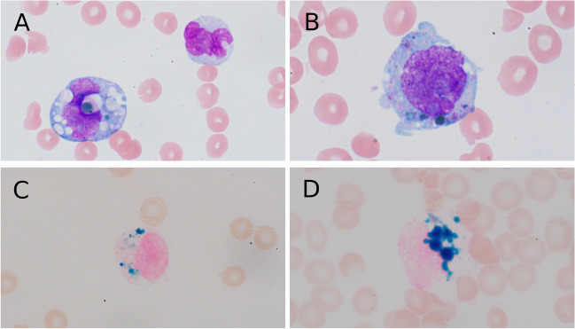
A complete blood count showed hemoglobin of 6.2 g/dL, MCV 89.6 fL, white blood cells 7.0 × 10⁹/L, and platelets 52 × 109/L. A Wright-Giemsa peripheral blood smear demonstrated 77% circulating blasts with overt monocytic differentiation and large green cytoplasmic granules detectable in approximately 15% of leukemic forms (Fig. 1A, B). Cytochemical staining revealed strong positivity with Perls’ Prussian blue (Fig. 1C, D), consistent with overt hemosiderin deposits within the cytoplasm of the monocytic leukemic blasts.
Fig. 1.
A, B The Wright-Giemsa stain of peripheral blood smear showing blasts with green cytoplasmic granules. C, D Perl’s Prussian blue stain of peripheral blood showing hemosiderin deposits in the cytoplasm of blasts
Hemosiderin deposits are cytoplasmic iron-storage complexes comprised of digested ferritin and lysosomes. They accumulate in benign macrophages after digestion of heme molecules (e.g., secondary to hemorrhage and erythrophagocytosis) [1] or in inflammatory states where secretion of hepatic hepcidin induces degradation of the macrophage iron exporter ferroportin and subsequent accumulation of cytoplasmic iron [2]. While the mechanism for leukemic hemosiderin accumulation, in this case, is unknown, the overt monocytic differentiation suggests a similar process to that of benign macrophages in response to inflammation. Previous peripheral blood smears and bone marrow biopsies demonstrated no evidence of erythrophagocytic blasts prior to the discovery of this finding. Unfortunately, 2 days after the discovery of these inclusions, the patient passed away.
Funding
No funding was received to assist with the preparation of this manuscript.
Declarations
Ethical approval
This article does not contain any studies with human participants or animals performed by any of the authors. The Mayo Clinic Institutional Review Board (IRB) acknowledges that based on the responses submitted for this new activity through the Mayo Clinic IRBe Human Subjects Research Wizard tool, and in accordance with the Code of Federal Regulations, 45 CFR 46.102, the above-noted activity does not require IRB review. The Principal Investigator is responsible for the accuracy and reliability of the information submitted through the Human Subjects Research Wizard tool, for following all applicable Federal, State, and local laws and/or regulations, and is also responsible for submitting research studies to the IRB when required.
Informed consent
For this type of study, informed consent is not required.
Consent for publication
For this type of study, consent for publication is not required.
Conflict of interest
The authors declare no competing interests.
Footnotes
Publisher's note
Springer Nature remains neutral with regard to jurisdictional claims in published maps and institutional affiliations.
Roeun Im and Pedro Horna contributed equally to this work.
References
- 1.Mahe E, Nakashima M, Olteanu H (2023) Macrophage with hemosiderin (siderophage). In: Glassy E (ed) Color atlas of hematology, 2nd Edn. Northfield, Illinois, pp 290–293
- 2.Girelli D, Nemeth E, Swinkels D. Hepcidin in the diagnosis of iron disorders. Blood. 2016;127(23):2809–2813. doi: 10.1182/blood-2015-12-639112. [DOI] [PMC free article] [PubMed] [Google Scholar]


