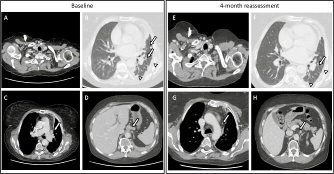
Figure 2.

CT-scan with contrast performed before (A-B-C-D) and after 4 months of entrectinib (E-F-G-H). In (A) and (E), residual thyroid tissue is highlighted with an asterisk; in (B) and (F), arrows and arrowheads point to lung and subpleural nodules and thickenings, respectively; in (C) and (G), an arrow points to a mediastinal node; in (D) and (H), an arrow points to an abdominal lymph node cluster.
