Abstract
Treatment options for patients with secondary acute myeloid leukemia (sAML) and AML with myeloid-related changes (AML-MRC) aged 60 to 75 years are scarce and unsuitable. A pivotal trial showed that CPX-351 improved complete remission with/without incomplete recovery (CR/CRi) and overall survival (OS) as compared with standard "3+7" regimens. We retrospectively analyze outcomes of 765 patients with sAML and AML-MRC aged 60 to 75 years treated with intensive chemotherapy, reported to the PETHEMA registry before CPX-351 became available. The CR/CRi rate was 48%, median OS was 7.6 months (95% confidence interval [CI]: 6.7-8.5) and event-free survival (EFS) 2.7 months (95% CI: 2-3.3), without differences between intensive chemotherapy regimens and AML type. Multivariate analyses identified age ≥70 years, Eastern Cooperative Oncology Group performance status ≥1 as independent adverse prognostic factors for CR/CRi and OS, while favorable/intermediate cytogenetic risk and NPM1 were favorable prognostic factors. Patients receiving allogeneic stem cell transplant (HSCT), autologous HSCT, and those who completed more consolidation cycles showed improved OS. This large study suggests that classical intensive chemotherapy could lead to similar CR/CRi rates with slightly shorter median OS than CPX-351.
Introduction
Treatment approaches in patients aged over 60 years diagnosed with acute myeloid leukemia (AML) are still showing unsatisfactory results.1-3 Many studies have shown poor prognosis features in this setting, as compared to younger patients, such as more adverse cytogenetics, worse performance status or higher rate of secondary AML (sAML).1-2 Frequency of sAML varies between 20-30% of all cases, showing dismal prognosis across different study cohorts.4-18
Several treatment options are now available for older sAML patients, such as hypomethylating agents (HMA) with or without venetoclax for those considered unfit for intensive therapy, and intensive chemotherapy (3+7 regimens or CPX-351) followed by an allogeneic hematopoietic stem cell transplant (allo-HSCT) for those considered fit enough. CPX-351 is a liposomal encapsulation of cytarabine and daunorubicin indicated for the treatment of patients diagnosed with AML after receiving cytotoxic, radiation or immunosuppressive treatments (t-AML) and those patients diagnosed with AML with myelodysplasia-related changes (AML-MRC),19 including this therapeutic indication a slightly different population than that enrolled in the pivotal phase III trial leading to its regulatory approval.20 In that study, CPX-351 showed improved complete remission with or without incomplete recovery (CR/CRi) rate and median overall survival (OS) compared with standard “3+7” regimens (47% vs. 33%, and 9.6 vs. 5.9 months, respectively). However, there is limited evidence regarding efficacy of classical intensive chemotherapy regimens in a similar real-life cohort of sAML and AML-MRC patients aged 60 to 75 years.
This study aims to analyze the main characteristics and therapeutic results of sAML and AML-MRC patients receiving classical intensive chemotherapy (before CPX-351 became available) in a large epidemiological registry of the “Programa Español para el Tratamiento de las Hemopatías Malignas” (PETHEMA) (clinicaltrials gov. Identifier: NCT02607059).
Methods
Eligibility
All patients with diagnosis of sAML or AML-MRC aged between 60 and 75 years who were treated with intensive chemotherapy (excluding CPX-351) and reported to the multinational PETHEMA AML registry until May, 31st 2020 were included in the study.17 Informed consent was a requisite for patients and the corresponding Research Ethics Board approved the study according to the Declaration of Helsinki.
Induction treatment
Patients receiving standard-dose cytarabine (100-200 mg/m2/day, days 1 to 7) with idarubicin (10-12 mg/m2/day, days 1 to 3) or daunorubicin (60 mg/m2/day, days 1 to 3) were classified in the “3+7” cohort. When the 3+7 regimen was dose-reduced or another drug was added to the induction schedule, therapy was considered not standard, and patients were considered as “other intensive therapy” group (Online Supplementary Table S1).
Study definitions and endpoints
According to the 2016 World Health Organization (WHO) classification,19 AML-MRC diagnosis includes AML with MDS-related cytogenetic abnormalities, those previously diagnosed with MDS (MDS-AML) or chronic myelomonocytic leukemia (CMML-AML) and AML with morphological multilineage dysplasia. Patients without information related to previous neoplastic antecedents but with MDS-related cytogenetic abnormalities were assessed in a different type of AML group (unknown). Similarly, those patients with dysplasia but without information regarding previous therapy, neoplastic antecedents or lack of cytogenetic diagnosis were also analyzed in the unknown group.19 MDS-AML, CMML-AML and t-AML were grouped as sAML. Cytogenetic results were stratified as per the Medical Research Council (MRC) classification.21
Complete remission (CR) and CR with incomplete recovery (CRi) required the absence of extramedullary disease, no blast cells in the peripheral blood (PB) and <5% of them in the bone marrow (BM).22 Neutrophil and platelet counts in PB should be >1x10⁹/L and >100x109/L, respectively to achieve CR. Patients not achieving these neutrophil or platelet counts in PB after chemotherapy were considered CRi.22 Reduction of BM blasts >50% compared to the basal value and a total count between 5-25% were necessary to reach partial remission (PR). Induction death was defined as death before the patient was assessed for response after starting the first cycle of induction. Resistance was the result if patients did not meet the aforementioned criteria. Relapse required an increase of ≥5% in BM blast cells or their resurgence in PB or as extramedullary AML after having achieved CR/CRi.
The main objective was to analyze the baseline characteristics in a real-life population and the response to intensive therapy in terms of CR/CRi rate, OS and event-free survival (EFS) in all registered patients. Other secondary endpoints were to compare different therapeutic approaches (“3+7” vs. “other intensive therapies”) and type of AML (sAML subgroups, de novo AML with MDS-related cytogenetic abnormalities or solely multilineage dysplasia). Causes to stop the intensive treatment cycles were also recorded, during induction and after remission. Postremission therapy was considered as completed when hemtopoietic stem cell transplantation (HSCT) was performed or when treatment was discontinued because of relapse before transplantation.
Statistical analysis
Patients’ baseline characteristics and response to induction therapy were expressed with the median and the interquartile range (IQR) and were analyzed with Χ2 with Yates’ correction, ANOVA, Wilcoxon, Kruskal-Wallis, ANOVA or Student’s t test, determined by the type of variable. OS and EFS prediction were computed from the date of AML diagnosis until the date of death for the first time-to-event element, whereas EFS was assessed until the date of treatment failure, relapse or death, depending on which occured first. These lifetime variables were estimated by the Kaplan-Meier estimator23 and they were compared with the log-rank test.24 In order to calculate medians and confidence intervals (CI), the corrected method of Kalbfleisch and Prentice was seleted.25 Multivariate analysis for OS was performed through a Cox proportional hazards model. Characteristics with clinically and statistically significant association in the univariate analysis (P<0.1) and those available variables with a possible relationship in previous studies were included in the analysis. Allogeneic HSCT (allo-HSCT) and autologous HSCT (auto-HSCT) were included as time-dependent variables, in a Cox proportional hazards regression with timedependent covariates. Afterwards, the Mantel-Byar test was performed and a Simon-Makuch plot was the method to represent them.26,27 Living patients were censored the last day they were alive before October, 31st 2021, when patients’ follow-up was updated. All mentioned P values are two-sided. Statistical computing and graphics were conducted using the R software.
Results
Accrual and patient characteristics
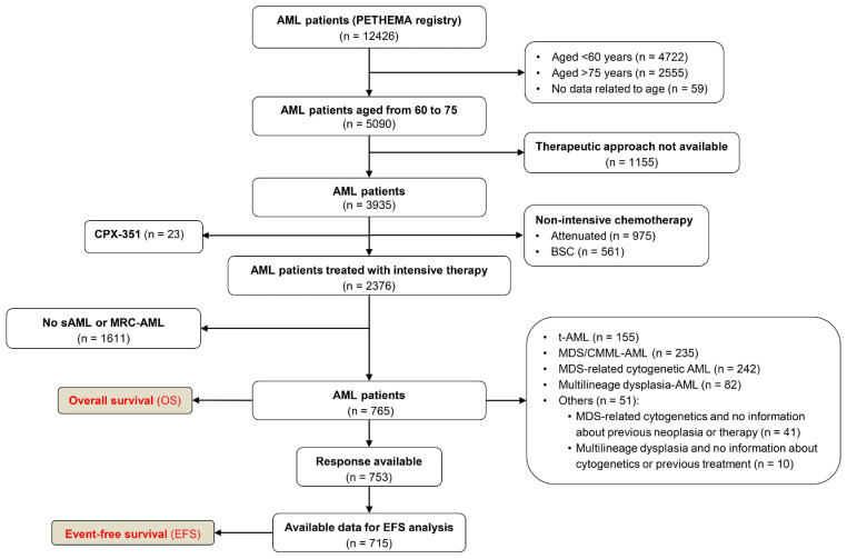
Until May 2020, 12,426 patients were registered and 5,090 (41%) were aged between 60 and 75 years. Of them, 2,376 (47%) had received intensive schedules as front-line therapy and 765 (15%) were sAML or AML-MRC as previously defined. A Consolidated Standards of Reporting Trials (CONSORT) diagram is detailed in Figure 1.
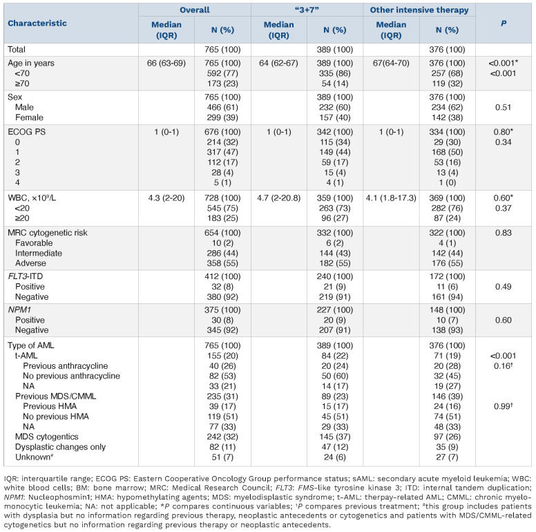
Among the 765 included patients, median age was 66 years (IQR, 63-69), median Eastern Cooperative Oncology Group performance status (ECOG PS) was 1 (IQR, 0-1) and 61% were male. White blood cell (WBC) count was ≥20×109/L in 25% of patients and median blast cells in BM at diagnosis was 48% (IQR, 31-70). Karyotype was not available in 102 (13%) patients, and it was normal in 174 (23%). When available, cytogenetic risk was adverse in 358 (55%) patients and favorable in ten (2%), NPM1 mutation was present in 30 (8%) patients, and 32 (8%) were positive for FLT3-internal tandem duplication (ITD) mutation.
Characteristics of “3+7” versus other intensive-therapy cohorts
Overall, 389 (51%) received 3+7 and 376 (49%) other intensive therapy (Online Supplementary Table S1), being the “3+7” cohort older (P<0.001) and with more t-AML (P<0.001) (Table 1; Online Supplementary Table S2).
Front-line therapy
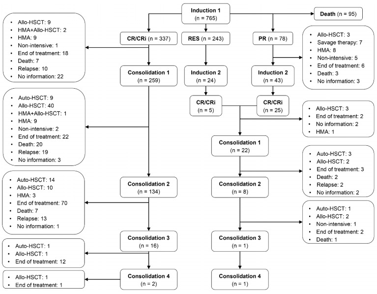
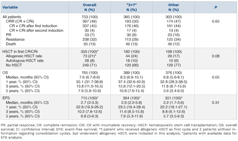
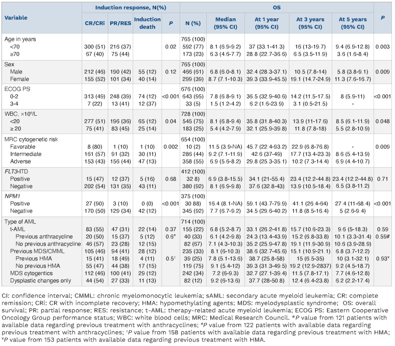
BM assessments were performed after every cycle of treatment. From 753 (98%) patients with available response assessment, 95 (13%) died during induction, CR/CRi was achieved after cycle 1 in 337 (45%), partial response (PR) in 78 (10%) and resistance in 243 (38%) (Figure 2). A second identical induction cycle was administered in 64 (8%) subjects, increasing the CR/CRi rate up to 49%. No differences in CR/CRi were observed between “3+7” and other intensive regimens (P=0.65) (Table 2). Factors associated with induction response and P<0.05 included age (P=0.02), ECOG PS (P<0.001), white blood cells (WBC) count (P=0.04), creatinine (P=0.04), uric acid (P=0.003), which were negative predictors, and albumin levels (P<0.001), MRC cytogenetic risk (P=0.002), and NPM1 mutation status (P<0.001), which were positively associated with response. No differences were found according to type of AML (therapy-related AML [t-AML] vs. secondary to MDS/CMML vs. de novo AML with MDS-cytogenetic vs de novo AML with dysplasia) or FLT3-ITD mutation status (P=0.37 and P=0.68, respectively; Table 3, Online Supplementary Table S3).
When patients were included in a multinomial logistic regression, older age (P=0.004), and higher ECOG PS (P<0.001) were independent adverse prognostic factors for CR/CRi; while presence of NPM1 mutation (P<0.001), and favorable (P=0.03) and intermediate cytogenetic risk (P<0.001) were favorable prognostic factors.
Post-remission treatment
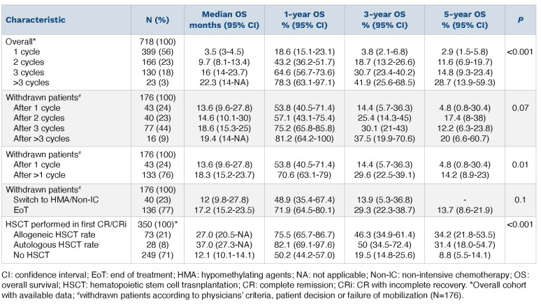
Data on HSCT was available in 350 of 367 patients who achieved CR/CRi after induction. Of them, 28 (8%) received autologous HSCT (auto-HSCT) and 73 (21%) allo-HSCT (Table 2). More patients in the “3+7” cohort received subsequent HSCT in first CR/CRi (statistical trend P=0.08). The total number of cycles of intensive chemotherapy during front-line therapy was available in 719 patients, with a mean number of 1.69 cycles (median: 1 cycle; IQR, 1-2). One cycle was administered in 399 (55%) patients, two cycles in 166 (23%), three cycles in 130 (18%) patients, four cycles in 20 (3%), five cycles in two (0.3%) and six cycles in one (0.1%) (Figure 2). Detailed information regarding second induction and consolidation regimens are shown in Online Supplementary Tables S4 and S5.
Causes of front-line treatment withdrawal
Overall, 143 patients completed post-remission therapy, 224 had incomplete post-remission schedule, and information on schedule completeness was not available in 30 patients. Causes of withdrawal were: 37 patients (11%) died in CR/CRi because of treatment complications, five (1%) rejected further intensive treatment, five (1%) failed to perform a planned auto-HSCT due to mobilization failure, 147 (44%) were considered unfit to receive further cycles (20 [6%] because of serious events during prior cycles, and 126 [38%] due to concomitant comorbidities, age or any other reason according to physicians’ criteria). In addition, three patients achieving PR after one cycle of induction therapy had no further information and 32 did not receive a second identical cycle: three died before starting it, 23 switched to alternative schedules, two suffered serious complications during the first cycle, and four were considered unfit to receive more therapies.
Overall survival
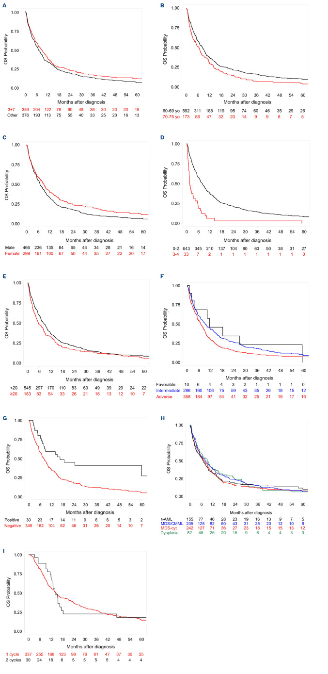
The median OS of the 765 patients included in the study was 7.6 months (95% CI: 6.7-8.5), 8.3 months (95% CI: 6.9-10.1) in the “3+7” cohort versus 6.8 months (95% CI: 5.6-8.1) in other intensive therapies group (P=0.05; Figure 3A; Table 2). In univariate analyses, prognostic factors for prolonged OS were: age <70 years (Figure 3B; P=0.003), female (Figure 3C; P=0.009), ECOG PS ≤2 (Figure 3D; P<0.001), WBC count <20×109/L (Figure 3E; P=0.048), platelet count >20×109/L (P=0.01), creatinine levels ≤1.3 mg/dL (P=0.001), uric acid ≤7 mg/dL (P=0.001), albumin >3.5 g/dL (P=0.003), lower percentage of blast cells in BM (P=0.04), intermediate and favorable cytogenetic risk (Figure 3F; P=0.009) and mutated NPM1 (Figure 3G; P<0.001). No statistical differences were found as per FLT3-ITD mutational status, type of AML (Figure 3H), CR/CRi after one or two cycles of induction (Figure 3I; P=0.75), or prior treatment with anthracyclines or HMA (Table 3; Online Supplementary Table S3).
Figure 1.
Consolidated Standards of Reporting Trials (CONSORT) diagram for adult patients aged between 60 and 75 years with secondary acute myeloid leukemia and acute myeloid leukemia with myeloid-related changes. AML: acute myeloid leukemia; sAML: secondary AML; MRC-AML: AML with myeloid-related changes; MDS: myelodisplastic syndrome; CMML: chronic myelomonocytic leukemia; t-AML: therapy-related AML; BSC: best supportive care.
Multivariate Cox regression showed that age ≥70 years (P=0.007), ECOG PS ≥1 (P=0.046) and higher WBC counts (P=0.04) were independent adverse prognostic factors for OS. Intermediate cytogenetic risk (P=0.016), and NPM1 mutation (P=0.007) were favorable prognostic factors (Online Supplementary Table S6). The Cox proportional hazard regression with time-dependent covariates showed ECOG PS 3 (hazard ratio [HR] =2.27; 95% CI: 1.50-3.42; P<0.001), ECOG PS 4 (HR=3.83; 95% CI: 1.42-10.38; P=0.008) and no allo- or auto-HSCT (HR=17.26; 95% CI: 7.74-38.47; P<0.001) as independent adverse prognostic factors.
Table 1.
Demographic and baseline characteristics of the study population (“3+7” vs. other intensive therapy).
Impact of post-remission therapy on overall survival
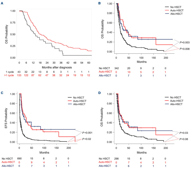
We analyzed the impact of post-remission schedule on OS (Table 4). Among 176 patients withdrawing post-remission schedule as per physicians’/patient decision or mobilization failure, statistical differences were found after stratifying between one cycle and >1 cycle, with longer OS in the later group in comparison to the former one i.e., 1 cycle: median and 5 years OS of 13.6 months; 95% CI: 8.9-23 and 5%; 95% CI: 0.8-30.4 versus >1 cycle: 18.3 months; 95% CI: 15.2-23.7 and 14%; 95% CI: 8.9-23; P=0.01, respectively (Figure 4A).
Median and 5 years OS of patients undergoing allo-HSCT after achieving first CR/CRi were 27 months (95% CI: 20.5-not applicable [NA]) and 34.2% (95% CI: 21.8-53.5) versus 37 months (95% CI: 27.3-NA) and 31.4% (95% CI: 18.0-54.7) in those receiving auto-HSCT, and 12.1 months (95% CI: 10.1-14.1) and 8.8% (95% CI: 5.5-14.1) in those not consolidated with HSCT (P<0.001), respectively. According to the Mantel-Byar test, patients receiving HSCT in first CR/CRi had better OS in comparison with those who did not (P<0.001), with a significant difference both allo-HSCT and auto-HSCT versus no HSCT (P=0.003 and P=0.008, respectively) (Figure 4B). No differences were observed in OS between allo-HSCT and auto-HSCT i.e., the median OS from the date of HSCT was 18.9 months (95% CI: 9.9-81.7) and 25.2 (95% CI: 13-54.1), respectively (P=0.97).
Event-free survival
The median EFS of the 715 AML patients with available data for this analysis was 2.7 months (95% CI: 2-3.3), with 1- and 5-year EFS of 22.8% (95% CI: 19.9-26.2) and 6.8% (95% CI: 5-9.3), respectively. No significant differences were observed between “3+7” and other intensive schedules cohorts, with 1- and 5-year EFS of 23.5% (95% CI: 19.4-28.4) and 7.9 (95% CI: 5.3-11.9) versus 22.2% (95% CI: 18.1-27.1) and 5.7 (95% CI: 3.5-9.3), respectively (P=0.31) (Online Supplementary Figure S1; Table 2). The median EFS according to HSCT in first CR/CRi from the 698 patients with available data (allo-HSCT vs. auto-HSCT vs. no HSCT) were 25.1 months (95% CI: 16.0-NA) versus 28.3 (95% CI: 15.2-94.1) versus 1.6 (95% CI: 1.5-1.9), respectively. From the date of diagnosis, the 5-year EFS were 35.3% (95% CI: 23.1-54.1) in the allo-HSCT group, 28.6% (95% CI: 15.9-51.3) in the auto-HSCT, and 2.9% (95% CI: 1.7-4.8) in the no-HSCT group (P<0.001). According to the Mantel-Byar test, patients receiving HSCT in first CR/CRi had better EFS in comparison with those who did not (P<0.001), with significant difference both allo-HSCT and auto-HSCT versus no HSCT (P<0.001 and P=0.02, respectively) (Figure 4C). No differences were observed in EFS between allo-HSCT and auto-HSCT (P=0.81).
Figure 2.
Induction response and post-remission therapy. Auto-HSCT: autologous stem cell transplant; Allo-HSCT: allogeneic HSCT; HMA: hypomethylating agent; CR: complete remission; CRi: complete remission with incomplete recovery.
CPX-351-like cohort
The so called “CPX-351-like” cohort included 673 patients, after removing 92 patients who did not fulfill disease characteristics for inclusion in the pivotal phase III trial leading to CPX-351 approval (i.e., those patients with multilineage dysplasia-AML as sole criterion for AML-MRC classification). Median age of this cohort was 66 years (IQR, 63-69), 264 (39%) patients were female, 28 (5%) had an ECOG PS >2, 466 (73%) had a WBC count <20x109/L, and 356 (62%) had an adverse MRC cytogenetic risk. From 661 with available data, 293 (44%) achieved CR/CRi after one cycle of induction cycle and 27 (4%) after two cycles with the same schedule (CR/CRi rate of 48%). Eighty-two patients (12%) died before assessment, 44 (7%) achieved partial response (PR) and 215 (33%) were considered resistant. Median OS was 7.6 months (95% CI: 6.5-8.4), without differences in CR/CRi rate and OS between “3+7” and other intensive therapy cohorts (P=0.6 and P=0.2, respectively). Median EFS was 2.0 months (95% CI: 1.9-3.3) and no differences were observed regarding induction therapy i.e., the median EFS in “3+7” cohort was 2.7 months (95% CI: 2.0-3.8) versus 2.0 (95% CI: 1.6-3.6) in the other group (Online Supplementary Figure S2A; P=0.66) or other type of AML i.e., 3.3 months in t-AML group (95% CI: 1.9-5.1) versus 2.5 in MDS/CMML-AML (95% CI: 1.9-4.5) versus 1.8 (95% CI: 1.5-3.2) in the MDS cytogenetics (Online Supplementary Figure S2B; P=0.38). Among those patients achieving CR/CRi, there were data related to HSCT in front-line therapy in 305 (64 underwent an allo-HSCT, 21 auto-HSCT, and 220 did not receive HSCT). Median OS was higher in patients receiving an auto-HSCT or an allo-HSCT versus no HSCT i.e., 35.8 months (95% CI: 14-NA) versus 27.0 (95% CI: 20.5-NA) versus 12.7 (95% CI: 10.3-14.6), with 1-year OS of 81% (95% CI: 65.8-99.6), 74.6% (95% CI: 64.2-86.6) and 52.1 (95% CI: 45.7-59.3), respectively (P<0.001). According to the Mantel-Byar test, patients receiving HSCT in first CR/CRi had better OS in comparison with those who did not (P=0.018), with a significant difference between allo-HSCT and no HSCT (P=0.029), and a trend between auto-HSCT and no HSCT (P=0.08) (Figure 4D). No differences were observed in OS between allo-HSCT and auto-HSCT i.e., the median OS from the date of HSCT were 18.9 months (95% CI: 9.9-81.7) and 29.4 (95% CI: 7.3-149.5), respectively (P=0.83). After selecting patients with ECOG PS 0-2, serum creatinine <2.0 mg/dL and serum total bilirubin <2.0 mg/dL, 368 were analyzed. Median age was 66 years (IQR, 63-69) and baseline characteristics were similar to patients included in the CPX-351-like cohort. The CR/CRi rate in this group was 51%, with a median OS and EFS of 7.7 months (95% CI: 6.6-9.1) and 2.8 months (95% CI: 2-3.9), respectively (Online Supplementary Table S7).
Table 2.
Induction response, hematopoietic stem cell transplantation rates in first complete remission/complete remission with incomplete recovery, overall survival and event-free survival according to therapeutic group.
Discussion
This study shows the baseline characteristics and the clinical outcomes in a real-life cohort of patients aged between 60 and 75 years, who were diagnosed with t-AML or MRC-AML and treated with intensive schedules. This cohort included 765 patients (roughly 7% of all AML registered patients in the study period), achieving a CR/CRi rate of 48% after one or two induction cycles, and median OS of 7.6 months (95% CI: 6.7-8.5), roughly 2 months better than observed in the 3+7 control arm of the pivotal phase III trial.20 In this study, we have analyzed outcomes of patients with AML and multilineage dysplasia at diagnosis according to the CPX-351 regulatory approval, although isolated morphological multilineage dysplasia was not among inclusion criteria for the pivotal phase III trial.20 When we take into account the similar study population to the pivotal phase III trial (CPX-351-like cohort), median OS remained unchanged at 7.6 months. We must underline that the CPX-351 data comes from a randomized trial, which is the gold standard to have new therapies for our patients. Thus, our comparison should be taken with a great deal of caution. We can speculate on reasons for non-comparable results between our real-life series and the control arm of the pivotal phase III trial: i) our cohort included patients with worse clinical features (i.e., deteriorated ECOG PS or organ function, more patients with adverse MRC cytogenetic risk or WBC count ≥20x109/L) as compared to those enrolled in clinical trial; ii) given the open label design of the phase III trial, control arm patients could withdraw earlier frontline therapy as they were not assigned to experimental arm; iii) although our study cohort comes from an epidemiologic registry, a positive or negative reporting bias could occur; and iv) a marked variability in cytarabine plus anthracycline induction regimens was noted in our series, where half of the patients received upfront “3+7”, and half received “other intensive therapies” with addition of a third drug, dose reductions or intensive clinical trials. On the other hand, we can also hypothesize that the adverse effect of FLT3-ITD mutations was not observed in our study due to: i) its association with higher WBC counts, which was shown as an independent factor for survival; and ii) our cohort shows lower prevalence of FLT3-ITD mutation (8%) in comparison with published series with de novo and younger patients. Interestingly, no significant differences in CR/CRi, OS or EFS were observed between both treatment groups, with the exception of a trend to better OS in the “3+7” group, probably related to older age observed in the “other intensive therapies” group.
Table 3.
Induction response and overall survival according to baseline characteristics.
Figure 3.
Impact of patient’s characteristics at diagnosis and number of cycles to achieve complete remission/complete remission with incomplete recovery. (A) Overall survival (OS) in secondary acute myeloid leukemia (sAML) and AML patients with myeloid-related changes (AML-MRC) according to therapeutic approach (“3+7” vs. other intensive therapy). (B) OS in sAML and AML-MRC according to age (60-69 vs. >70 years). (C) OS in sAML and AML-MRC according to sex. (D) OS in sAML and AML-MRC according to Eastern Cooperative Oncology Group performance status (ECOG PS). (E) OS in sAML and AML-MRC according to value of white blood cell (WBC) count. (F) OS in sAML and AML-MRC according to cytogenetic risk. (G) OS in sAML and AML-MRC according to NPM1 mutation. (H) OS according to the type of AML (t-AML, sAML myelodysplastic syndrome/chronic myelomonocytic leukemia [MDS/CMML], MDS cytogenetics and multilineage dysplasia). (I) OS sAML and AML-MRC between patients who achieved complete remission/complete remission with incomplete recovery (CR/CRi) after 1 or 2 cycles of induction; yo: years old.
Table 4.
Overall survival according to cycles of intensive chemotherapy and post-remission approach.
The main clinical outcomes in our CPX-351-like cohort were better as compared with the “3+7” control arm of the pivotal phase III trial of CPX-351.19 Although similar baseline characteristics were observed in both studies regarding age (66 vs. 67.8 years) and sex (39% vs. 38.6% female), some differences were observed in WBC count <20x109/L (73% vs. 85.6%) and ECOG PS >2 (5% vs. 0%).19 Our CPX-351-like cohort achieved similar CR/CRi (48% vs. 47%) and EFS (2.4 vs. 2.5 months), and worse OS than experimental CPX-351 arm (7.6 vs. 9.6 months), but our cohort showed better outcomes than the “3+7” comparator of phase III trial (CR/CRi 33%, EFS 1.3 months, OS 5.9 months).19 Early mortality rates with CPX-351 were 13.7% through day 60, while our cohort showed 13% mortality during the first induction cycle.
Regarding HSCT, 29.4% received allo-HSCT in phase III trial (34% CPX-351 cohort vs. 25% 3+7 cohort) compared to 21% of patients in our series. As in the Lancet et al. study19 and a recent population-based study,26 we show that patients undergoing allo-HSCT after induction had significantly better OS and EFS. Moreover, we were able to show that performing auto-HSCT or administering more consolidation cycles increased OS, highlighting the inherent favorable selection bias of AML patients completing their planned consolidation and intensification schedules.
Recent real-world studies analyzed relatively small cohorts of t-AML and AML-MRC older patients treated with CPX-351.29-32 A German31 series showed 47% CR/CRi rate, but Italian29 and French30 cohorts reported higher responses (70% and 59%). Interestingly, several real-life studies with CPX-351 have reported better median OS as compared to the phase III trial (e.g., 16.130 2131 1532 months and median not reached29). Several factors could explain better results observed in real world evidence studies with CPX-351: i) they included adult patients without limit of age, and ii) they analyzed more recent periods were allo-HSCT could be improved and more available as compared with the phase III and our historical cohort.
We found that older age, higher ECOG PS and WBC count were independent adverse factors for CR/CRi and OS, whereas favorable/intermediate cytogenetic risk and NPM1 mutation were favorable factors. Similarly, Lancet et al. described ECOG PS, cytogenetic risk classification, and WBC count as prognostic factors after multivariate analysis.20 Recent series of patients treated with CPX-351 showed that non-spliceosome,30 TP53, and PTPN11 mutations30 were adverse risk factors, but we could not analyze their impact due to the limited information on these mutations in our series. We revealed the favorable impact of NPM1 mutations, while others failed probably due to the scarce number of NPM1-mutated patients among the targeted population. As expected, there were differences in age and type of AML among patient subgroups according to HSCT (Online Supplementary Table S8). After performing the Mantel-Byar test to analyze the impact of HSCT on survival, only ECOG PS ≥3 and not performing a HSCT in first CR/CRi remained as independent adverse factors for OS.
Figure 4.
Impact of post-remission therapy. (A) Overall survival (OS) according to cycles of intensive treatment (1 cycle vs. >1 cycle) in those patients withdrawn according to physicians’ criteria, patient decision or failure of mobilization (n=176). (B) Simon-Makuch plot of OS according to hematopoietic stem cell transplantation (HSCT) in first complete remission/complete remission with incomplete (CR/CRi) (allogeneic HSCT [allo-HSCT] vs. autologous HSCT [auto-HSCT] vs. no HSCT). (C) Simon-Makuch plot of event-free survival (EFS) according to HSCT in first CR/CRi (allo-HSCT vs. auto-HSCT vs. no HSCT). (D) Simon-Makuch plot of OS in the CPX-351-like cohort according to HSCT in first CR/CRi (allo-HSCT vs. auto-HSCT vs. no HSCT).
Some limitations should be addressed: i) this is a real-life analysis with great heterogeneity and not a truly population-based registry; ii) missing data and retrospective analyses; and iii) secondary-AML type mutations (e.g., TP53, ASXL1, RUNX1, IDH) were not available. However, the Lancet study did not show these genetic characteristics as well, so the mutational landscape of both series cannot justify similar results in OS between our historical cohort and the CPX-351 phase III.
In conclusion, this large study in older patients with t-AML or MRC-AML suggests that classical intensive chemotherapy could lead to similar CR/CRi rates with slightly shorter median OS than CPX-351. We confirm improved OS after allo-HSCT in this setting, and we suggest the role of intensification cycles to improve long-term outcomes. The advantage of CPX-351 has been established in a phase III pivotal trial, but this should be confirmed in larger registry studies focusing on baseline mutational status, measurable residual disease, and toxicities.
Supplementary Material
Acknowledgments
The authors would like to thank María D. García, Carlos Pastorini, Asier Laria, Yolanda Mendizábal, and Teresa Martinez Sena for data collection and management.
Funding Statement
Funding: This study was partially supported by the Jazz Pharmaceuticals and Cooperative Research Thematic Network (RTICC) grant RD12/0036/014 (ISCIII & ERDF).
References
- 1.Döhner H, Estey E, Grimwade D, et al. Diagnosis and management of AML in adults: 2017 ELN recommendations from an international expert panel. Blood. 2017;129(4):424-447. [DOI] [PMC free article] [PubMed] [Google Scholar]
- 2.Tallman MS, Wang ES, Altman JK, et al. Acute myeloid leukemia, version 3.2019, NCCN Clinical Practice Guidelines in Oncology. J Natl Compr Canc Netw. 2019;17(6):721-749. [DOI] [PubMed] [Google Scholar]
- 3.Daver N, Wei AH, Pollyea DA, Fathi AT, Vyas P, DiNardo CD. New directions for emerging therapies in acute myeloid leukemia: the next chapter. Blood Cancer J. 2020;10(10):107. [DOI] [PMC free article] [PubMed] [Google Scholar]
- 4.Leone G, Mele L, Pulsoni A, Equitani F, Pagano L. The incidence of secondary leukemias. Haematologica. 1999;84(10):937-945. [PubMed] [Google Scholar]
- 5.Granfeldt Østgård LS, Medeiros BC, Sengeløv H, et al. Epidemiology and clinical significance of secondary and therapy-related acute myeloid leukemia: a national population-based cohort study. J Clin Oncol. 2015;33(31):3641-3649. [DOI] [PubMed] [Google Scholar]
- 6.Hulegårdh E, Nilsson C, Lazarevic V, et al. Characterization and prognostic features of secondary acute myeloid leukemia in a population-based setting: a report from the Swedish Acute Leukemia Registry population-based study of secondary AML. Am J Hematol. 2015;90(3):208-214. [DOI] [PubMed] [Google Scholar]
- 7.Szotkowski T, Rohon P, Zapletalova L, Sicova K, Hubacek J, Indrak K. Secondary acute myeloid leukemia - a single center experience. Neoplasma. 2010;57(2):170-178. [DOI] [PubMed] [Google Scholar]
- 8.Xu X-Q, Wang J-M, Gao L, et al. Characteristics of acute myeloid leukemia with myelodysplasia-related changes: A retrospective analysis in a cohort of Chinese patients: characteristics of AML-MRC in Chinese patients. Am J Hematol. 2014;89(9):874-881. [DOI] [PubMed] [Google Scholar]
- 9.Aström M, Bodin L, Nilsson I, Tidefelt U. Treatment, long-term outcome and prognostic variables in 214 unselected AML patients in Sweden. Br J Cancer. 2000;82(8):1387-1392. [DOI] [PMC free article] [PubMed] [Google Scholar]
- 10.Schoch C, Schnittger S, Klaus M, Kern W, Hiddemann W, Haferlach T. AML with 11q23/MLL abnormalities as defined by the WHO classification: incidence, partner chromosomes, FAB subtype, age distribution, and prognostic impact in an unselected series of 1897 cytogenetically analyzed AML cases. Blood. 2003;102(7):2395-2402. [DOI] [PubMed] [Google Scholar]
- 11.Wheatley K, Brookes CL, Howman AJ, et al. Prognostic factor analysis of the survival of elderly patients with AML in the MRC AML11 and LRF AML14 trials. Br J Haematol. 2009;145(5):598-605. [DOI] [PubMed] [Google Scholar]
- 12.Ostgård LSG, Kjeldsen E, Holm MS, et al. Reasons for treating secondary AML as de novo AML. Eur J Haematol. 2010;85(3):217-226. [DOI] [PubMed] [Google Scholar]
- 13.Kayser S, Döhner K, Krauter J, et al. The impact of therapy-related acute myeloid leukemia (AML) on outcome in 2853 adult patients with newly diagnosed AML. Blood. 2011;117(7):2137-2145. [DOI] [PubMed] [Google Scholar]
- 14.Oran B, Weisdorf DJ. Survival for older patients with acute myeloid leukemia: a population-based study. Haematologica. 2012;97(12):1916-1924. [DOI] [PMC free article] [PubMed] [Google Scholar]
- 15.Gangatharan SA, Grove CS, P’ng S, et al. Acute myeloid leukaemia in Western Australia 1991-2005: a retrospective population-based study of 898 patients regarding epidemiology, cytogenetics, treatment and outcome: AML in Western Australia. Intern Med J. 2013;43(8):903-911. [DOI] [PubMed] [Google Scholar]
- 16.Medeiros BC, Satram-Hoang S, Hurst D, Hoang KQ, Momin F, Reyes C. Big data analysis of treatment patterns and outcomes among elderly acute myeloid leukemia patients in the United States. Ann Hematol. 2015;94(7):1127-1138. [DOI] [PMC free article] [PubMed] [Google Scholar]
- 17.Martínez-Cuadrón D, Megías-Vericat JE, Serrano J, et al. Treatment patterns and outcomes of 2310 patients with secondary acute myeloid leukemia: a PETHEMA registry study. Blood Adv. 2022;6(4):1278-1295. [DOI] [PMC free article] [PubMed] [Google Scholar]
- 18.Bertoli S Tavitian S, BoriesPet al. Outcome of patients aged 60-75 years with newly diagnosed secondary acute myeloid leukemia: a single-institution experience. Cancer Med. 2019;8(8):3846-3854 [DOI] [PMC free article] [PubMed] [Google Scholar]
- 19.Arber DA, Orazi A, Hasserjian R, et al. The 2016 revision to the World Health Organization classification of myeloid neoplasms and acute leukemia. Blood. 2016;127(20):2391-2405. [DOI] [PubMed] [Google Scholar]
- 20.Lancet JE, Uy GL, Cortes JE, et al. CPX-351 (cytarabine and daunorubicin) liposome for injection versus conventional cytarabine plus daunorubicin in older patients with newly diagnosed secondary acute myeloid leukemia. J Clin Oncol. 2018;36(26):2684-2692. [DOI] [PMC free article] [PubMed] [Google Scholar]
- 21.Grimwade D, Walker H, Harrison G, et al. The predictive value of hierarchical cytogenetic classification in older adults with acute myeloid leukemia (AML): analysis of 1065 patients entered into the United Kingdom Medical Research Council AML11 trial. Blood. 2001;98(5):1312-1320. [DOI] [PubMed] [Google Scholar]
- 22.Cheson BD, Bennett JM, Kopecky KJ, et al. Revised recommendations of the International Working Group for diagnosis, standardization of response criteria, treatment outcomes, and reporting standards for therapeutic trials in acute myeloid leukemia. J Clin Oncol. 2003;21(24):4642-4649. [DOI] [PubMed] [Google Scholar]
- 23.Kaplan EL, Meier P. Nonparametric estimation from incomplete observations. J Am Stat Assoc. 1958;53(282):457. [Google Scholar]
- 24.Mantel N. Evaluation of survival data and two new rank order statistics arising in its consideration. Cancer Chemother Rep 1966;50(3):163-170. [PubMed] [Google Scholar]
- 25.Kalbfleisch JD, Prentice RL. The statistical analysis of failure time data. New York: John Wiley & Sons. 1980. [Google Scholar]
- 26.Mantel N, Byar D: Evaluation of response-time data involving transient states: an illustration using heart transplant data. J Am Stat Assoc. 1974;69(345):81-86. [Google Scholar]
- 27.Simon R, Makuch RW. A non-parametric graphical representation of the relationship of an event: application to responser versus non-responder bias. Stat Med. 1984;3(1):35-44. [DOI] [PubMed] [Google Scholar]
- 28.Nilsson C, Hulegårdh E, Garelius H, et al. Secondary acute myeloid leukemia and the role of allogeneic stem cell transplantation in a population-based setting. Biol Blood Marrow Transplant. 2019;25(9):1770-1778. [DOI] [PubMed] [Google Scholar]
- 29.Guolo F, Fianchi L, Minetto P, et al. CPX-351 treatment in secondary acute myeloblastic leukemia is effective and improves the feasibility of allogeneic stem cell transplantation: results of the Italian compassionate use program. Blood Cancer. 2020;10(10):96. [DOI] [PMC free article] [PubMed] [Google Scholar]
- 30.Chiche E, Rahmé R, Bertoli S, et al. Real-life experience with CPX-351 and impact on the outcome of high-risk AML patients: a multicentric French cohort. Blood Adv. 2021;5(1):176-184. [DOI] [PMC free article] [PubMed] [Google Scholar]
- 31.Rautenberg C, Stölzel F, Röllig C, et al. Real-world experience of CPX-351 as first-line treatment for patients with acute myeloid leukemia. Blood Cancer. 2021;11(10):164. [DOI] [PMC free article] [PubMed] [Google Scholar]
- 32.Lee D, Jain AG, Deutsch Y, et al. CPX-351 yields similar response and survival outcome in younger and older patients with secondary acute myeloid leukemia. Clin Lymphoma Myeloma Leuk. 2022;22(10):774-779. [DOI] [PubMed] [Google Scholar]
Associated Data
This section collects any data citations, data availability statements, or supplementary materials included in this article.


