Abstract
Background
Endometrial cancer (EC) has the characteristics of high mortality and poor prognosis in the advanced stage, which seriously threatens women’s health. Killer cell lectin-like receptor B1 (KLRB1) is a promising immune checkpoint of which the expression level can regulate the killing effect on tumor cells of the immune system, thereby affecting the survival and prognosis of tumor patients. However, it is still unclear whether KLRB1 is associated with survival and prognosis in patients with EC. Therefore, our study focused on the relationship between KLRB1 and immune cells to explore the role of KLRB1 on the immune microenvironment, and to further explore its feasibility as a prognostic marker in EC.
Methods
In this study, The Cancer Genome Atlas (TCGA) and Gene Expression Omnibus (GEO) databases were used to analyze the messenger RNA (mRNA) expression level of KLRB1 in normal endometrial and EC tissues. The University of Alabama at Birmingham Cancer data analysis Portal (UALCAN) database was used to determine the correlation between KLRB1 mRNA expression and clinical features among the EC patients. KLRB1 expression levels were investigated in the Tumor IMmune Estimation Resource (TIMER) database to reveal its relationship with immune cell infiltration of EC. Finally, using the R package clusterProfiler, enrichment analysis was performed on KLRB1 to study its potential function.
Results
The results suggested that KLRB1 expression varied in different tumor tissues, and the EC group had lower mRNA expression levels than did the control group. It was also found that patients with high expression of KLRB1 had a better prognosis. According to further enrichment and immune infiltration analyses, KLRB1 expression had a closed relationship with the level of infiltration of some immune cell types, such as B cells memory, eosinophils, and Tregs, among others.
Conclusions
KLRB1 expression is associated with the infiltration of immune cells and can be used as a prognostic biomarker in EC.
Keywords: Endometrial cancer (EC), KLRB1, immune infiltration, prognostic biomarker
Highlight box.
Key findings
• KLRB1 expression is associated with the infiltration of immune cells and can be used as a prognostic biomarker in endometrial cancer (EC).
What is known and what is new?
• KLRB1 is a promising immune checkpoint of which the expression level can regulate the killing effect of the immune system on tumor cells, thereby affecting the survival and prognosis of tumor patients.
• The results of our study suggested that the EC group had lower KLRB1 mRNA expression levels than those of the control group. KLRB1 also has close correlation with cancer stage, ethnicity, weight, histological subtypes, and immune infiltration. High expression of KLRB1 was associated with a better prognosis.
What is the implication, and what should change now?
• KLRB1 is a potential biomarker for EC prognosis.
Introduction
Endometrial cancer (EC) has the characteristics of high mortality and poor prognosis in the advanced stage, with 417,000 new diagnoses made globally in 2020 (1). Unfortunately, the number of patients diagnosed with EC continues to increase. Based on the best-fitting model, by 2030 there will be 42.13 new cases of EC annually for every 100,000 women (2). Although early detection of EC is ideal and some progress has been made in early treatment, the current reality is that the majority of clinically aggressive EC subtypes are usually diagnosed at an advanced stage, and female EC still has an increased mortality rate (3). In addition, advanced EC is predisposed to occur in 3–13% of cases and has a poor prognosis (4). Furthermore, women who are cured of EC still face a significant risk of cardiovascular death due to the mostly unrecognized and undertreated risk factors (5). To improve the risk stratification system and the quality of care for women with EC, the latest European (ESGO/ESTRO/ESP 2020) guidelines proposed a novel risk stratification model including The Cancer Genome Atlas (TCGA) molecular groups to assess the prognosis of EC and the role of the molecular subtypes of EC as prognostic factors independent from classic types (6,7). Although molecular signature-characterized studies are innovative and precise in diagnosing EC patient characteristics, they are complex and costly. Therefore, new biomarkers are needed to predict the EC prognosis and provide new targets for the treatment.
Killer cell lectin-like receptor B1 (KLRB1) is a gene which encodes CD161 and is expressed on CD4 T cells, CD8 T cells, and natural killer (NK) cells (8). In general, KLRB1 makes great contributions to lymphocyte differentiation (9). A previous study suggested that in most tumors, high expression of KLRB1 can suppress tumor development, helping to improve the quality of life and extend the lifespan of patients (10). Besides, the role of KLRB1 in lung adenocarcinoma, glioma, and colon cancer has been investigated, and the results have shown that the immune function of KLRB1 is strongly associated with the development of those tumors (11-13). Another study also suggested that knockdown of KLRB1 inhibits tumor cell growth in esophageal squamous cell carcinoma (14). Meanwhile, KLRB1 can interact with LLT1 and actively participate in anti-tumor immune response in non-small cell lung cancer (15). Therefore, KLRB1 is considered an indispensable gene in tumor immunomodulation. However, little research has been conducted on KLRB1 in patients with EC.
In this study, pan-cancer analysis of KLRB1 expression was performed. Furthermore, the expression of KLRB1 in normal endometrium and EC tissues was investigated using TCGA and the Gene Expression Omnibus (GEO) databases. We also analyzed the relationship between KLRB1 expression levels and various clinical features of EC patients by using the University of Alabama at Birmingham Cancer data analysis Portal (UALCAN) database. Finally, we focused on the relationship between KLRB1 and immune cells to explore the role of KLRB1 on the tumor immune microenvironment (TIME) and to further explore its feasibility as a prognostic marker in EC. We present this article in accordance with the REMARK reporting checklist (available at https://tcr.amegroups.com/article/view/10.21037/tcr-23-697/rc).
Methods
Materials
The data used in this study were all from public databases on the Internet. We followed the methods of the previous study to conduct the data analysis (16). The following are links to the website of each database that we used: TCGA database (https://cancergenome.nih.gov/), Tumor IMmune Estimation Resource (TIMER) database (http://timer.cistrome.org/), GEO database (https://www.ncbi.nlm.nih.gov/), The Human Protein Atlas (HPA) database (https://www.proteinatlas.org/), and UALCAN database (http://ualcan.path.uab.edu). This study was conducted in accordance with the Declaration of Helsinki (as revised in 2013).
Messenger RNA sequencing data collection and analysis
Pan-cancer analysis of KLRB1 was performed using the HPA and TIMER databases. Next, the KLRB1 expression in tumor tissue and normal or paraoncological tissue was analyzed using samples from the TCGA and GEO databases. Besides, by using the UALCAN database, we obtained the clinical data of EC patients and analyzed the correlation between KLRB1 expression and these clinical features.
Survival prognosis analysis
The “survminer” package in R studio (version 4.3.1; R Foundation for Statistical Computing, Vienna, Austria) was used to analyze the data in TCGA to explore the correlation between overall survival (OS) and progression-free survival (PFS) in KLRB1 and EC patients.
Correlation and gene set enrichment analysis
In order to further explore the relationship between KLRB1 and EC, we used TCGA data to study the correlation between KLRB1 and other genes associated with EC. An enrichment analysis of the top 50 genes positively correlated with KLRB1 was conducted to determine the function of KLRB1. A Gene Ontology (GO) enrichment analysis, including biological process (BP), cellular component (CC), and molecular function (MF), was performed using the EnrichGO function in the R package “clusterProfiler”. The EnrichKEGG function from the R package clusterProfiler was utilized to conduct Kyoto Encyclopedia of Genes and Genomes (KEGG) analysis.
Immune cell infiltration analysis
Tumor-immune interactions in EC were performed using Cell-type Identification by Estimating Relative Subsets of RNA Transcripts (CIBERSORT). Gene expression profiling data were used to investigate the correlation between KLRB1 expression on immune cell infiltration and the abundance of tumor-infiltrating immune cells. Finally, KLRB1 expression levels were investigated in the TIMER database in relation to immune cells infiltration of EC.
Statistical analysis
The abovementioned datasets were generated for all statistical analyses and differences between normal and tumor groups using a t-test. Survival curves were generated using the Kaplan-Meier method. A P value less than 0.05 was considered statistically significant.
Results
KLRB1 gene expression in various human cancers
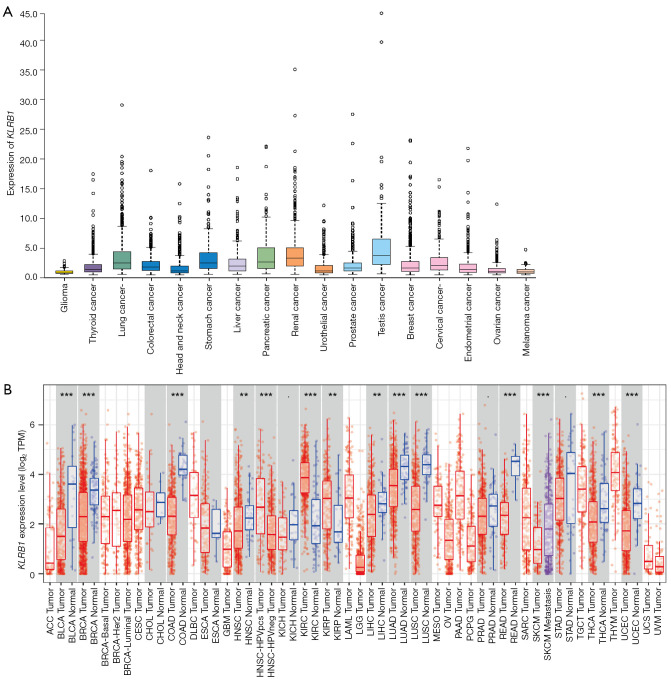
The expression of KLRB1 in pan-carcer is shown in Figure 1A,1B. The results showed that the expression of KLRB1 was low in a majority of cancers, including bladder urothelial carcinoma (BLCA), liver hepatocellular carcinoma (LIHC), thyroid carcinoma (THCA), uterine corpus endometrial carcinoma (UCEC), and others, whereas in the other tumors such as kidney renal clear cell carcinoma (KIRC), KLRB1 mRNA was highly expressed.
Figure 1.
KLRB1 expression levels in pan-cancer. (A) Expression of KLRB1 in different tumors in HPA database. These images are available from v21.0.proteinatlas.org. (https://www.proteinatlas.org/ENSG00000111796-KLRB1/pathology). (B) KLRB1 expression in different types of tumors in the TIMER database (dark spots were automatically generated by the software and is equivalent to splitting characters. It has no practical meaning. For example, DLBC. Tumor represents lymph cancer). **, P<0.01; ***, P<0.001. KLRB1, killer cell lectin-like receptor B1; HPA, Human Protein Atlas; TIMER, Tumor IMmune Estimation Resource; DLBC, diffuse large B cell lymphoma; TPM, transcripts per million .
Relationship between KLRB1 expression and patients’ status of UCEC
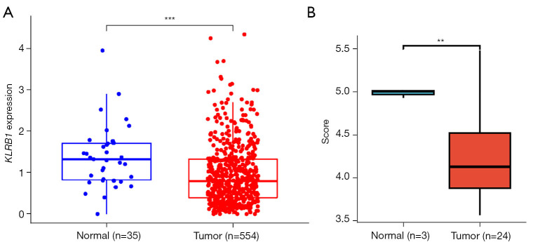
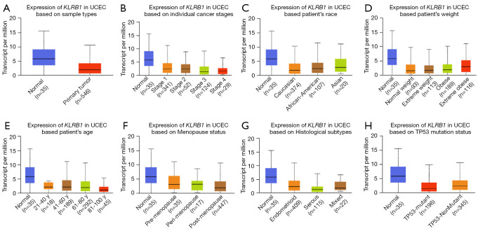
According to our results, the expression level of KLRB1 was generally low in tumor patients (Figure 2A,2B). Further sub-group analysis of the UALCAN database showed low KLRB1 transcription in 546 EC samples. The expression of KLRB1 were lower in EC patients in subgroup analyses based on cancer stage, ethnicity, race, weight, menopausal status, histological subtypes, and TP53 mutation status (Figure 3).
Figure 2.
The relative expression of KLRB1 between normal individuals and EC patients. (A) The relative expression of KLRB1 in normal and EC samples in TCGA. (B) The relative expression of KLRB1 in normal and EC samples in GSE115810. **, P<0.01; ***, P<0.001. KLRB1, killer cell lectin-like receptor B1; EC, endometrial cancer; TCGA, The Cancer Genome Atlas.
Figure 3.
KLRB1 transcription in subgroups of patients with endometrial cancer, stratified based on cancer stages, patient’s age, patient’s weight, patient’s race, and other criteria (UALCAN) (A-H). KLRB1, killer cell lectin-like receptor B1; UALCAN, University of Alabama at Birmingham Cancer data analysis Portal; UCEC, uterine corpus endometrial carcinoma.
Survival and prognostic value of KLRB1 in EC
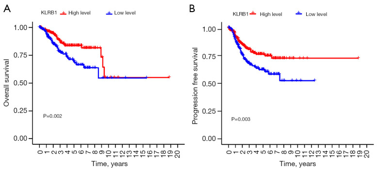
TCGA data were analyzed to further investigate KLRB1’s value as a prognostic marker for UCEC. The results shown in Figure 4A,4B indicated that high expression of KLRB1 can improve the clinical survival outcome and prolong OS and PFS in patients of EC. Therefore, we suggested that KLRB1 may be a tumor suppressor gene in EC, contributing to the improvement of patients’ survival cycle, and may be a promising prognostic biomarker in EC.
Figure 4.
Prognostic value of KLRB1 mRNA expression in EC patients. (A) Low KLRB1 expression was associated with poor OS in EC patients in TCGA database. (B) Low KLRB1 expression was associated with poor PFS in EC patients in TCGA database. KLRB1, killer cell lectin-like receptor B1; EC, endometrial cancer; TCGA, The Cancer Genome Atlas; OS, overall survival; PFS, progression-free survival.
Correlation and KLRB1-related gene enrichment analysis
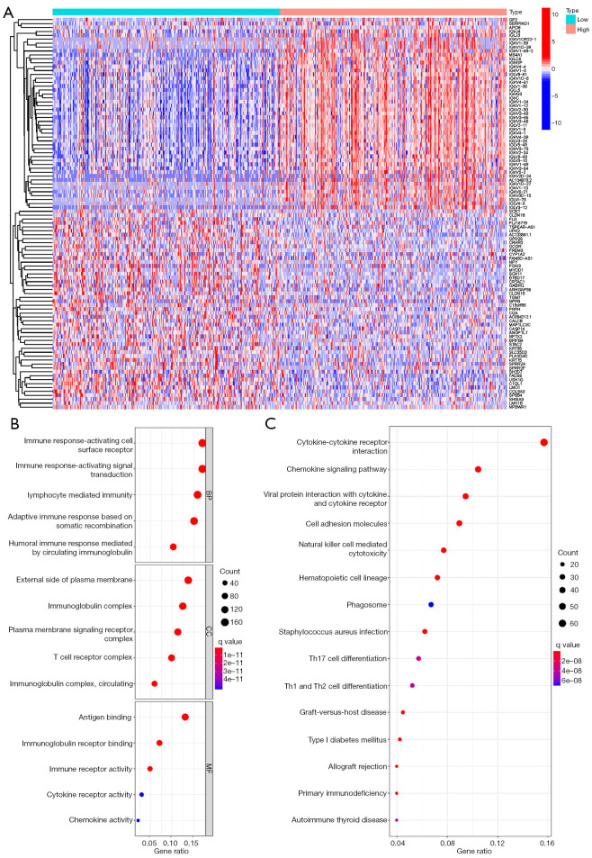
In order to clarify how KLRB1 works, we searched TCGA for genes associated with KLRB1 expression for subsequent pathway analysis. Based on the heat map, we identified the top 50 significant gene sets related to KLRB1 both positively and negatively (Figure 5A). By using the “clusterProfiler” R package, we obtained the KLRB1 positively correlated genes using GO and KEGG enrichment analyses. GO enrichment analysis consisted of three parts: BP, CC, and MF. First, in BP, the results showed that those co-expressed genes were related to immune function, including lymphocyte-mediated immunity and humoral immune response mediated by circulating immunoglobulin. Moreover, the co-expressed genes were found to be enriched in the immunoglobulin complexes, T cell receptor complexes, and the immunoglobulin complexes in the CC. Finally, in MF, the genes were enriched in antigen binding, immunoglobulin receptor binding, immune receptor activity (Figure 5B). KEGG enrichment analysis (Figure 5C) showed that co-expressed genes were mainly concentrated in cytokine-cytokine receptor interaction, chemokine signaling pathway, NK cell-mediated cytotoxicity, and so on.
Figure 5.
Function and pathway enrichment analysis of KLRB1 in EC from TCGA. (A) Heat map of differentially expressed genes associated with KLRB1 in EC. (B) GO annotations of KLRB1 in EC. (C) Significant KEGG pathway associated with KLRB1 in EC. KLRB1, killer cell lectin-like receptor B1; EC, endometrial cancer; TCGA, The Cancer Genome Atlas; GO, Gene Ontology; KEGG, Kyoto Encyclopedia of Genes and Genomes; CC, cellular component; MF, molecular function.
Relationship between KLRB1 expression and immune cell infiltration
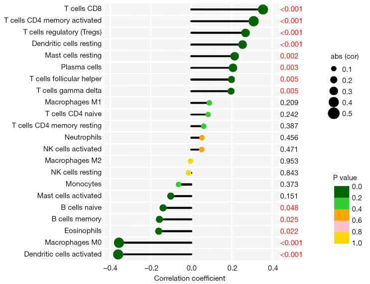
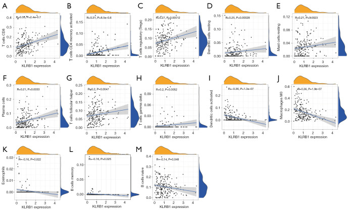
The above enrichment analysis showed that KLRB1 was associated with immune response. Therefore, we explored whether KLRB1 expression was associated with immune cell infiltration. Our study suggested that KLRB1 had significant correlation with T cells CD8, Tregs, dendritic cells (DC) resting, plasma cells, T cells gamma delta, macrophage M0, eosinophils, and B cells naïve, among others (Figure 6). Further research showed that KLRB1 expression was positively correlated with infiltration levels of T cells CD8 (Figure 7A, R=0.35, P=2.4e−07), T cells CD4 memory activated (Figure 7B, R=0.31, P=8.5e−06), Tregs (Figure 7C, R=0.27, P=0.00012), DC resting (Figure 7D, R=0.25, P=0.00028), mast cells resting (Figure 7E, R=0.21, P=0.0023), plasma cells (Figure 7F, R=0.21, P=0.0033), T cells follicular helper (Figure 7G, R=0.2, P=0.0047), and T cells gamma delta (Figure 7H, R=0.2, P=0.0052). In contrast, KLRB1 expression was negatively correlated with that of DC activated (Figure 7I, R=−0.36, P=1.3e−07), macrophage M0 (Figure 7J, R=−0.36, P=1.9e−07), eosinophils (Figure 7K, R=−0.16, P=0.022), B cells memory (Figure 7L, R=−0.16, P=0.025), and B cells naive (Figure 7M, R=−0.14, P=0.048).
Figure 6.
Lollipop chart of KLRB1 expression level in 22 immune cells. KLRB1, killer cell lectin-like receptor B1; NK, natural killer.
Figure 7.
Correlation between KLRB1 expression and immune cell infiltration. (A-M) T cells CD8, T cells CD4 memory activated, Tregs, DC resting, mast cells resting, plasma cells, T cells follicular helper, T cells gamma delta, DC activated, macrophage M0, eosinophils, B cells memory, and B cells naive. KLRB1, killer cell lectin-like receptor B1; DC, dendritic cells.
Prognosis analysis of KLRB1 expression with immune infiltration status
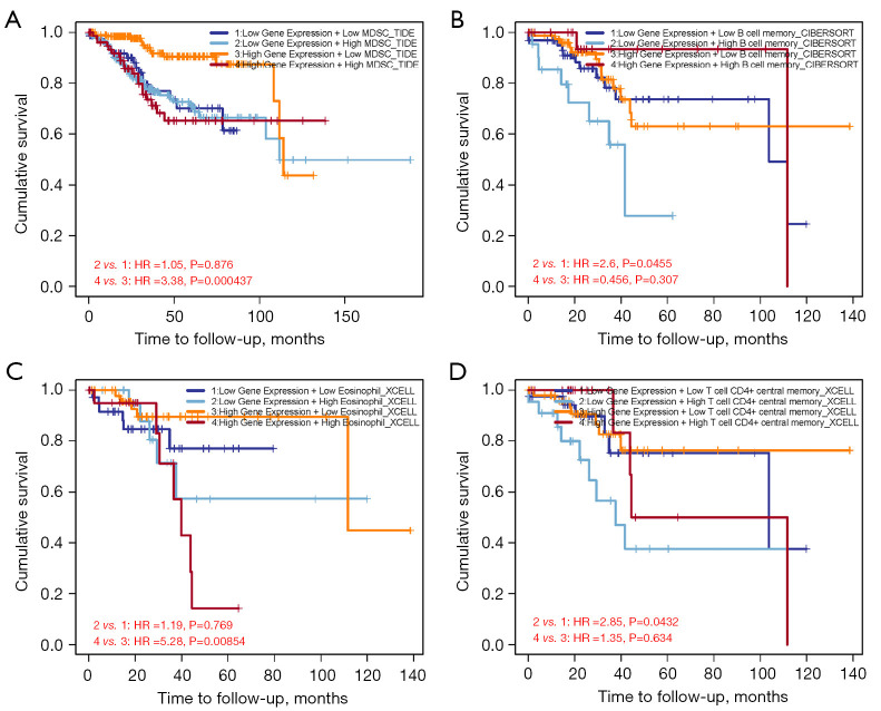
Using the TIMER database, we assessed how immune cell infiltration impacts clinical survival outcomes among patients with EC. The results showed that with the greater expressions of myeloid-derived suppressor cells (MDSC), B-cell memory, eosinophils, and T-cell CD4+ in the tumor immune microenvironment, the worse prognosis patients had (Figure 8A-8D).
Figure 8.
Impact of immune cell infiltration on prognosis in EC patients. (A) Clinical survival outcome of EC patients in the high-myeloid-derived suppressor cells group. (B) Clinical survival outcome of EC patients in the high-B cell memory group. (C) Clinical survival outcome of EC patients in the high-eosinophils group. (D) Clinical survival outcome of EC patients in the high T cell CD4 memory group. EC, endometrial cancer; HR, hazard ratio; CIBERSORT, Cell-type Identification by Estimating Relative Subsets of RNA Transcripts.
Discussion
EC has the characteristics of high mortality and poor prognosis in the advanced stage, which seriously threatens women’s health, and the incidence is expected to further increase in the next 10 years (17). There are various treatment options for EC at earlier stages; most of them are curable with surgery and the prognosis is generally good. However, there are limited treatment options for EC at advanced stages (III or IV) and it usually comes with poor prognosis (18). Based on the current situation, how to monitor the progress of EC and predict its survival and prognosis is still a major problem in modern medicine. The 2020 ESGO/ESTRO/ESP guidelines stratify the prognosis of EC patients combining TCGA molecular signature and pathological factors, including lymphovascular space invasion (LVSI) (19). It is well known that clinicopathological factors of EC (such as myometrial invasion, histotype, or lympho space invasion) are a very hot topic. Research from another research group revealed that a high accuracy could be achieved with one-step nucleic acid amplification detection of EC lymph node metastasis (20). A recent study showed that, in EC patients, LVSI has a prognostic value independent of TCGA groups, age, and adjuvant treatment. In particular, the presence of LVSI increased the risk of all-cause mortality, EC death, and recurrent or progressive disease by 1.5–2 times (21). However, how to precisely integrate molecular features with classical pathological factors still needs more basic theoretical and clinical exploration. Therefore, a reliable biomarker is needed to guide us towards more precise diagnosis and treatment. According to the previous studies, KLRB1 is recognized as a potential prognosis biomarker in breast cancer (22), human esophageal squamous cell carcinoma (14), liver cancer (23), lung adenocarcinoma (24), and bladder cancer (25). The correlation of EC and KLRB1 expression had not been studied until now. Therefore, our study aimed to investigate the correlation between KLRB1 and immune cell infiltration in EC and whether it affects the occurrence and development of EC, so as to explore its guiding effect on survival and prognosis of patients with EC.
Recently, studies have suggested that the expression of the gene and protein of KLRB1 are downregulated to some extent in most tumors (10,26). Tumor immunity is strongly influenced by the KLRB1 gene and its encoded protein, CD161. A study showed that tumor cell immune escape was associated with the downregulation of CD161 expression in oropharyngeal squamous cell carcinoma, which may further exacerbate tumor progression (27). If CD161 is highly expressed in T cells, the tumor burden will be significantly reduced, which can improve the survival rate of tumor patients (28). In addition, a relevant study (10) showed that the sensitivity of tumors to chemotherapy drugs has a close relationship with the expression of KLRB1. In most tumors, the lower KLRB1 expression, the better the clinical effect of chemotherapy. These findings suggest that KLRB1 may serve as a biomarker to help guide clinical drug selection and patient outcomes prognosis. However, the role of CD161 also varies between different tumors. In most tumors, CD161 is associated with a better prognosis, whereas in a small number of cancer types, CD161 has shown a role in promoting adverse tumor outcomes. For example, in glioma, blocking or inactivating CD161 can enhance the killing of gliomas by T cells, which is more conducive to controlling the growth of tumors in vivo (24).
In our study, the results indicated that KLRB1 was downregulated in majority of tumors including EC through pan-cancer analysis which is consistent with previous research (10,26). The correlation of different clinical features with KLRB1 expression in EC patients was also assessed. The results showed that in the subgroup analysis based on criteria such as patient weight, patient ethnicity, cancer stage, and patient age, the KLRB1 transcription level of the EC group was significantly increased compared to that of the healthy controls. As compared with all other cancers, the incidence of EC had the strongest association body weight. A previous study showed that 57% of all EC cases are attributable to obesity in America (29). Ethnic differences are also significantly associated with the incidence of EC. A survey study from the United States comparing invasive and non-endometrioid tumors by race showed a significant increase in the prevalence of EC in black women, suggesting that biological racial differences may influence the prevalence of EC (30). Further results showed that patients with higher KLRB1 expression tended to have better OS than those with low KLRB1 expression. Above all of these results, KLRB1 may influence tumor development in multiple dimensions, and can be used as a biomarker to predict the survival and prognosis of EC patients. Furthermore, the majority cell types in the tumor microenvironment (TME) are immune cells, which can interact with each other to control tumor growth and metastasis, and make great contributions to tumor progression, metastasis, and treatment resistance (31). As a very important checkpoint in the immune microenvironment, CD161 has been shown to cooperate with other immune checkpoints in regulating the TME. These findings might provide great support for the development of new immunotherapy drugs (32). To further explore the correlation between KLRB1 expression and immune cell infiltration, we conducted the following studies to explore the relationship between KLRB1 and CD8, Tregs, DC, macrophages, T cells, and so on. Thus, we speculate that the different expression levels of KLRB1 can change the proportion of some immune cell types in the TME and further affect the occurrence and development of tumors. In summary, our study suggested that the downregulation of KLRB1 expression in EC was strongly associated with poor survival outcomes, KLRB1 can be used as a biomarker, and its expression level can predict the tumor development trend and survival prognosis of patients. Unfortunately, the study was conducted only by analyzing information from public databases. Although our research shows that the expression level of KLRB1 can affect the immune cell types in the TME and can be used as a prognostic marker in EC, the study was only conducted by analyzing information from public databases. How KLRB1 affects immune cell infiltration in TME and the specific mechanism affecting tumor progression still need to be verified by further clinical and basic experiments.
Conclusions
We found that the downregulation of KLRB1 expression in EC was strongly associated with poor survival outcomes. The effect of KLRB1 on the development and prognosis of EC may be related to its influence on the level of immune cell infiltration in the TME. Our research determined that KLRB1 can be used as a biomarker, and its expression level can predict the tumor development trend and survival prognosis of patients to a certain extent.
Supplementary
The article’s supplementary files as
Acknowledgments
We extend our thanks to TCGA, GEO, TIMER, and other public databases that provide many valuable data resources for free.
Funding: This study was supported by the National Famous Old Chinese Medicine Expert Inheritance Studio of Peijuan Wang (grant No. 22QGGZS1), National Natural Science Foundation of China (grant No. 82074487), and Graduate Student Training Innovation Project in Jiangsu Province (grant No. SJCX23-0818).
Ethical Statement: The authors are accountable for all aspects of the work in ensuring that questions related to the accuracy or integrity of any part of the work are appropriately investigated and resolved. The study was conducted in accordance with the Declaration of Helsinki (as revised in 2013).
Footnotes
Reporting Checklist: The authors have completed the REMARK reporting checklist. Available at https://tcr.amegroups.com/article/view/10.21037/tcr-23-697/rc
Peer Review File: Available at https://tcr.amegroups.com/article/view/10.21037/tcr-23-697/prf
Conflicts of Interest: All authors have completed the ICMJE uniform disclosure form (available at https://tcr.amegroups.com/article/view/10.21037/tcr-23-697/coif). The authors have no conflicts of interest to declare.
References
- 1.Crosbie EJ, Kitson SJ, McAlpine JN, et al. Endometrial cancer. Lancet 2022;399:1412-28. 10.1016/S0140-6736(22)00323-3 [DOI] [PubMed] [Google Scholar]
- 2.Sheikh MA, Althouse AD, Freese KE, et al. USA endometrial cancer projections to 2030: should we be concerned? Future Oncol 2014;10:2561-8. 10.2217/fon.14.192 [DOI] [PubMed] [Google Scholar]
- 3.Urick ME, Bell DW. Clinical actionability of molecular targets in endometrial cancer. Nat Rev Cancer 2019;19:510-21. 10.1038/s41568-019-0177-x [DOI] [PMC free article] [PubMed] [Google Scholar]
- 4.Vitale SG, Valenti G, Gulino FA, et al. Surgical treatment of high stage endometrial cancer: current perspectives. Updates Surg 2016;68:149-54. 10.1007/s13304-015-0340-1 [DOI] [PubMed] [Google Scholar]
- 5.Moss EL, Gorsia DN, Collins A, et al. Utility of Circulating Tumor DNA for Detection and Monitoring of Endometrial Cancer Recurrence and Progression. Cancers (Basel) 2020;12:2231. 10.3390/cancers12082231 [DOI] [PMC free article] [PubMed] [Google Scholar]
- 6.Concin N, Matias-Guiu X, Vergote I, et al. ESGO/ESTRO/ESP guidelines for the management of patients with endometrial carcinoma. Int J Gynecol Cancer 2021;31:12-39. 10.1136/ijgc-2020-002230 [DOI] [PubMed] [Google Scholar]
- 7.Cuccu I, D'Oria O, Sgamba L, et al. Role of Genomic and Molecular Biology in the Modulation of the Treatment of Endometrial Cancer: Narrative Review and Perspectives. Healthcare (Basel) 2023;11:571. 10.3390/healthcare11040571 [DOI] [PMC free article] [PubMed] [Google Scholar]
- 8.Konduri V, Oyewole-Said D, Vazquez-Perez J, et al. CD8(+)CD161(+) T-Cells: Cytotoxic Memory Cells With High Therapeutic Potential. Front Immunol 2021;11:613204. 10.3389/fimmu.2020.613204 [DOI] [PMC free article] [PubMed] [Google Scholar]
- 9.Poggi A, Rubartelli A, Moretta L, et al. Expression and function of NKRP1A molecule on human monocytes and dendritic cells. Eur J Immunol 1997;27:2965-70. 10.1002/eji.1830271132 [DOI] [PubMed] [Google Scholar]
- 10.Cheng X, Cao Y, Wang X, et al. Systematic Pan-Cancer Analysis of KLRB1 with Prognostic Value and Immunological Activity across Human Tumors. J Immunol Res 2022;2022:5254911. 10.1155/2022/5254911 [DOI] [PMC free article] [PubMed] [Google Scholar]
- 11.Liu L, Xu S, Huang L, et al. Systemic immune microenvironment and regulatory network analysis in patients with lung adenocarcinoma. Transl Cancer Res 2021;10:2859-72. 10.21037/tcr-20-2275 [DOI] [PMC free article] [PubMed] [Google Scholar]
- 12.Di W, Fan W, Wu F, et al. Clinical characterization and immunosuppressive regulation of CD161 (KLRB1) in glioma through 916 samples. Cancer Sci 2022;113:756-69. 10.1111/cas.15236 [DOI] [PMC free article] [PubMed] [Google Scholar]
- 13.Mo J, Zheng L, Gao Z, et al. The Study of Mucosal-Associated Invariant T Cells in Colon Cancer and Roles in Immune Activities. Onco Targets Ther 2021;14:5263-73. 10.2147/OTT.S332822 [DOI] [PMC free article] [PubMed] [Google Scholar]
- 14.Zhang G, Liu Y, Dong F, et al. Transcription/Expression of KLRB1 Gene as A Prognostic Indicator in Human Esophageal Squamous Cell Carcinoma. Comb Chem High Throughput Screen 2020;23:667-74. 10.2174/1386207323666200517114154 [DOI] [PubMed] [Google Scholar]
- 15.Braud VM, Biton J, Becht E, et al. Expression of LLT1 and its receptor CD161 in lung cancer is associated with better clinical outcome. Oncoimmunology 2018;7:e1423184. 10.1080/2162402X.2017.1423184 [DOI] [PMC free article] [PubMed] [Google Scholar]
- 16.Zhao K, Ma Z, Zhang W. Comprehensive Analysis to Identify SPP1 as a Prognostic Biomarker in Cervical Cancer. Front Genet 2022;12:732822. 10.3389/fgene.2021.732822 [DOI] [PMC free article] [PubMed] [Google Scholar]
- 17.Lee YC, Lheureux S, Oza AM. Treatment strategies for endometrial cancer: current practice and perspective. Curr Opin Obstet Gynecol 2017;29:47-58. 10.1097/GCO.0000000000000338 [DOI] [PubMed] [Google Scholar]
- 18.Ulrich LS. Endometrial cancer, types, prognosis, female hormones and antihormones. Climacteric 2011;14:418-25. 10.3109/13697137.2010.550974 [DOI] [PubMed] [Google Scholar]
- 19.Golia D'Augè T, Cuccu I, Santangelo G, et al. Novel Insights into Molecular Mechanisms of Endometrial Diseases. Biomolecules 2023;13:499. 10.3390/biom13030499 [DOI] [PMC free article] [PubMed] [Google Scholar]
- 20.Raffone A, Travaglino A, Santoro A, et al. Accuracy of One-Step Nucleic Acid Amplification in Detecting Lymph Node Metastases in Endometrial Cancer. Pathol Oncol Res 2020;26:2049-56. 10.1007/s12253-019-00727-9 [DOI] [PubMed] [Google Scholar]
- 21.Raffone A, Travaglino A, Raimondo D, et al. Lymphovascular space invasion in endometrial carcinoma: A prognostic factor independent from molecular signature. Gynecol Oncol 2022;165:192-7. 10.1016/j.ygyno.2022.01.013 [DOI] [PubMed] [Google Scholar]
- 22.Ye Q, Han X, Wu Z. Bioinformatics analysis to screen key prognostic genes in the breast cancer tumor microenvironment. Bioengineered 2020;11:1280-300. 10.1080/21655979.2020.1840731 [DOI] [PMC free article] [PubMed] [Google Scholar]
- 23.Pan L, Fang J, Chen MY, et al. Promising key genes associated with tumor microenvironments and prognosis of hepatocellular carcinoma. World J Gastroenterol 2020;26:789-803. 10.3748/wjg.v26.i8.789 [DOI] [PMC free article] [PubMed] [Google Scholar]
- 24.Ma C, Luo H, Cao J, et al. Identification of a Novel Tumor Microenvironment-Associated Eight-Gene Signature for Prognosis Prediction in Lung Adenocarcinoma. Front Mol Biosci 2020;7:571641. 10.3389/fmolb.2020.571641 [DOI] [PMC free article] [PubMed] [Google Scholar]
- 25.Tang F, Li Z, Lai Y, et al. A 7-gene signature predicts the prognosis of patients with bladder cancer. BMC Urol 2022;22:8. 10.1186/s12894-022-00955-3 [DOI] [PMC free article] [PubMed] [Google Scholar]
- 26.Pleshkan VV, Zinov’eva MV, Vinogradova TV, et al. Transcription of the KLRB1 gene is suppressed in human cancer tissues. Mol Gen Mikrobiol Virusol 2007:3-7. [PubMed] [Google Scholar]
- 27.Welters MJP, Ma W, Santegoets SJAM, et al. Intratumoral HPV16-Specific T Cells Constitute a Type I-Oriented Tumor Microenvironment to Improve Survival in HPV16-Driven Oropharyngeal Cancer. Clin Cancer Res 2018;24:634-47. 10.1158/1078-0432.CCR-17-2140 [DOI] [PubMed] [Google Scholar]
- 28.Kesselring R, Thiel A, Pries R, et al. The number of CD161 positive Th17 cells are decreased in head and neck cancer patients. Cell Immunol 2011;269:74-7. 10.1016/j.cellimm.2011.03.026 [DOI] [PubMed] [Google Scholar]
- 29.Makker V, MacKay H, Ray-Coquard I, et al. Endometrial cancer. Nat Rev Dis Primers 2021;7:88. 10.1038/s41572-021-00324-8 [DOI] [PMC free article] [PubMed] [Google Scholar]
- 30.Dubil EA, Tian C, Wang G, et al. Racial disparities in molecular subtypes of endometrial cancer. Gynecol Oncol 2018;149:106-16. 10.1016/j.ygyno.2017.12.009 [DOI] [PubMed] [Google Scholar]
- 31.Usui T, Sakurai M, Enjoji S, et al. Establishment of a Novel Model for Anticancer Drug Resistance in Three-Dimensional Primary Culture of Tumor Microenvironment. Stem Cells Int 2016;2016:7053872. 10.1155/2016/7053872 [DOI] [PMC free article] [PubMed] [Google Scholar]
- 32.Zhou X, Du J, Liu C, et al. A Pan-Cancer Analysis of CD161, a Potential New Immune Checkpoint. Front Immunol 2021;12:688215. 10.3389/fimmu.2021.688215 [DOI] [PMC free article] [PubMed] [Google Scholar]
Associated Data
This section collects any data citations, data availability statements, or supplementary materials included in this article.
Supplementary Materials
The article’s supplementary files as


