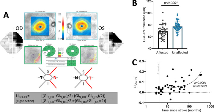
Figure 1.
A: Computation of LI for the GCL-IPL using the nasal (N) and temporal (T) sector GCL-IPL (GI) thickness values of both eyes, excluding superior (S) and inferior (I) sectors since they overlapped the vertical meridian. B: Plot comparing GCL-IPL thicknesses in the affected or unaffected hemiretinas (paired t-test, CI95 =5.212 to 10.60, t42 =5.921, p = <0.0001). C: Plot of LIGCL-IPL against time since stroke (linear regression, R2=0.2703, CI95(y-intercept)=0.018 to 0.057, p=0.0004).

