Endoscopic submucosal dissection (ESD) is recommended for colorectal tumors that cannot be completely removed by snare-based techniques. Endoscopic resection for appendiceal orifice lesions (Toyonaga’s type 3: lesion entirely covers the orifice) is technically challenging because the appendiceal portion cannot be incised accurately 1 . Such lesions can be endoscopically removed if the patient has undergone an appendectomy, but submucosal fibrosis is a potential issue. Several reports have shown that ESD using a traction device or underwater strategies are useful for such lesions 2 3 4 . We demonstrate ESD for a lesion using the water pressure method ( Video 1 ), which is a technique that uses the waterjet function of an endoscope to secure the working space 5 .
Endoscopic submucosal dissection using the water pressure method for a large cecal adenoma covering the appendiceal orifice after appendectomy.
Video 1
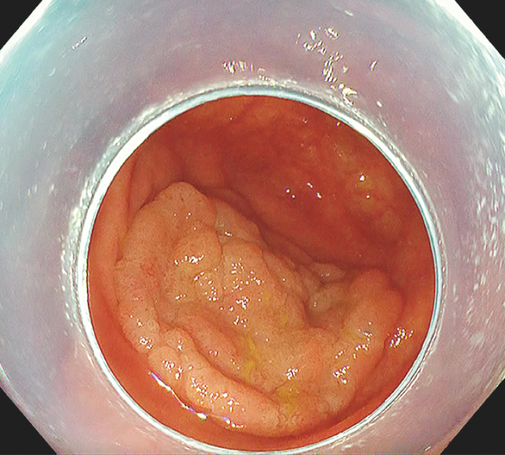
A 62-year-old woman, who had undergone appendectomy for acute appendicitis 45 years previously, was diagnosed with a 35-mm laterally spreading tumor in the cecum on colonoscopy ( Fig. 1 ). The scar of the appendiceal orifice could not be identified because the lesion was covering it. We performed ESD for the lesion with a FlushKnife BT-S (1.5 mm, DK2620J; Fujifilm Medical, Tokyo, Japan).
Fig. 1.
A 35-mm laterally spreading tumor covering the appendiceal orifice after appendectomy.
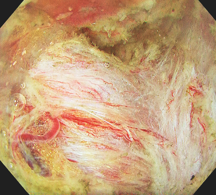
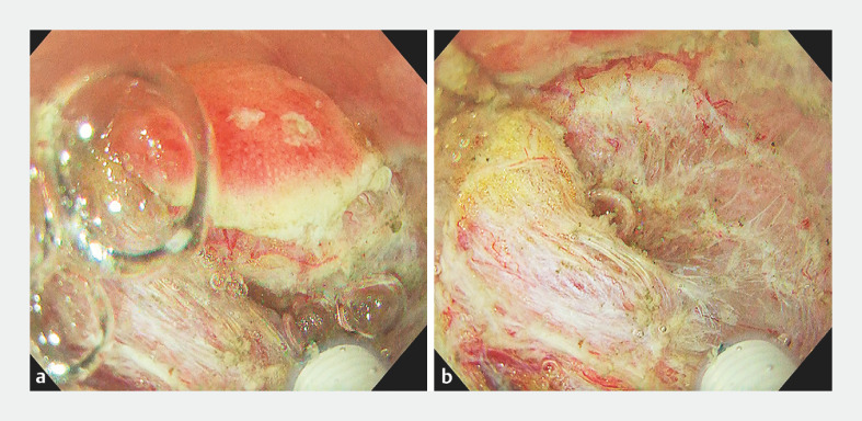
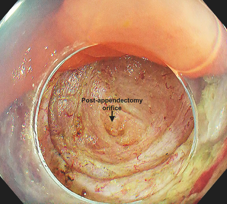
A solution of sodium alginate was injected into the submucosa for thickening. The endoscope faced the lesion perpendicularly, and endoscope stability was poor due to respiratory fluctuations. To secure a stable field of view, the water pressure method was used while sucking air from the cecum. Severe fibrosis was partly visible in the submucosa during ESD ( Fig. 2 ). En bloc resection was achieved while maintaining a good visual field using the water pressure method ( Fig. 3 , Fig. 4 ). The procedure was completed without any adverse events.
Fig. 2.
Severe fibrosis was partly visible in the submucosa during submucosal dissection.
Fig. 3.
The water pressure method. a Before water injection. b Just after water injection.
Fig. 4.
The appendiceal orifice was identifiable in the resection wound.
The patient was discharged on postoperative day 4. Histopathological examination revealed a high grade tubulovillous adenoma with negative margins.
A large cecal adenoma covering the appendiceal orifice after appendectomy could be removed by ESD with the water pressure method.
Endoscopy_UCTN_Code_TTT_1AQ_2AD
Acknowledgement
We would like to thank Editage (www.editage.jp) for editing and reviewing this manuscript in the English language.
Footnotes
Conflict of Interest T. Kanesaka has received lecture honoraria from Olympus Corporation, AI Medical Service Inc., and AstraZeneca. R. Ishihara has received lecture honoraria from Olympus Corporation, FUJIFILM Medical Co., Ltd., Daiichi Sankyo Co., Ltd., Miyaras Pharmaceutical Co., Ltd., AI Medical Service Inc., AstraZeneca, MSD, and Ono Pharmaceutical Co., Ltd. Y. Asada, T. Michida, H. Satomi, and K. Honma declare that they have no conflict of interest.
Endoscopy E-Videos https://eref.thieme.de/e-videos .
E-Videos is an open access online section of the journal Endoscopy , reporting on interesting cases and new techniques in gastroenterological endoscopy. All papers include a high-quality video and are published with a Creative Commons CC-BY license. Endoscopy E-Videos qualify for HINARI discounts and waivers and eligibility is automatically checked during the submission process. We grant 100% waivers to articles whose corresponding authors are based in Group A countries and 50% waivers to those who are based in Group B countries as classified by Research4Life (see: https://www.research4life.org/access/eligibility/ ). This section has its own submission website at https://mc.manuscriptcentral.com/e-videos .
References
- 1.Jacob H, Toyonaga T, Ohara Y et al. Endoscopic submucosal dissection of cecal lesions in proximity to the appendiceal orifice. Endoscopy. 2016;48:829–836. doi: 10.1055/s-0042-110396. [DOI] [PubMed] [Google Scholar]
- 2.Iacopini F, Gotoda T, Montagnese F et al. Underwater endoscopic submucosal dissection of a nonpolypoid superficial tumor spreading into the appendix. VideoGIE. 2017;2:82–84. doi: 10.1016/j.vgie.2017.01.007. [DOI] [PMC free article] [PubMed] [Google Scholar]
- 3.Oung B, Rivory J, Chabrun E et al. ESD with double clips and rubber band traction of neoplastic lesions developed in the appendiceal orifice is effective and safe. Endosc Int Open. 2020;8:E388–E395. doi: 10.1055/a-1072-4830. [DOI] [PMC free article] [PubMed] [Google Scholar]
- 4.Figueiredo M, Yzet C, Wallenhorst T et al. Endoscopic submucosal dissection of appendicular lesions is feasible and safe: a retrospective multicenter study (with video) Gastrointest Endosc. 2023;98:634–638. doi: 10.1016/j.gie.2023.06.024. [DOI] [PubMed] [Google Scholar]
- 5.Kato M, Takatori Y, Sasaki M et al. Water pressure method for duodenal endoscopic submucosal dissection (with video) Gastrointest Endosc. 2021;93:942–949. doi: 10.1016/j.gie.2020.08.018. [DOI] [PubMed] [Google Scholar]


