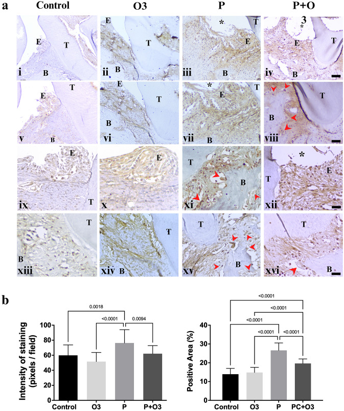
Fig. 7.
MMP-9 immunohistochemistry of murine periodontal tissues. a The representative images of the slides stained with the MMP-9 antibody. Microphotographs show that positive cells were mainly detected in periodontal ligament infiltrated with inflammatory cells near the oral epithelium (E), enlarged fibroblasts in the underlying connective tissue of the ligature area, and osteoclast-like cells (red arrows) near the bone (B). Osteoclast-like cells near the bone destruction showed stronger positive staining in the Periodontitis group (P) than in the Periodontitis group treated with Omega-3 (P + O3). Stronger staining was observed in the P group (Fig. 7iii, vii, xi, and xv); by contrast, mild or moderate staining was observed in the Periodontitis group treated with Omega-3 (P + O3) (Fig. 7iv, viii, xii and xvi), the Control (Fig. 7i, v, ix, xiii), and the Omega-3- supplemented diet group (Fig. 7ii, vi, x, and ixv). b Graphic representation of the staining intensity and the percentage of the total area that was positive in all the examined groups stained with the MMP-9 antibody. a (*) Ligature region, (T) tooth, (B) bone, (E) epithelium. (i–iv) magnification 10 × , scale bar 200 µm; (Fig. 7v–viii), magnification 20 × , scale bar 100 µm; (Fig. 7ix–xvi) magnification 40 × , scale bar 20 µm. b Control compared with the periodontitis-induced group; periodontitis-induced group compared with the treatment group, and the Omega-3-supplemented group. The bracket indicates the p value of the ordinary-one-way ANOVA test, the graphs represent the mean and standard error of the mean of 20 fields per group at 400 × magnification

