

Abstract
Intramedullary nailing is the gold standard for long bone fractures in the lower limbs. Fractures of the tibia with bending of the intramedullary nail is a very uncommon complication. Removing a bent nail is an unpredictable and challenging procedure for any trauma surgeon. We report the case of a 34-year-old man presenting a refractured tibia with a bent intramedullary nail following a motor vehicle accident. We describe the management of this patient as well as a substantial review of the literature to depict the peculiarities of this entity compared to its more commonly reported femoral counterpart. When approaching a deformed tibia nail, standard extraction techniques should be attempted and are more likely to be successful than in deformed femoral nails.
Keywords: Intramedullary nail, tibia, bent, removal, case report, review
Introduction
Modern intramedullary nailing procedure, as we know it today, has first been described by G. Küntscher during World War II (1939). Ever since, intramedullary nailing has become the gold standard for lower limbs long bones fracture fixation. 1 The first intramedullary devices were made of ivory, a material that proved to be inappropriate to the biomechanics of bone healing. 2 This eventually led to the experimentation of different metallic nails until the wide adoption of stainless-steel nails. However, with the popularization of these nails came some new complications such as fatigue failure and nail breakage. These complications are increasingly uncommon with modern nailing techniques and new nail designs. 3 Bending of the nail in situ following a new trauma with subsequent fracture is a rarely described complication, only reported as case reports in the literature. 4 Since the first case of bent tibia nail reported in 1996 by Yip et al., 5 the published cases were few and far in between. Because of the relatively small number of reported cases, there is no real consensus regarding the best surgical technique to adopt to be successful in treating this unusual condition. A multitude of techniques have been described in the literature. Based on the extensive review published by Kose et al. 6 in 2016, decisional flowcharts have been drawn to try to develop a standardized approach to this complication.
The authors report a case of a bent tibia intramedullary nail secondary to a new trauma with a review of the literature. The aim of this report is to focus on the tibial location of bent intramedullary nails, describing its peculiarities compared to its more commonly reported femoral counterpart.
Case report
A otherwise healthy male patient in his thirties presented to the emergency room following a motor vehicle accident (motorcyclist hit by a car) with the main complaint of an isolated painful and deformed lower leg. The patient’s medical history revealed a previous trauma to the same leg 19 months ago resulting in a midshaft tibia and fibula fracture that went through to full union after being treated in the same hospital by locked intramedullary nailing. On clinical examination, there was an obvious deformity of the right leg (Figure 1) highly suggestive of a broken tibia. However, surprisingly, there was little crepitus at the fracture site with the patient spontaneously moving his leg without much pain. The skin was intact, and there was no neurovascular fallout.
Figure 1.
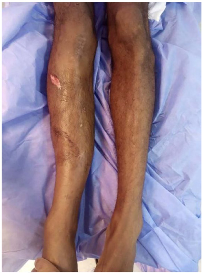
Valgus deformity of the right leg.
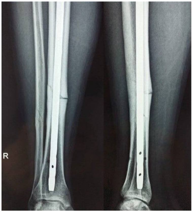
Radiologic assessment revealed an intramedullary tibial nail in situ. The nail was bent with a subsequent tibia and fibula fracture. An overall valgus deformity of approximately 25° in the frontal plane was measured on the antero-posterior view of the leg (Figure 2).
Figure 2.

Radiologic assessment showing a fractured tibia and fibula with the bent intramedullary nail in situ. An overall valgus deformity of approximately 25° in the frontal plane was measured on the antero-posterior view of the leg.
The decision to remove the nail and exchange it with a straight one was easily taken by the surgeons; however, the surgical procedure to follow was less agreed upon. After pre-operative planning, the decision was to try to straighten the nail in situ before removing it. In prevision of failure of this first plan, the surgeons have prepared industrial metal cutting drill bits that were kept sterile.
Under spinal anesthesia, with the patient in the supine position, a well-padded bolster was fixed to the edge of the operating table at the level of the apex of the deformity. A second support against the lateral aspect of the right knee was used to counter the reducing forces. A vigorous deforming force was applied on the lateral aspect of the ankle to try to straighten the bent nail (Figure 3). A substantial amount of force was necessary to reduce the uniplanar deformity of the leg. The procedure was controlled both clinically and fluoroscopically. Fortunately, the deformity could be reduced without, however being able to completely straighten the nail. Neurovascular examination following the reduction procedure was without abnormalities.
Figure 3.

A well-padded bolster was fixed to the edge of the operating table at the level of the apex of the deformity. A second support against the lateral aspect of the right knee was used to counter the reducing forces (yellow arrows). A vigorous deforming force was applied on the lateral aspect of the ankle to try to straighten the bent nail (white arrow).
After sterile draping of the right lower limb, a routine removal of the nail was carried on without major difficulties. A 9-mm diameter stainless steel tibial nail was removed followed by the insertion of a similar nail. After intramedullary nailing, the construct felt stable and no added locking screws were deemed necessary.
Control X-rays showed an acceptable reduction with a good alignment (Figure 4).
Figure 4.
Control X-rays showing an acceptable reduction with good alignment.
On examination of the removed nail, it was found that the deformity was brought down from 25° initially to 7° (Figure 5). Postoperative follow-up was uneventful with no skin necrosis. Fracture consolidation was obtained at 4 months after surgery.
Figure 5.

On examination of the removed nail, it was found that the deformity was brought down from 25° initially to 7° in the frontal plane (a), and there was no angulation in the sagittal plane (b).
Discussion
Removing a bent nail, be it in the tibia or in the femur, represents a real challenge for the orthopedic surgeon. It has been referred to as a “Man versus Metal” contest by some authors. 3 Unlike routine hardware removal, failing to remove a bent intramedullary nail is not an option.5,7 This complication has been scarcely reported. To the best of our knowledge and after a thorough review of the literature, about 32 cases of bent femoral nails have been published since the first case in 1970. On the other hand, there were only 10 cases of tibial localization of this complication with our case being the 11th.5–13 The reported cases are summarized in Table 1. Kose et al., 6 in 2016, published an extensive review of the literature based on 25 articles reporting femoral fractures and only five cases of tibia fractures with intramedullary nails in situ. Overall, they compiled six techniques to remove the bent nail, two of them having the advantage to preserve the fracture hematoma, whereas the remaining four necessitate exposing the fracture site with all the subsequent drawbacks. These techniques are: (1) simple extirpation of the nail, (2) in situ straightening of the nail using manual external maneuvers followed by classic nail removal, (3) weakening partially the nail, straightening, and then removal in one piece, (4) completely severing the nail and removal in two pieces, (5) corticotomy followed by removal of the nail, and (6) straightening the nail using a plate applied to the bone with bone forceps. In the most recent review of the literature published in 2020 by Dunleavy et al., 4 the success rate of closed removal of bent femoral nail (techniques 1 and 2 according to Kose et al.) was about 12.5% of all cases with a mean angulation of 36.2° (18° to 85°). However, our review of the literature focused on bent tibial intramedullary nails following a new trauma with subsequent tibia fracture found a success rate of closed extirpation of the nail either with or without prior manipulation of about 45% of the cases with a mean angulation of 33.8° (13°–60°). The difference in closed removal rates was statistically significant (p = 0.029), whereas the two groups were comparable in terms of angulation (p = 0.69). This is most probably due to the fact that intramedullary nails used for tibia fractures are usually thinner than those used to nail femur fractures. In fact, it has been shown that if the diameter of the nail increases by just 2 mm (as is generally the case in tibia and femoral nails: 9 mm versus 11 mm respectively), the nail resistance to the deforming forces is multiplied by a factor of 82%. 14 But the diameter is not the only parameter influencing the nail stiffness. In fact, the material of the nail determines its flexibility with stainless-steel nails being more resistant to straightening forces compared to the titanium nails. 11 McChesney et al. 11 succeeded to remove a bent intramedullary 8.3-mm hollow titanium tibial nail with standard extraction technique without any additional procedures despite an initial angulation of 38°. A deformity-based, broad therapeutic strategy for bent nail removal in both the tibia and femur was formulated by Kose et al. 6 However, following undiscerningly these guidelines may result in ineffective or harmful practices as a multitude of other factors can play an important role in the ease in which a nail can be removed such as the size of the nail, the material, the fracture comminution, and the orientation of the deformity.3,11 McChesney et al. 11 found that the direction of the curve is a more important factor to consider than its degree. He supports that apex-posterior angulated nails are much easier to remove and recommends rotating anteriorly angulated nails to facilitate extirpation. Yip et al. 5 reported two cases of bent tibia nails (13° and 26°) that were easily removed with standard technique even though they were apex-anteriorly angulated.
Table 1.
Review of the literature with a summary of the reported bent tibial intramedullary nails.
| Author | Year | Age, sex | Type of implant | Deformity | Removal technique | Equipment |
|---|---|---|---|---|---|---|
| Yip et al. 5 | 1996 | 23, M | Stainless steel | 26° apex-anterior ; 30° valgus | 1 | — |
| 34, M | Stainless steel | 13° apex-anterior ; 20° valgus | 1 | — | ||
| Kelsch et al. 8 | 2003 | 51, M | Stainless steel | 40° valgus | 4 | NR |
| Wierer et al. 9 | 2010 | 17, M | NR | 60° apex-anterior | 4 | Midas Rex |
| Aggerwal et al. 7 | 2011 | 30, M | Stainless steel | 26° varus; 28° apex-anterior | 3 | Metal cutting drill bit |
| Buunaaim et al. 10 | 2012 | 28, F | NR | 25° apex-posterior | 1 | — |
| Kose et al. 6 | 2016 | 39, M | Stainless steel | 32° valgus | 1 | — |
| McChesney et al. 11 | 2019 | 65, M | Titanium | 38° apex-posterior ; 7° valgus | 3 | Metal cutting drill bit |
| Pathak et al. 12 | 2019 | 45, M | Stainless steel | 42° apex-anterior | 4 | Metal cutting drill bit |
| Arif et al. 13 | 2023 | 18, M | Stainless steel | ?? | Osteotomy + 3 | Metal cutting drill bit |
| Our case | 2023 | 34, M | Stainless steel | 25° valgus | 2 | — |
M: male; F: female; NR: not reported.
Removal techniques: (1) standard extraction, (2) closed in situ nail straightening + extraction, (3) partial cutting of the nail + straightening + standard removal, and (4) full section of the nail + extraction in two pieces.
The previously reported bent tibial nail removals did not require any closed reduction of the deformity. In our case, and in accordance with the algorithm proposed by Kose et al., 6 we opted for a preliminary attempt to reduce the angulation of the nail curve to minimize the risks of a difficult hardware removal. To do so, a modification of the Patterson and Ramser technique 15 was improvised by the operating team resulting in a substantial decrease in the angulation from 25° to 7° rendering the nail removal a straightforward procedure. However, when using this technique, all measures should be taken to prevent further soft tissue damage (well-padded bolster, counter pressure at the knee to avoid ligament injury) and possible additional fracture lines.
If closed removal of the nail fails, then other techniques should be attempted such as weakening the nail using metal drill bits or metal cutters before straightening the nail and removing it in one piece, or completely cutting the nail and removing it in two pieces. Both techniques expose to soft tissue necrosis due to the heat generated and complications related to metal debris. These two techniques were used in 37.5% and 25% of femoral nails, respectively, and in 27.2% and 27.2% of tibial nails, respectively. These techniques can be difficult to apply in limited resources countries where sophisticated surgical metal cutting devices can be difficult to supply. In these circumstances, many authors have advised using regular metal-cutting hacksaw blade or drill-bits to section the nail before removal. 3
Conclusion
Removing a bent intramedullary nail can be a challenging situation for trauma surgeons where several scenarios should be prepared. When approaching a deformed tibia nail, standard extraction techniques should be attempted and are more likely to be successful than in deformed femoral nails. In all cases, a thorough knowledge of the nail’s attributes (material, thickness, design, etc.) is paramount before any attempt is made. Decisional algorithms should serve as a guide with a case-by-case approach. In low-income countries where sophisticated surgical metal cutting devices are not readily available, low-cost industrial devices can be a problem-solving alternative.
Acknowledgments
Not applicable.
Footnotes
Author contributions: All the authors participated in the design, performance, analysis and drafting of this manuscript.
The authors declared no potential conflicts of interest with respect to the research, authorship, and/or publication of this article.
Funding: The authors received no financial support for the research, authorship, and/or publication of this article.
Ethical approval: Ethical approval for the study was granted from the Ethics Committee at the MT Maamouri Hospital, Nabeul; Tunisia (Reference: 61/2023).
Informed consent: Written informed consent was obtained from the patient for the anonymized information to be published in this article.
ORCID iDs: Walid Bouaicha  https://orcid.org/0000-0003-4837-1954
Mohamed Jlidi
https://orcid.org/0000-0002-8862-2722
References
- 1. Wong Wei Kang N, Tan WPJ, Phua YMC, et al. Intramedullary nail: the past, present and the future—A review exploring where the future may lead us. Orthop Rev (Pavia) 2021; 13(2): 25546. [DOI] [PMC free article] [PubMed] [Google Scholar]
- 2. Meccariello L, Bisaccia M, Caraffa A, et al. From the down to modern era: the history of the nailing. Can Open Orthop Traumatol J 2016; 3: 10–17. [Google Scholar]
- 3. Mencia MM, Moonsie R. Removing a bent femoral nail—man versus metal: a case report. Int J Surg Case Rep 2022; 99: 107679. [DOI] [PMC free article] [PubMed] [Google Scholar]
- 4. Dunleavy ML, Burton A, Reid JS, et al. Surgical management of angulated femoral intramedullary nails associated with closed fractures: a systematic review of the literature. J Orthop 2020; 21: 314–320. [DOI] [PMC free article] [PubMed] [Google Scholar]
- 5. Yip KM, Leung KS. Treatment of deformed tibial intramedullary nail: report of two cases. J Orthop Trauma 1996; 10(8): 580–583. [DOI] [PubMed] [Google Scholar]
- 6. Kose O, Guler F, Kilicaslan OF, et al. Removal of a bent intramedullary nail in lower extremity: report of two cases and review of removal techniques. Arch Orthop Trauma Surg 2016; 136(2): 195–202. [DOI] [PubMed] [Google Scholar]
- 7. Aggerwal S, Soni A, Saini UC, et al. Removal of a bent tibial intramedullary nail: a rare case report and review of the literature. Chin J Traumatol 2011; 14(2): 107–110. [PubMed] [Google Scholar]
- 8. Kelsch G, Kelsch R, Ulrich C. Unreamed tibia nail (UTN) bending: case report and problem solution. Arch Orthop Trauma Surg 2003; 123(10): 558–562. [DOI] [PubMed] [Google Scholar]
- 9. Wierer M, Biberthaler P, Mutschler W, et al. Verbogener intramedullärer Tibiamarknagel nach erneutem Trauma. Fallbeschreibung und Literaturübersicht [Removal of a bent intramedullary tibia nail. Case report and review of literature]. Unfallchirurg 2011; 114(7): 629–633. [DOI] [PubMed] [Google Scholar]
- 10. Buunaaim A, Sekimpi P. Anatomic approach to the removal of a bent intramedullary nail in a refractured tibia minimising soft tissue and bone injury: a rare case report. Intern J Orthop Surg 2012; 19(2):1–5. [Google Scholar]
- 11. McChesney GR, Gurbani BN, Hagedorn JC, 2nd. Extraction of a bent tibial nail after refracture: a case report. JBJS Case Connect 2019; 9(3): e0385. [DOI] [PubMed] [Google Scholar]
- 12. Pathak SK, Gautam RK, Anjum R, et al. Bent tibial nail with refracture: a cost-effective removal method: a case report. JBJS Case Connect 2020; 10(2): e19.00542. [DOI] [PubMed] [Google Scholar]
- 13. Arif H, Molina S, LeBrun C. Removal of a bent tibial intramedullary nail through osteotomy and partial sectioning: a case report. JBJS Case Connect 2023; 13(3). [DOI] [PubMed] [Google Scholar]
- 14. Bong MR, Kummer FJ, Koval KJ, et al. Intramedullary nailing of the lower extremity: biomechanics and biology. J Am Acad Orthop Surg 2007; 15(2): 97–106. [DOI] [PubMed] [Google Scholar]
- 15. Patterson RH, Ramser JR., Jr. Technique for treatment of a bent Russell-Taylor femoral nail. J Orthop Trauma 1991; 5(4): 506–508. [DOI] [PubMed] [Google Scholar]