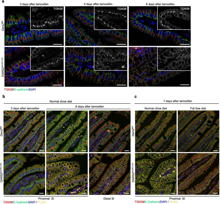
Extended Data Fig. 10. DARS2 depletion causes gradual Golgi disorganisation in proximal enterocytes that precedes LD formation and requires the presence of fat in the diet.
a, Representative fluorescence microscopy images from the proximal SI of 8-12-week-old Dars2fl/fl (n = 6) and DARS2tamIEC-KO (n = 6) mice sacrificed 3, 5 and 8 days upon the last tamoxifen injection and immunostained with antibodies against TGN38 (red) and E-cadherin (green). Insets shows only TGN38 staining in white. b, Representative fluorescence microscopy images from the proximal and distal SI of 8-12-week-old Dars2fl/fl (n = 6) and DARS2tamIEC-KO (n = 6) mice fed with NCD sacrificed 3 and 8 days upon the last tamoxifen injection and immunostained with antibodies against TGN38 (red), E-cadherin (green) and PLIN2 (yellow). c, Representative fluorescence microscopy images from the proximal SI of 8-12-week-old Dars2fl/fl (n = 6) and DARS2tamIEC-KO (n = 6) mice under NCD and FFD sacrificed 7 days upon the last tamoxifen injection and immunostained with antibodies against TGN38 (red), E-cadherin (green) and PLIN2 (yellow). Nuclei stained with DAPI (blue). Scale bars, 50 μm. Confocal images shown are representative of the number of mice analysed as indicated in Supplementary Table 4.

