Abstract
Background:
Ecthyma contagiosum, sometimes referred to as human orf, is a zoonotic disease caused by the orf virus that is mostly acquired by coming into contact with diseased animals such as sheep or goats. The orf virus, a DNA virus belonging to the Poxviridae family, infects epidermal keratinocytes via breaking down the skin barrier, which can be caused by burns or wounds. The accompanying characteristic skin lesions can take on a range of morphologies depending on the infection’s stage; lesions that are crusted, papillomatous, maculopapular, targetoid, and nodular can occur before clearing up. In addition to the lips and corners of the mouth, infected animals may also have lesions on the neck, vulva, and teeth. Skin sores caused by Ecthyma contagiousum discharge the orf virus into the environment.
Case presentation:
A 4-month-old male infant with no medical history brought himself to the dermatology clinic with a minor fever and a skin lesion on his nose. An orf virus infection was discovered in the newborn through blood culture and PCR testing. For a subsequent infection, the patient received fusidic acid cream, an antibiotic, and an antipyretic. Following a follow-up of 3 months, the lesion vanished entirely.
Conclusion:
Rarely, as in our instance, are orf nodules seen somewhere else than the hands. In order to appropriately treat a patient without fear, clinicians should keep this in mind, especially if they come up with a history similar to that of our patient.
Keywords: antibiotics, fucidin, nose, orf
Inrtoduction
Highlights
Ecthyma contagiosum, sometimes referred to as human orf, is a zoonotic disease caused by the orf virus that is mostly acquired by coming into contact with diseased animals such as sheep or goats.
Ecthyma contagiosum skin lesions release the virus into the environment, which can lead to animal-to-animal or animal-to-human transmission. Rarely occurs human-to-human transfer.
Depending on the stage of the infection, the accompanying distinctive skin lesions take on a variety of morphologies.
Ecthyma contagiosum, sometimes referred to as human orf, is a zoonotic disease caused by the orf virus that is mostly acquired by coming into contact with diseased animals such as sheep or goats. A DNA virus from the Poxviridae family called the orf virus infects epidermal keratinocytes by disrupting the skin barrier (such as through wounds or burns)1. Depending on the stage of the infection, the accompanying distinctive skin lesions take on a variety of morphologies, with maculopapular, targetoid, nodular, papillomatous, and crusted lesions developing before clearance. Lesions can occur on the neck, vulva, and tongue in addition to the lips and mouth corners in infected animals2. Ecthyma contagiousum skin lesions release the virus into the environment, which can lead to animal-to-animal or animal-to-human transmission. Rarely occurs human-to-human transfer3. Although there have been cases of infections on the face or perianal region, human infections are mostly restricted to sores on the arms and hands. Additionally, the patient may develop lymphadenopathy and a fever. In the human body, orf lesions appear in a distinct pattern. It is possible for the illness to start off as an erythematous maculopapular lesion before developing into a target-shaped lesion with a red center. It might appear as a dry lesion with black spots during the regeneration stage, turn papillomatous, and develop a dry crust during the next stage4. The presence of clinical lesions, history of interaction with infected animals, and viral cultures are used to make the diagnosis. The illness often has a sense of self-limitation5. Topical antibiotics are used as supportive therapy, and systemic antibiotics may also be used if the wound is bacterially infected. In 2–3 weeks, patients often recover6. Surgery may be necessary for larger lesions. Additionally, recommended treatments include ribavirin and imiquimod2. We present a case of a 4-month-old male baby with an orf lesion on his nose, which makes it a very rare case.
Presentation of case
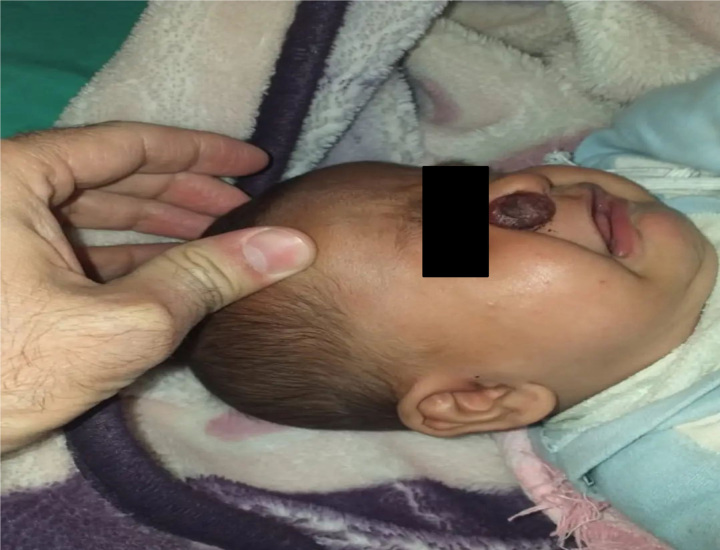
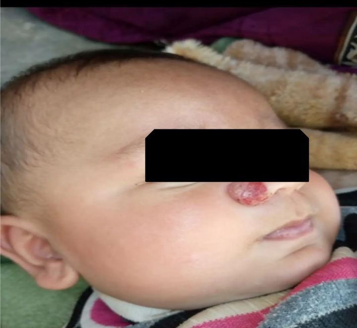
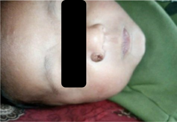
A 4-month-old boy with no medical history was brought by his parents to the dermatology clinic with a mild fever and a skin lesion on his nose (Fig. 1). It was a big red nodule, perhaps one centimeter in size. The baby’s overall health was somehow good, and no lymphadenopathy was noticed. While the parents were being questioned, it was discovered that his parents kept animals in the same home and that a sheep had approached the sleeping child, petting and sniffing him. That lesion on the youngster manifested itself after a week. An orf virus infection was discovered in the newborn through blood culture and PCR testing. For a subsequent infection, the patient received fusidic acid cream, an antibiotic, and an antipyretic. Two weeks following the initiation of therapy, the lesion improved a lot (Fig. 2), and after a month of follow-up and continued symptomatic treatment, the lesion improved further and became smaller (Fig. 3). After 3 months of follow-up, the lesion completely disappeared. The diagnosis of orf was confirmed by following up on the case, and no biopsy was needed.
Figure 1.
The nodule on the nose when the patient presented the the dermatology clinic.
Figure 2.
The nodule after a week of the treatment.
Figure 3.
The nodule after a month of follow-up and treatment.
Discussion
The typical manifestation of an orf infection is a single, limited 2–3-cm lesion that progresses into six different clinical stages, each lasting around a week: (1) maculopapular stage: an elevated red macule forms; (2) targetoid stage: the red core of the lesion is surrounded by a white ring and an outside red halo; (3) acute stage: a red, weepy nodule forms; (4) stage of regeneration: the lesion dries up and black, pyknotic, cell-filled patches start to show up on the surface. (5) The lesion may go through two stages: the papillomatous stage, in which the lesion may resemble an enlarged wart with dry, verrucous papillations on its surface; and the regressive stage (6), in which the lesion flattens and a dry crust eventually forms and peels off6. Lesions are most commonly detected in the hands, but they have also been reported in the face and perianal area7. A few known cases of multiple lesions, which are uncommon in immunocompetent individuals, include malaise, fever, lymphangitis, and lymphadenitis. Well-defined nodules with a core crust, structureless white areas, and sparkling white streaks are visible during dermoscopy of orf lesions. Around lesions, there are hairpin veins, dotted lines, and a fine peripheral scale8. The orf illness reached its climax in New Zealand in 1983 and in Norway in 1975. A study found that among 15 012 individuals over 20 who attended an Iranian dermatology clinic between 1991 and 1996, the prevalence of orf was 0.4%. A patient with a history of cutting meat with an infected knife developed orf disease. Five cases of orf disease in children have been reported over a 10-year period in agricultural households in western Ireland9. The disease is primarily found in goats, sheep, and cows, and humans contract it through contact with infected animals. The double-standard DNA virus that causes orf disease is a member of the parapox family10. Human orf disease is primarily caused by occupational hazards and typically affects veterinarians, abattoir workers, and farm workers11,12. The illness often manifests in those who interact with animals as isolated lesions that resemble tiny papules on the hand, finger, or forearm7. In reference to our instance, the patient acquired the infection from the sheep as he slept in his yard. On his face, the lesion was developing next to his nostril. The history of interaction with infected animals, the appearance of clinical lesions, and viral cultures are used to make the diagnosis. In our instance, there were several differential diagnoses, including misnomer, leishmaniasis, and hemangioma. However, an orf virus infection was discovered in the newborn through blood culture and PCR testing. Usually, orf illness is self-limited. In addition to topical antibiotics for supportive care, systemic antibiotics may also be used if the wound is bacterially infected. Most patients recover in 2–3 weeks. Occasionally, larger lesions require surgery6. Ribavirin and imiquimode therapy have also been proposed2. Since the wound was contaminated by microorganisms, our patient was treated with fusidic acid cream, an antibiotic, and an antipyretic. The lesion lessened 2 weeks after the medicine was started. The lesion disappeared completely after 3 months, which supported the diagnosis.
Conclusion
Ecthyma contagiosum, often known as human orf, is a zoonotic illness mostly contracted via contact with sick animals like sheep or goats. It is caused by the orf virus. Burns or other wounds can cause the orf virus, a DNA virus that is a member of the Poxviridae family, to breach the skin barrier and infect epidermal keratinocytes. Depending on the stage of the infection, the accompanying distinctive skin lesions can have a variety of morphologies, including crusted, papillomatous, maculopapular, targetoid, and nodular lesions that may appear before clearing up. Animals with the infection may also have lesions on their necks, vulva, and teeth, in addition to the lips and corners of their mouths. Orf nodules can rarely be located anywhere other than the hands, as in our case. Therefore, doctors should keep such a place in their minds, especially if they come up with a history like our patient, so that they can treat it properly without fear.
Ethical approval
It is not applicable because all dates belong to the authors of this article.
Consent
Parental consent for minors: Written informed consent was obtained from the patient’s parents/legal guardian for publication and any accompanying images. A copy of the written consent is available for review by the Editor-in-Chief of this journal on request.
Sources of funding
None.
Author contribution
M.S.: wrote most of the manuscript, performed data analysis or interpretation, and designed the study; B.S., H.Als., M.A., M.Alz., M.K., and A.A.: wrote a part of the manuscript; H.A.: wrote a part of the manuscript and designed the study. All authors reviewed the final manuscript.
Conflicts of interest disclosure
There are no conflicts of interest.
Research registration unique identifying number (UIN)
None.
Guarantor
Not applicable. All data belongs to the authors.
Data availability statement
None.
Provenance and peer review
Not commissioned, externally peer-reviewed.
Assistance with the study
None.
Methods
The work has been reported in line with the SCARE criteria.
Acknowledgements
None.
Footnotes
Sponsorships or competing interests that may be relevant to content are disclosed at the end of this article.
Published online 2 December 2023
Contributor Information
Mouhammed Sleiay, Email: www.abdmouh1234mouhmouh@gmail.com.
Mohammed Alqreea, Email: abokhaledqreea@gmail.com.
Hadi Alabdullah, Email: dr.alabdullah.md@gmail.com.
Ahmad Almohamed, Email: ahmad.al.mhamd.715850@gmail.com.
Hasan Alsmoudi, Email: adda85517@gmail.com.
Mulham Al-Zahran, Email: mulhamz2000@gmail.com.
Mouhammed Rabee Katth, Email: rabiekatta@gmail.com.
Bilal Sleiay, Email: Bilalsleiay123@gmail.com.
References
- 1.Vellucci A, Manolas M, Jin S, et al. Orf virus infection after Eid al-Adha. IDCases 2020;30:21–854. [DOI] [PMC free article] [PubMed] [Google Scholar]
- 2.Alinejad F, Momeni M, Keyvani H, et al. Introduction to a case of orf disease in a burn wound at Motahari Hospital. Ann Burns Fire Disasters 2018;31:243–245. [PMC free article] [PubMed] [Google Scholar]
- 3.Haddock ES, Cheng CE, Bradley JS, et al. Extensive orf infection in a toddler with associated id reaction. Pediatr Dermatol 2017;34:337–340. [DOI] [PMC free article] [PubMed] [Google Scholar]
- 4.Becher P, König M, Müller G, et al. Characterization of sealpox virus, a separate member of the parapoxviruses. Arch Virol 2002;147:1133–1140. [DOI] [PubMed] [Google Scholar]
- 5.Uzel M, Sasmaz S, Bakaris S, et al. A viral infection of the hand commonly seen after the feast of sacrifice: human orf (orf of the hand). Epidemiol Infect 2005;133:653–657. [DOI] [PMC free article] [PubMed] [Google Scholar]
- 6.Parker S, Handley L, Buller RM. Therapeutic and prophylactic drugs to treat orthopoxvirus infections. Future Virol 2008;3:595–612. [DOI] [PMC free article] [PubMed] [Google Scholar]
- 7.Shirazi MR, Pedram N. Orf: report of eleven cases in five Iranian families. Iran J Clin Infect Dis 2007;2:83–85. [Google Scholar]
- 8.Bergqvist C, Kurban M, Abbas O. Orf virus infection. Rev Med Virol 2017;27:10–1002. [DOI] [PubMed] [Google Scholar]
- 9.Alinejad F, Momeni M, Keyvani H, et al. Introduction to a case of orf disease in a burn wound at Motahari Hospital. Ann Burns Fire Disasters 2018;31:243–245. [PMC free article] [PubMed] [Google Scholar]
- 10.Mavridou K, Bakola M. Orf (ecthyma contagiosum). Pan Afr Med J 2021;1:38–322. [DOI] [PMC free article] [PubMed] [Google Scholar]
- 11.Johannessen JV, Krogh HK, Solberg I, et al. Human orf. J Cutan Pathol 1975;2:265–283. [DOI] [PubMed] [Google Scholar]
- 12.Shahmoradi Z, Abtahi-Naeini B, Pourazizi M, et al. Orf disease following “Eid ul-Adha”: a rare cause of erythema multiforme. Int J Prev Med 2014;5:912–914. [PMC free article] [PubMed] [Google Scholar]
Associated Data
This section collects any data citations, data availability statements, or supplementary materials included in this article.
Data Availability Statement
None.


