Abstract
We have investigated the capacity of live attenuated Shigella flexneri strains to act as vectors for the induction of local and systemic antibody responses against heterologous epitopes. The S. flexneri IpaC antigen was selected as a carrier protein into which the C3 neutralizing epitope of the poliovirus VP1 protein was inserted in eight sites distributed along IpaC. The resulting IpaC-C3 hybrid proteins were expressed from recombinant plasmids in the S. flexneri 2a vaccine candidate, SC602. Their production was similar to that of wild-type IpaC. All of the hybrid proteins but one were secreted as efficiently as wild-type IpaC. Immunization of mice with each of the recombinant SC602 derivatives reveals that one construct is able to induce serum and local anti-C3 antibodies, showing that at least one permissive site of insertion within IpaC can be defined. Furthermore, mouse-to-mouse variability in the anti-C3 response indicates that the amount of hybrid proteins produced in the host by SC602 should be improved for optimal use of S. flexneri live attenuated strains as mucosal vectors for foreign epitopes.
Live attenuated vectors are one of the most efficient delivery systems for stimulation of the mucosa-associated immune system (for a review, see reference 10). They have therefore been extensively used to express foreign antigens and epitopes selected from pathogens against which the induction of a local immune response is required for protection (for a review, see reference 23). Usually, foreign epitopes are inserted within a carrier protein that is expressed in the live vector.
Shigella flexneri live attenuated strains have been developed as candidates for vaccines against shigellosis, an invasive disease of the human colon (22). The capacity of such strains to act as mucosal vectors has been recently reported (16). Local and systemic antibody responses to fimbriae and CS3 fibrillae of enterotoxigenic Escherichia coli were generated in guinea pigs or mice following immunization with these antigens expressed in CVD1203, an S. flexneri 2a live attenuated strain that confers protection against Shigella keratoconjunctivitis in the guinea pig model (15).
The purpose of the present study was to investigate whether S. flexneri vaccine strains could be used as immunization vectors to express heterologous epitopes of eukaryotic origin and, in turn, elicit local and systemic antibody responses to foreign sequences. The IpaC antigen, previously reported as a potential carrier protein (1), was selected for the insertion of the neutralizing C3 epitope of the VP1 protein of poliovirus (26). This 11-residue-long sequence has been previously used as a reporter epitope (5, 25). As immunogenicity of a B-cell epitope depends on its flanking sequences within the hybrid protein (25), the C3 epitope was inserted into eight different sites within the IpaC coding sequence. SC602, an S. flexneri 2a vaccine strain attenuated both in its capacity to move intra- and intercellularly and in its survival in tissues (1), was used as vector. This strain is safe and protective in the macaque monkey model (8) as well as in human volunteers (9). The IpaC-C3 hybrid proteins were expressed from recombinant plasmids within SC602 to retain the functionality of the wild-type (wt) IpaC, thus maintaining the invasiveness of the live attenuated vector and ensuring efficient stimulation of local immunity. Immunogenicity of the IpaC-C3 proteins expressed in SC602 was assessed following an immunization protocol that allows the induction of local and systemic anti-IpaC antibody responses in mice (1).
MATERIALS AND METHODS
Bacterial strains and growth media.
E. coli MC1061, GM48, and ZK501 (21) were used for construction of plasmids and for demethylation of StuI and BclI restriction sites, respectively. The S. flexneri 2a derivative SC602 [ΔicsA Δ(iuc iut)] (1) was used for expression of the hybrid proteins, and the S. flexneri 5 derivative SF635 [Δ(ipaB ipaC ipaD ipaA)] (19) was used to recover high amounts of hybrid proteins in the culture supernatant. Bacteria were grown in Trypticase casein soy broth supplemented with ampicillin (100 μg/ml) when necessary.
Construction of hybrid proteins.
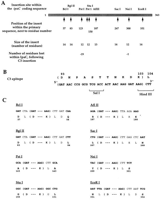
Synthetic oligonucleotides corresponding to the C3 coding sequence were inserted within each of the selected ipaC restriction sites. Plasmid pC1, a pUC derivative containing the ipaC gene (14), was used for C3 insertion within the StuI and AflII sites. Plasmid pC2, generated by removing the EcoRI site present in the pC1 polylinker sequence, was used for C3 insertion within EcoRI site as well as the BclI, PstI, SacI, and NsiI sites. Finally, plasmid pC3, constructed by removing the BglII site located upstream from the IpaC coding sequence in pC2, was used for C3 insertion within the BglII site. The resulting plasmids were designated pC57, pC83, pC123, pC150, pC187, pC267, pC300, and pC351, with the number indicating the position of the insert within the IpaC sequence (Fig. 1A). For the corresponding hybrid proteins, “pC” was replaced by “IpaC”. Transformations were performed as previously described (21).
FIG. 1.
Construction of IpaC-C3 hybrid proteins. (A) C3 insertion sites (the restriction site used for cloning and the resulting position of C3 within the IpaC sequence are indicated by arrows), size of the insert, and modification of IpaC sequence resulting from genetic manipulation. (B) Amino acid residues and corresponding DNA sequence of the C3 epitope. (C) Amino acid residues and corresponding DNA sequences of IpaC-C3 hybrid proteins for each insertion site, indicated as boldface characters inside parentheses for C3, boldface characters outside parentheses for IpaC, and lightface characters for additional sequences resulting from cloning. IpaC residue numbers are indicated.
Protein analysis.
Proteins present in whole-cell extracts and culture supernatants of SC602 derivatives were analyzed by sodium dodecyl sulfate-polyacrylamide gel electrophoresis (SDS-PAGE) and immunoblotting. Those present in culture supernatants of SF635 derivatives were analyzed by SDS-PAGE and enzyme-linked immunosorbent assay (ELISA). Culture dilutions were performed considering that for an optical density at 600 nm (OD600) of 1, the bacterial concentrations were 5 × 10⁸ and 3 × 10⁸ CFU/ml for S. flexneri and E. coli, respectively. Exponential-phase (OD600 = 1.5) or stationary-phase (OD600 = 7) bacterial cultures were harvested, and whole-cell bacterial extracts were obtained after solubilization of bacterial pellets with Laemmli buffer (11). Supernatants from exponential-growth cultures of SC602 derivatives were recovered, filtered, and concentrated 100-fold in Laemmli buffer as already described (2). Supernatants of SF635 derivatives were prepared similarly and were used without being concentrated for ELISA and diluted twice in Laemmli buffer for SDS-PAGE analysis.
SDS-PAGE (10% gel) was performed with either bacteria (3 × 10⁷/well), concentrated culture supernatant from SC602 derivatives (7 μl/well), or supernatant from SF635 derivatives (20 μl/well). Proteins were either stained with Coomassie brilliant blue or transferred onto nitrocellulose membranes. After transfer, IpaC and IpaC-C3 hybrid proteins were detected with the anti-IpaC monoclonal antibody (MAb) K24 (2 μg/ml) (20) or with an anti-C3 mouse polyclonal serum (diluted 1/1,000) kindly given by C. Leclerc, Institut Pasteur, Paris, France. Horseradish peroxidase-labeled goat anti-mouse antibodies were used as secondary antibodies and were visualized by enhanced chemiluminescence (Amersham International, Buckinghamshire, England).
For epitope accessibility studies of native hybrid proteins, ELISA was performed on SF635 derivative culture supernatants as follows. Undiluted and serial twofold dilutions of culture supernatants were used for coating ELISA microtiter plates. The reactivity of each hybrid protein with the anti-C3 MAb (dilution at use of ascitic fluid, 1/300; a kind gift of R. Crainic, Institut Pasteur, Paris, France) and the anti-IpaC K24 and J22 MAbs (3 μg/ml) (20) was determined by using alkaline phosphatase-labeled goat anti-mouse antibodies (dilution at use, 1:4,000; Sigma) as secondary antibodies. The titer was defined as the last dilution of supernatant leading to an OD405 twice that of the negative control. The amount of hybrid proteins in culture supernatants was also evaluated by SDS-PAGE.
Immunization of mice and analysis of the antibody responses.
Seven-week-old BALB/c female mice (Janvier, Le Genest, France) were immunized intravenously (i.v.) with formaldehyde-killed SC602 bacteria (at stationary phase) expressing each of the IpaC-C3 proteins (108 CFU/mouse, three times at 15-day intervals). Intranasal immunizations with live bacteria (5 × 10⁶ CFU/mouse, three injections at 15-day intervals) were performed beginning 15 days after the last i.v. immunization. Wild-type SC602 and SC602(pC1) were used as controls. Ten mice were used per group, and experiments were repeated three times. Immunizations with the IpaC300 protein, purified by using a protocol previously described for the purification of IpaC (7), were performed as follows. Nineteen 7-week-old BALB/c female mice were immunized subcutaneously with 25 μg of IpaC300 or IpaC (used as a control) in the presence of incomplete Freund’s adjuvant, followed by two i.v. immunizations without adjuvant at 15-day intervals.
Sera were collected from the tail artery, and bronchoalveolar washes were obtained by injecting 1 ml of sterile 0.9% NaCl, twice intratracheally, in mice sacrificed by cervical dislocation. A total volume of 1.5 ml was obtained per mouse. Samples were stored at −20°C until tested.
Anti-C3 and anti-IpaC IgG antibody responses were analyzed by ELISA using as antigens the C3 peptide (12) (100 ng/well) and purified IpaC (20 ng/well), respectively. The C3 peptide consists of amino acid residues 95 to 104 of capsid protein VP1 of poliovirus type 1, flanked by additional Tyr-Gly-Cys-Gly residues at the N terminus and by the Gly-Cys residues at the C terminus. Antibody titers were defined as the last dilution of samples leading to an OD405 twice that of the negative control. In vitro poliovirus neutralization tests using pooled sera were performed as previously described (25).
RESULTS AND DISCUSSION
Construction of the hybrid proteins.
Hybrid ipaC-C3 genes were constructed by inserting synthetic oligonucleotides corresponding to the C3 coding sequence within each of the following ipaC restriction sites: BclI, BglII, PstI, StuI, AflII, SacI, NsiI, and EcoRI (Fig. 1A). All of the C3-encoding oligonucleotides, except those used for cloning within the StuI and AflII sites, were designed with a HindIII restriction site located at the 3′ end (Fig. 1B). Therefore, the size of the inserted segment slightly exceeds the size of C3 coding sequence as a result of genetic manipulations and introduction of an additional leucine codon at the end of the C3 coding sequence. This codon is present in all constructs except that in which the C3 epitope was inserted within the StuI site (Fig. 1C). All of the hybrid proteins contain the entire sequence of IpaC except for IpaC123, which lacks residues 124 to 142, and IpaC300, which lacks residue 300 (Fig. 1A).
Production and secretion of IpaC-C3 proteins in SC602.
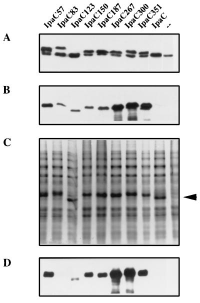
The production and stability of the IpaC-C3 proteins expressed within the vaccine candidate strain SC602 during exponential growth, in stationary phase, and after formaldehyde killing were analyzed by SDS-PAGE (11) and immunoblotting of whole bacterial extracts as described in Materials and Methods. All hybrid proteins were produced at a level comparable to that of wt IpaC expressed in SC602(pC1) (Fig. 2A). Using the anti-IpaC MAb, we detected two bands that correspond to the hybrid protein (upper band) and to the wt IpaC (lower band). Since the molecular weights of IpaC123 and IpaC are almost identical, the two bands were not separated in SC602(pC123) whole bacterial extract. However, production of this recombinant protein was confirmed by using the anti-C3 serum (Fig. 2B). Comparison of the relative intensities of the signals obtained for each protein with the anti-IpaC and anti-C3 sera indicated that recognition of the C3 epitope within the IpaC-C3 hybrid proteins depends on the insertion site (Fig. 2B). Overproduction of the hybrid proteins from the recombinant plasmids led to their accumulation within bacteria harvested in stationary phase (Fig. 2C). Similarly, the hybrid proteins remained stable after formaldehyde treatment of the bacteria (data not shown).
FIG. 2.
Production of IpaC-C3 hybrid proteins in SC602. (A, B, and D) Detection of IpaC-C3 hybrid proteins in whole-cell extracts with the anti-IpaC K24 MAb (A) or anti-C3 polyclonal serum (B) and in culture supernatants with anti-C3 serum (D) by immunoblot analysis of exponentially grown cultures. (C) SDS-PAGE and Coomassie blue staining of whole-cell extracts of stationary-phase cultures after formaldehyde treatment. The arrowhead indicates the position of the hybrid proteins.
Secretion of the hybrid proteins was analyzed by SDS-PAGE and immunoblotting of proteins present in culture supernatants of bacteria in the exponential phase of growth and prepared as described in Materials and Methods. As shown in Fig. 2D, with the anti-C3 mouse polyclonal serum used for detection, all of the hybrid proteins except IpaC83 were secreted. By detection of the hybrid proteins with the K24 anti-IpaC MAb, we observed that their level of secretion was comparable to that of wt IpaC (data not shown). As previously reported for secretion of IpaC83 by an ipaC mutant (2), the low recovery of IpaC83 from the culture medium of the SC602 derivative was probably due to its inefficient secretion rather than to a decreased stability of the protein in the extracellular medium. The maintenance of IpaC secretion even after epitope insertion emphasizes the potential of IpaC as a carrier protein in Shigella vectors. Actually, production of the antigen in a soluble form is required for the induction of an efficient antibody response to a T-cell-dependent antigen expressed in a bacterial strain (12, 13).
Antigenicity of the IpaC-C3 hybrid proteins.
Western blot experiments performed with anti-C3 antibodies suggested that the hybrid proteins exhibited variable antigenicity with respect to the C3 epitope (Fig. 2). Therefore, we studied the accessibility of this epitope in comparison of two linear epitopes of IpaC, J22 and K24, which are located between residues 25 to 33 and 300 to 349, respectively. The accessibility of C3, J22, and K24 epitopes was assessed by ELISA as described in Materials and Methods, using each of the hybrid proteins for coating. To easily obtain large amounts of the hybrid proteins in the culture supernatant, we expressed each of them in S. flexneri SF635 (19). In this strain, in contrast to SC602, secretion of IpaC and consequently IpaC-C3 proteins is not controlled by IpaB and IpaD, which regulate the Mxi-Spa secretion machinery. Therefore, each hybrid protein, even IpaC83, which is not efficiently secreted in SC602, is found in the culture supernatant.
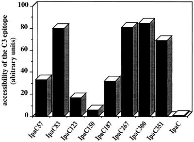
Coomassie blue staining following SDS-PAGE of proteins present in the culture supernatants revealed that for all of the hybrid proteins, anti-K24 and anti-J22 titers were both proportional to the amount of IpaC-C3 proteins present in culture supernatants (data not shown). This result indicated that the accessibility of both K24 and J22 epitopes was not affected by the C3 insertion. To take into account the variability which might arise from differences in the amount of hybrid protein, each anti-C3 titer was reported relative to the corresponding anti-K24 titer. As shown in Fig. 3, antigenicity of the C3 epitope within the native hybrid proteins exhibited high variability that correlated with recognition of the C3 epitope in immunoblot experiments (Fig. 2B). Insertion sites in the middle of IpaC (positions 123, 150, and 187) allowed minimal accessibility, while those located in the C-terminal third of IpaC were clearly recognized by anti-C3 MAb; IpaC300 appeared to be the most accessible construct. These results correlate with predictions derived from analysis of the IpaC hydropathy profile which suggest that the most antigenic sites (positions 87, 267, 300, and 351) are located within hydrophilic regions of IpaC (4), very close to or within domains containing B-cell epitopes, and nonantigenic sites are located within hydrophobic regions of IpaC containing no B-cell epitope (20, 24).
FIG. 3.
Antigenicity of C3 within IpaC-C3 hybrid proteins. Accessibility of the C3 epitope is expressed as arbitrary units corresponding to the ratio (anti-C3 antibody titer/anti-K24 antibody titer) × 100.
Identification of permissive insertion sites within IpaC that allow induction of anti-C3 humoral responses.
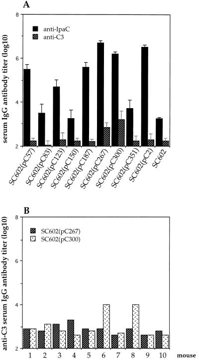
To identify insertion sites within IpaC that allow induction of a systemic anti-C3 antibody response, mice were immunized i.v. with killed (formaldehyde-treated) SC602 expressing each of the IpaC-C3 as described in Materials and Methods. SC602 and SC602(pC2) were used as controls. Antibody responses were assessed by ELISA (see Materials and Methods). The results are shown in Fig. 4. As expected, anti-IpaC antibodies were induced following immunization with each of the recombinant strains. The titer obtained following immunization with SC602(pC2) was higher than that obtained following immunization with SC602 (Fig. 4A). This result is consistent with the higher production of IpaC in SC602(pC2), in which IpaC is expressed from both the virulence plasmid and a recombinant plasmid (Fig. 2C). Five of the eight strains induced anti-IpaC antibody titers similar to that induced upon immunization with SC602(pC2). In contrast, anti-IpaC antibody titers raised upon immunization with SC602(pC83), SC602(pC150), and SC602(pC351) were similar to that of wild-type SC602. Differences in the secretion efficiency or in the amount of proteins could not account for the differences observed in the titers, as priming was achieved with killed bacteria, and all strains produced similar levels of hybrid proteins (Fig. 2A). Differences in the ability to induce anti-IpaC antibodies have also been observed when another foreign epitope was inserted within two different sites of IpaC (3). Since no major conformational modifications of IpaC could be detected following C3 insertion (2), these results suggest that insertions within a protein carrier may influence its own immunogenicity without significantly affecting its overall conformation.
FIG. 4.
Anti-C3 and anti-IpaC serum IgG antibody titers in mice immunized with SC602 producing IpaC-C3 hybrid proteins. (A) Mean antibody titers in serum samples are shown for each group of mice immunized with each of the IpaC-C3 hybrid proteins expressed in SC602. SC602(pC2) and SC602 were used as controls. SD is indicated. The results are representative of three experiments, each using 10 mice per group. (B) Individual anti-C3 serum IgG antibody titers in SC602(pC267)- and SC602(pC300)-immunized mice.
In contrast to the anti-IpaC response, serum anti-C3 antibodies were raised with only SC602(pC267) and SC602(pC300) (Fig. 4A). Interestingly, these two strains are among those inducing the highest anti-IpaC antibody titer. This finding is consistent with previous results demonstrating a correlation between the antibody titers to the carrier protein and to the foreign sequence. For instance, following immunization with MalE-C3 proteins either purified or expressed in a Salmonella vector, an anti-C3 antibody response is detected only if the anti-MalE antibody titer is at least 5 to 6 (log10) (17). In addition, the immunogenicity of C3 also depends on its accessibility within IpaC. As shown in Fig. 3, the accessibility of C3 is significantly reduced in IpaC57, -123, -150, and -187. In this case, whatever the level of the anti-IpaC response, no anti-C3 antibody response is detected (Fig. 4A). The highest accessibility of C3 is observed for IpaC83, -267, -300, and -351 (Fig. 3). Among these proteins, IpaC83 and -351 do not induce a high anti-IpaC antibody titer, and therefore no significant anti-C3 response is detected (Fig. 4A). Thus, only IpaC267 and -300, in which C3 is accessible and which induce a high anti-IpaC antibody titer, elicit an anti-C3 antibody response. It is noteworthy that considerable mouse-to-mouse variability in the antibody response to C3 was observed (Fig. 4B). Only 20 to 40% of mice immunized with SC602(pC267) or SC602(pC300) responded to C3, while all of the immunized mice elicited comparable anti-IpaC antibody responses.
To assess the utility of SC602 as a mucosal vector, we next studied the local antibody response to the foreign epitope expressed in this strain. We have previously established that a local anti-IpaC antibody response occurs after local boosting only in mice that mount a high serum anti-IpaC antibody response upon systemic immunization (1). We also show, in the present work, that the induction of anti-C3 antibodies is related to the anti-IpaC antibody titer. Therefore, only the two groups of mice, immunized with SC602(pC267) and SC602(pC300), that mounted significant antibody responses were boosted intranasally (see Materials and Methods). The local response was analyzed by ELISA of bronchoalveolar washes. Local immunoglobulin A (IgA) and IgG responses to C3 were induced in mice boosted with SC602(pC300) but not SC602(pC267). In IpaC300-immunized mice, only those previously eliciting systemic anti-C3 antibodies responded, with anti-C3 IgA and IgG antibody titers (log10) that varied from 1.6 to 2 and from 1.2 to 1.8, respectively. Local anti-IpaC IgA and IgG antibody responses were similar for both groups, with antibody titers (log10) of 1.8 and 2.8, respectively.
Altogether, these data show that IpaC contains at least one permissive site of insertion for foreign sequences that allows the induction of local and systemic antibody responses to the inserted sequence. Interestingly, this site allows insertion of 45- and 75-residue-long sequences without significantly affecting the stability of the resulting proteins (3). SC602 seems to be a potential mucosal vector for heterologous epitopes. However, the mouse-to-mouse variability in the response to C3 remains a limitation, reminiscent of that observed in the case of immunization with MalE-C3 proteins expressed in a Salmonella attenuated strain: some mice failed to respond, while others mounted high anti-C3 antibody titers (18). This failure has been observed for many different antigens expressed in Salmonella attenuated strains used as vectors (for a review, see reference 6). The main difficulty is to obtain stable expression of the heterologous epitope or antigen at a level which is sufficient to elicit a protective immune response in the host. Strategies have been developed to increase both the plasmid stability and the level of antigen expression. Attempts have been made to integrate several copies of the gene into the chromosome or to use promoters that allow optimal expression in the relevant host compartment (6). In our system, it does not seem that the variability in the response to C3 is related to plasmid instability, since both the virulence plasmid and the expression vector pC1 are stable in SC602 when the strain is administered in mice (1).
To explain the variability in the C3 antibody response, we further investigated the immunogenicity of C3 in IpaC300. For that purpose, purified IpaC300 was used as the immunogen as described in Materials and Methods. This amount of protein was fivefold higher than that administered in mice upon immunization with SC602(pC300) or SC602(pC1). Seventy percent of the IpaC300-immunized mice exhibited a serum anti-C3 IgG antibody response (mean titer ± standard deviation [SD] [log10] = 3.78 ± 0.14). All the IpaC300- or IpaC-immunized mice exhibited an anti-IpaC antibody response (mean titer ± SD [log10] = 5.66 ± 0.05 for both groups). An anti-C3 neutralizing antibody titer of 1:16 was obtained with pooled sera on cultured cells as already described (25). These data confirmed that C3 inserted in IpaC300 retains its immunogenicity. In addition, they are in accordance with previous data showing that immunization by the i.v. route with C3 expressed in a soluble protein is the most efficient way to induce protective anti-C3 antibodies, i.e., antibodies of the IgG2a isotype. The route of immunization indeed influences the isotypic distribution and the biologic activity of the antipoliovirus antibodies (12). Therefore, the limited availability of C3 (the epitope represents only 3% of the hybrid protein which is expressed in SC602 in an amount of about 5 μg per 108 bacteria) probably explains the variability of the antibody response to C3 in mice.
In conclusion, our data are consistent with those previously reported and emphasize some of the limitations in the use of live attenuated vaccine strains as vectors. Improvements in antigen expression for efficient induction of immune responses are thus required for the use of SC602 as a mucosal vector. Moreover, the macaque monkey model will be further used to assess the ability of S. flexneri live attenuated strains to deliver foreign epitopes to the gut-associated lymphoid tissues and therefore to elicit protective immune responses to the pathogens from which the epitopes have been selected.
ACKNOWLEDGMENTS
We thank Claude Leclerc for kindly providing the anti-C3 polyclonal serum and the C3 synthetic peptide and for helpful discussions. We are pleased to acknowledge Véronique Cabiaux, Claude Parsot, and Anne-Marie Gilles for advice and Michelle Rathman for careful reading of the manuscript.
This work was supported by Association Nationale pour la Recherche sur le SIDA contract 5901296.
REFERENCES
- 1.Barzu S, Fontaine A, Sansonetti P J, Phalipon A. Induction of a local anti-IpaC antibody response in mice by use of a Shigella flexneri 2a vaccine candidate: implications for use of IpaC as a protein carrier. Infect Immun. 1996;64:1190–1196. doi: 10.1128/iai.64.4.1190-1196.1996. [DOI] [PMC free article] [PubMed] [Google Scholar]
- 2.Barzu S, Benjelloun-Touimi Z, Phalipon A, Sansonetti P J, Parsot C. Functional analysis of the Shigella flexneri IpaC invasin by insertional mutagenesis. Infect Immun. 1997;65:1599–1605. doi: 10.1128/iai.65.5.1599-1605.1997. [DOI] [PMC free article] [PubMed] [Google Scholar]
- 3.Barzu, S. Unpublished data.
- 4.Baudry B, Kaczorek M, Sansonetti P J. Nucleotide sequence of the invasion plasmid antigen B and C genes (ipaB and ipaC) of Shigella flexneri. Microb Pathog. 1988;4:354–357. doi: 10.1016/0882-4010(88)90062-9. [DOI] [PubMed] [Google Scholar]
- 5.Charbit A, Ronco J, Michel V, Werts C, Hofnung M. Permissive sites and the topology of an outer membrane protein with a reporter epitope. J Bacteriol. 1991;173:262–275. doi: 10.1128/jb.173.1.262-275.1991. [DOI] [PMC free article] [PubMed] [Google Scholar]
- 6.Chatfield S N, Dougan G, Roberts M. Progress in the development of multivalent oral vaccines based on live attenuated Salmonella. In: Kurstak E, editor. Modern vaccinology. New York, N.Y: Plenum Medical; 1994. pp. 55–86. [Google Scholar]
- 7.De Geyter C, Vogt B, Benjelloun-Touimi Z, Sansonetti P J, Ruysschaert J M, Parsot C, Cabiaux V. Interaction of IpaC, a protein involved in entry of Shigella flexneri into epithelial cells, with lipid membranes. FEBS Lett. 1997;400:149–154. doi: 10.1016/s0014-5793(96)01379-8. [DOI] [PubMed] [Google Scholar]
- 8.Etcheverria, P. 1995. Unpublished data.
- 9.Hale, L., and P. J. Sansonetti. Unpublished data.
- 10.Kraehenbühl J P, Neutra M. Molecular and cellular basis of immune protection of mucosal surfaces. Physiol Rev. 1992;72:853–879. doi: 10.1152/physrev.1992.72.4.853. [DOI] [PubMed] [Google Scholar]
- 11.Laemmli U K. Cleavage of structural proteins during the assembly of the head of bacteriophage T4. Nature (London) 1970;227:680–685. doi: 10.1038/227680a0. [DOI] [PubMed] [Google Scholar]
- 12.Leclerc C, Martineau P, Van der Werf S, Deriaud E, Duplay P, Hofnung M. Induction of virus-neutralizing antibodies by bacteria expressing the C3 poliovirus epitope in the periplasm. J Immunol. 1990;144:3174–3182. [PubMed] [Google Scholar]
- 13.Leclerc C, Lo-Man R, Fayolle C, Charbit A, Clement J M, Martineau P, O’Callagnan D, Hofnung M. Recombinant vectors in vaccine developement. Dev Biol Stand. 1994;82:193–199. [PubMed] [Google Scholar]
- 14.Ménard R, Sansonetti P J, Parsot C. Nonpolar mutagenesis of the ipa genes defines IpaB, IpaC, and IpaD as effectors of Shigella flexneri entry into epithelial cells. J Bacteriol. 1993;175:5899–5906. doi: 10.1128/jb.175.18.5899-5906.1993. [DOI] [PMC free article] [PubMed] [Google Scholar]
- 15.Noriega F R, Wang J Y, Losonsky G, Maneval D R, Hone D M, Levine M M. Construction and characterization of attenuated ΔaroA ΔvirG Shigella flexneri 2a strain CVD1203, a prototype live oral vaccine. Infect Immun. 1994;62:5168–5172. doi: 10.1128/iai.62.11.5168-5172.1994. [DOI] [PMC free article] [PubMed] [Google Scholar]
- 16.Noriega F R, Losonsky G, Wang J Y, Formal S, Levine M M. Further characterization of ΔaroA Δ virG Shigella flexneri 2a strain CVD 1203 as a mucosal Shigella vaccine and as a live-vector vaccine for delivering antigens of enterotoxigenic Escherichia coli. Infect Immun. 1996;64:23–27. doi: 10.1128/iai.64.1.23-27.1996. [DOI] [PMC free article] [PubMed] [Google Scholar]
- 17.O’Callaghan D, Charbit A, Martineau P, Leclerc C, Van der Werf S, Nauciel C, Hofnung M. Immunogenicity of foreign peptide epitopes expressed in bacterial envelope proteins. Res Microbiol. 1990;141:963–969. doi: 10.1016/0923-2508(90)90136-e. [DOI] [PubMed] [Google Scholar]
- 18.O’Callaghan D, Martineau P, Fayolle C, Leclerc C, Guillet J G, Charbit A, Hofnung M. Immune responses to hybrid maltose-binding proteins. Vaccine. 1993;11:140–142. doi: 10.1016/0264-410x(93)90009-m. [DOI] [PubMed] [Google Scholar]
- 19.Parsot C, Ménard R, Gounon P, Sansonetti P J. Enhanced secretion through the Shigella flexneri Mxi-Spa translocon leads to assembly of extracellular proteins into macromolecular structures. Mol Microbiol. 1995;16:291–300. doi: 10.1111/j.1365-2958.1995.tb02301.x. [DOI] [PubMed] [Google Scholar]
- 20.Phalipon A, Arondel J, Nato F, Rouyre S, Mazié J C, Sansonetti P J. Identification and characterization of B-cell epitopes of IpaC, an invasion-associated protein of Shigella flexneri. Infect Immun. 1992;60:1919–1926. doi: 10.1128/iai.60.5.1919-1926.1992. [DOI] [PMC free article] [PubMed] [Google Scholar]
- 21.Sambrook J, Fritsch E F, Maniatis T. Molecular cloning: a laboratory manual. 2nd ed. Cold Spring Harbor, N.Y: Cold Spring Harbor Laboratory Press; 1989. [Google Scholar]
- 22.Sansonetti P J. Molecular mechanisms of cell and tissue invasion by Shigella flexneri. Infect Agents Dis. 1994;2:201–206. [PubMed] [Google Scholar]
- 23.Schödel F, Curtiss R., III Salmonellae as oral vaccine carriers. Dev Biol Stand. 1995;84:245–253. [PubMed] [Google Scholar]
- 24.Turbyfill K R, Joseph S W, Oaks E V. Recognition of three epitopic regions on invasion plasmid antigen C by immune sera of rhesus monkeys infected with Shigella flexneri 2a. Infect Immun. 1995;63:3927–3935. doi: 10.1128/iai.63.10.3927-3935.1995. [DOI] [PMC free article] [PubMed] [Google Scholar]
- 25.Van der Werf S, Charbit A, Leclerc C, Mimic V, Ronco J, Girard M, Hofnung M. Critical role of neighbouring sequences on the immunogenicity of the C3 poliovirus neutralization epitope expressed at the surface of recombinant bacteria. Vaccine. 1990;8:269–277. doi: 10.1016/0264-410x(90)90057-s. [DOI] [PubMed] [Google Scholar]
- 26.Wychowski C, Van der Werf S, Siffert O, Crainic R, Bruneau P, Girard M. A poliovirus type 1 neutralization epitope is located within amino acid residues 93 to 104 of viral capsid polypeptide VP1. EMBO J. 1983;2:2019–2023. doi: 10.1002/j.1460-2075.1983.tb01694.x. [DOI] [PMC free article] [PubMed] [Google Scholar]


