Abstract
Parkinson’s disease (PD) is one of the most common and complex Neurodegeneration, with an inherited metabolic disorder. Fibroblast growth factor 21 (FGF21), an endocrine hormone that belongs to the fibroblast growth factor superfamily, plays an extensive role in metabolic regulation. However, our understandings of the specific function and mechanisms of FGF21 on PD are still quite limited. Here, we aimed to elucidate the actions and the underlying mechanisms of FGF21 on dopaminergic neurodegeneration using cellular models of parkinsonism. To investigate the effects of FGF21 on dopaminergic neurodegeneration in vitro, proteasome impairment models of PD were utilized. Human dopaminergic neuroblastoma SH-SY5Y cells were treated with the proteasome inhibitor lactacystin (5 μmol/L) for 12 h, then with 50 ng/ml FGF-21 with or without 5 mmol/L of 3-methyladenine.The cells were dissected to assess alterations in autophagy using immunofluorescence, immunoblotting and electron microscopy assays. Our data indicate that FGF21 prevents dopaminergic neuron loss and shows beneficial effects against proteasome impairment induced PD syndrome, indicating it might be a potent candidate for developing novel drugs to deal with PD.
Supplementary Information
The online version contains supplementary material available at 10.1007/s00221-023-06709-3.
Keywords: Fibroblast growth factor 21, Parkinson’s disease, Neurodegeneration, Autophagy, Proteasome, Ubiquitin–proteasome system
Introduction
Parkinson’s disease (PD) is one of the most common neurodegenerative diseases, which mainly affects the dopaminergic (DA) neuron system in the substantia nigra (SN) of the brain, and causes degeneration over time (Braak et al. 2003). This deterioration will lead to abnormal motor function of patients affected by PD, which will lead to tremor, bradykinesia, dyskinesia, postural instability and slurred speech. Recent studies also suggest a strong association with the development of non-motor dysfunction (Cuervo et al. 2004). The main pathological features of PD are the formation of inclusion bodies in the cell bodies of neurons (LBs) and the aggregation of α-syn folded incorrectly in the process of neurons, which leads to the formation of Lewis neurites (LNs) (Lansbury and Lashuel 2006). α-Syn, a presynaptic protein involved in neurotransmission, is usually degraded by ubiquitin–proteasome system (UPS) and Autophagosome Degradation System (ALS). The main function of UPS is the selective degradation of short-term proteins, while ALS is mainly responsible for the specific proteins elimination (Dawson and Dawson 2003; Lansbury and Lashuel 2006). The dysfunction of UPS can lead to the activation of ALS and enhance the removal of abnormal proteins. The unusually high affinity of mutant α-syn prevented the uptake of lysosomes and inhibited their degradation by ALS (Massey et al. 2006).This shows that ALS dysfunction is an important mechanism of neurodegenerative diseases, especially PD. Subsequently, Blockage of ALS may exacerbate various factors and further complicate PD (Shen et al. 2013).
In recent years, Many studies suggests that metabolic disorders are widely related to neurodegenerative diseases (Chen et al. 2014; Moran et al. 2019; Ristow 2004). Therefore, metabolic regulatory molecules such as insulin (Nelson and Alkon 2005), glucagon-like peptide 1 (Chen et al. 2014, 2019) and fibroblast growth factor 21(FGF21) (Chen et al. 2019) may represent potential pharmacotherapies for neurodegenerative diseases. FGF21 belongs to FGF superfamily, which is composed of 23 members and widely participates in many cellular procession: cell growth, differentiation, wound healing, neuron development and angiogenesis (Beenken and Mohammadi 2009). FGF21 is a secreted protein consisting of 210 amino acids and a hydrophobic amino terminal. FGF21 mainly works through its classic co-receptors β-klotho (β-Klotho) and FGF21 receptor 1 (FGFR1). FGF21 is highly expressed in liver, brain and adipose tissue, and can pass through the blood–brain barrier with high permeability (Luo and McKeehan 2013). FGF21 was first considered as an effective hypoglycemic hormone, which can increase the formation of ketone bodies and the oxidation of fatty acids, thus promoting the increase of glucose and triglyceride levels in obese mice (Cantó and Auwerx 2012). FGFR is expressed at low level in different regions of brain and liver. The role of FGF21 is mainly mediated by the brain, especially in the process of gluconeogenesis. The main function of FGF21 is to increase sympathetic nerve activity, thus increasing energy consumption. In a study, FGF21 transgenic mouse model (β-Klotho floxed/Camk2a-Cre) showed a decrease in energy consumption and an increase in body weight even with standard diet. In addition, mice lacking brain β-Klotho reported a decrease of sympathetic nerve activity in brown adipose tissue (BAT), which could be reversed by intracerebroventricular (ICV) injection of FGF21 (Owen et al. 2014). In another study, the central administration of low-dose FGF21 found that sympathetic nerve activated BAT activation and inguinal fat browning were measured by norepinephrine transformation. These evidences indicate that the existence of FGF21 receptor in brain is related to all central metabolism of FGF21. FGF21 can promote the development of PD pathology through proper autophagy. This may be an attractive field for future research. In addition to FGF21, the other two members who recently showed correlation in PD pathology are FGF2 and FGF20.FGF20 is a neurotrophic factor of DA neurons in rat midbrain. When monkey stem cells differentiated into DA in vitro and transplanted into primate PD model, exogenous FGF20 and FGF2 were used to treat neurons, and the relief of PD-related symptoms was observed (Jiang et al. 2004). FGF20 shows a promising answer in stem cell biology, although it shows some negative results in PD etiology in vivo (Ohmachi et al. 2003). In the classical FGF signal axis, FGFR stimulates FGFR tyrosine kinase. In addition, the activation mode of protein kinase B (AKT) and mitogen-activated protein kinase (MAPK) pathway depends on FGFR substrate 2α (FRS2α) (Kakoty et al. 2020). Autophagy leads to inhibition of differentiation of cardiac progenitor cells. However, it has not been determined whether all other members of FGF superfamily are involved in autophagy regulation in PD pathology. The anti-diabetes and weight loss effects of FGF21 in obesity have been fully proved. Recent animal studies have shown that FGF21 has a strong neuroprotective effect in classic AD and PD models (Takagi et al. 2005; Taliyan et al. 2019). Whether FGF21 has neuroprotective effect due to autophagy of dysfunction deserves further study, which is still an attractive research field.
In this study, we used the UPS injury model of PD to study the effect of FGF21 on dopaminergic neurodegeneration and its potential mechanism in vitro. In this study, the administration of recombinant FGF21 (rFGF21) protein were conducted. We studied the effect of FGF21 on degeneration and autophagy of dopaminergic neurons and its potential mechanism.
Materials and methods
Cell culture
SH-SY5Y cells (Cell Bank of Chinese Academy of Sciences, Shanghai, China) were maintained in a humidified incubator with 5% CO2 at 37 °C, was supplemented with 10% fetal bovine serum (FBS; HyClone; Thermofisher Scientific, Inc) and 1% penicillin/streptomycin Dulbecco modified Eagle medium (DMEM; Hyclone, Thermofisher Scientific, Inc.). When the cells grow to 70–80% confluence, they were treated with serum-free DMEM for 12 h to synchronize. Protease inhibitor lactacystin (Calbiochem, San Diego, California) was prepared in dimethyl sulfoxide (DMSO), the concentration of the stock solution was 1 mmol, then it was added into the culture to a final concentration of 5μmol/L and treated for 12 h. Drug carrier (0.1% DMSO) was used as treatment control. Then, an experiment was conducted (Shen et al. 2013).
Drug administration
Proteasome injury SH-SY5Y cells were rinsed three times with PBS and Hanks balanced salt solution were added. Then, the solution was replaced by DMEM with 10% FBS, treated with 50 ng/ml FGF 21(ProteTech Group, Inc., Chicago, IL USA) with or without 5 mmol/L 3-methyladenine (3-MA; MCE, USA), in a humidified incubator at 37 °C, and supplied with 5% CO2.
Viability assays
The viability of SH-SY5Y cells was measured by CCK-8 (Yeasen Biotech Co., Shanghai, China). After treatment, a total of 2 × 10³ cells were inoculated into each well of a 96-well plate. According to the manufacturer's plan, 10 μl CCK-8 solution was used to incubate the cells at 37 °C for 4 h, and then the optical density (OD) was measured by microplate reader (λ = 450 nm). The average OD of five wells was recorded and the cell viability was calculated. This process is repeated at least three times, The cells in the control group were considered to be 100% viable.
Ad green fluorescent protein (GFP) microtubule-associated proteins 1A/1B light chain 3β (LC3B) autophagy fluorescence labeling adenovirus autophagy assay.
Ad GFP LC3B autophagy fluorescent adenovirus reagent (Beyotime Institute of Biotechnology) with multiplicity of infection of 80 was added to SH-SY5Y cells cultured in 24-well plates (50 μl/well)0.12 h after transfection of 8 × 10⁶ pfu (plaque forming unit) adenovirus. After treatment, the expression of GFP was observed by confocal fluorescence microscope at × 100 magnification. The autophagy flux was evaluated by calculating the number of green spots (Yu et al. 2017), using Image J (version 1.8.0; National Institutes of Health, Bethesda, MD, USA).
Western blot analysis
Cultured SH-SY5Y cells were rinsed with cold PBS twice and were sonicated in ice-cold RIPA lysis buffer (all from Beyotime Institute of Biotechnology). Cell protein concentrations were measured by BCA method (Beyotime Institute of Biotechnology) according to the manufacturer's plan. A total of 40 µg of protein was loaded on 12% SDS–PAGE gel and then transferred to 0.45 μm polyvinylidene fluoride membrane (Roche Diagnostics Co., Shanghai, China). Following blocking in 5% non-fat milk 1 h at room temperature, the membrane was incubated overnight with primary antibodies (all dilutions were 1:1000) at 4 °C; the following primary antibodies were used: anti-LC3 (microtubule-associated protein 1A/1B light chain 3) (ProteTech Group, Inc.), anti-Beclin-1(ProteTech Group, Inc.), anti-P62(Abcam, Cambridge, UK), anti-phosphatidylinositol 3-kinase (PI3K) catalytic subunit type 3(Vps34; Abcam, Cambridge, UK.), Phospho-mTOR (Ser2448) antibody (CST, Danvers, MA, USA), mTOR antibody (CST) and anti-β-actin (ProteinTech Group, Inc).Then the membrane was incubated with the secondary antibodies at 37 °C for 2 h (HRP conjugated Affinipure goat anti-rabbit IgG;1:10,000; Sigma). Use the SuperSignal Detection Kit (Pierce, USA) to visualize the bands. An automatic chemiluminescence imaging analysis system was used to capture images.
Electron microscope
Cells were fixed with 2.5% glutaraldehyde in 100 mmol/L PBS, then fixed with 1% OsO4, dehydrated, then stained with uranyl acetate and lead citrate, and evaluated by electron microscope (Hatachi TEM system, Japan).
Statistical analysis
Immunoblots were quantified with ImageJ quantification software. Use GraphPad Prism 5.0 software (graphpad software, Inc., La Jolla, CA, USA) for statistical analysis. All experimental data are expressed as mean standard deviation and p < 0.05 was considered as statistically significant. Use one-way analysis of variance (ANOVA) and Bonferroni’s multiple comparison tests to determine statistical significance. Using the Shapiro–Wilk test to check the validity of the distribution assumptions.
Results
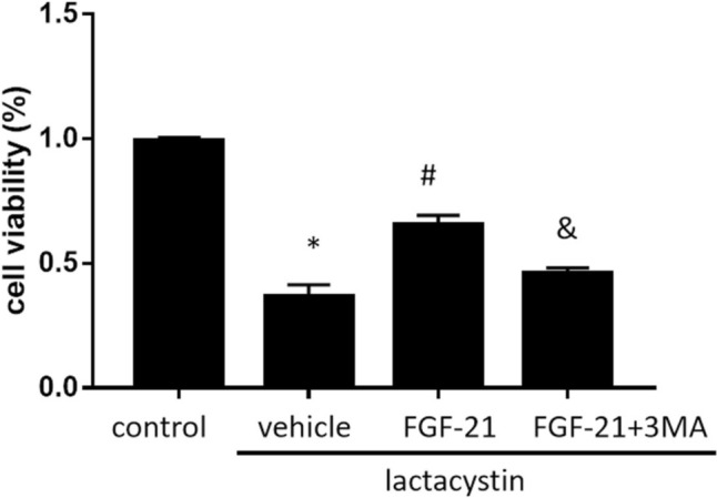
Protective effects of FGF21 on proteasome injury. After 12 h of lactacystin treatment, SH-SY5Y cells were cultured in 50ng/ml FGF-21 with/without 5 mmol/l 3MA for 1 h, and CCK-8 assay was performed to measure the cell viability. The results showed that the cell viability of lactacystin group [lactacystin vehicle group = 36.81% ± 4.60%; p = 0.000 < 0.05] was significantly lower than that of the control group. FGF-21 treatment attenuated the decrease of cell viability induced by proteasome injury. [Lactacystin + FGF-21(50 ng/ml) group = 65.37% ± 3.88%; p = 0.002 < 0.05]. After adding autophagy inhibitor 3-MA, the cell viability was reduced Compared with lactacystin + FGF-21 group [Lactacystin + FGF-21(50 ng/ml) + 5 mmol/l 3MA group = 45.97% ± 5.83%; p = 0.000 < 0.05] (Fig. 1). These results indicated that FGF21 significantly alleviates SH-SY5Y proteasome injury induced by lactacystin. 3-MA partially eliminated the effect of FGF-21 on the cell viability of lactacystin + FGF-21 group (Fig. 1). These results indicate that 3-MA may partially reverse the protective effect of FGF-21 on the survival of SH-SY5Y cells during proteasome injury.
Fig. 1.

Effects of FGF-21 on cell survival. Effects of FGF-21 (50 ng/ml) on the viability of cultured SH-SY5Y cell during proteasome injury with/without 5 mmol/l 3MA, as determined by the Cell Counting Kit-8 assay.*P<0.05 vs. control group, #P<0.05 vs. lactacystin group and &P<0.05 vs. lactacystin + FGF-21 group (n=5)
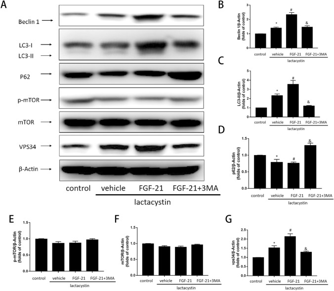
FGF21 enhances autophagy flux of SH-SY5Y cells in proteasome injury model. Western blot analysis indicated that, Compared with the control group, Beclin-1 protein [p = 0.001 < 0.05]and the formation of LC3-II [p = 0.000 < 0.05] in lactacystin group was significantly increased. Compared with the control group, the expression of p62 [p = 0.017 < 0.05] decreased significantly. P62 is a kind of receptor, which can maintain cell homeostasis by interacting with autophagy cargo and LC3 protein at the same time, thus promoting selective autophagy (Pankiv et al. 2007). Compared with lactacystin group, in lactacystin + FGF-21 group, the level of Beclin-1 protein [p = 0.000 < 0.05] and the formation of LC3-II increased [p = 0.011 < 0.05], and P62 [p = 0.036 < 0.05]expression decreased correspondingly. However, compared with lactacystin + FGF-21 group, after 3-MA treatment, the expression of Beclin-1 protein [p = 0.001 < 0.05] and the formation of LC3-II [p = 0.000 < 0.05] were down-regulated and the expression of p62 [p = 0.000 < 0.05] was up-regulated (Fig. 2B, C, D), which indicated that 3-MA partially inhibited FGF-21-induced autophagy.
Fig. 2.
Effects of FGF-21 on autophagy makers in proteasome-damaged SH-SY5Y. A Western blot analysis of autophagy makers. B Densitometric analysis of Beclin-1 levels. C Densitometric analysis of LC3-II levels. E Densitometric analysis of p62 levels. F Densitometric analysis of mTOR levels. F Densitometric analysis of p-mTOR levels. G Densitometric analysis of Vps34 levels. *P < 0.05 vs. control group, #P < 0.05 vs. lactacystin group and &P < 0.05 vs. lactacystin + FGF-21 group. Values are presented as mean ± standard deviation. Experiments were repeated in triplicate. FGF 21 fibroblast growth factor 21, 3 MA 3 methyladenine, LC3 II lipid modified microtubule-associated proteins 1A/1B light chain, p62 + sequestosome 1, mTOR mechanistic target of rapamycin, p phosphorylated (n = 5)
The effect of FGF21 is related to the change of mTOR and Vps34 signals induced by lactacystin. Acetylcholine can activate autophagy through AMPK–mTOR pathway and increase the tolerance of cells to proteasome injury. A preliminary study showed that, FGF21 plays a neuroprotective role in ApoE-KO mice with long-term calorie restriction by prolonging the activation of AMPK–mTOR signaling pathway (Rühlmann et al. 2016). Then, no significant difference was observed among the control group, lactacystin and lactacystin + FGF-21 group (Fig. 2E, F), indicating that FGF-21-induced autophagy may not occur via mTOR signaling pathway. The Beclin-1/Vps34 complex formed by Vps34 and Beclin-1 can regulate the formation of autophagy membrane (Yang et al. 2019). This study demonstrated that FGF-21 increased the expression level of Beclin-1 protein in proteasome injury cell (Fig. 2B). The autophagy inhibitor 3-MA used in this study is the inhibitor of Vps34. Therefore, the expression levels of Vps34 protein were examined in each group. The results showed that the expression level of Vsp34 protein [p = 0.001 < 0.05] in lactacystin group was significantly higher than that in control group. While lactacystin + FGF-21 group [p = 0.000 < 0.05]increased further. However, compared with lactacystin + FGF-21 group, the co-treatment of FGF-21 and 3-MA reduced the expression level of Vsp34 protein [p = 0.005 < 0.05]. In addition, compared with lactacystin group, the expression level of Vsp34 protein [p = 0.001 < 0.05] in lactacystin + 3-MA group was significantly reduced (Fig. 2G). These results indicate that Vps34 protein plays a role in FGF-21 enhancing autophagy of proteasome-damaged cells.
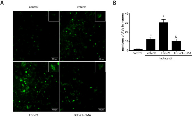
To monitor autophagy flux, serial fluorescence GFP-LC3B was performed on SH-SY5Y cells (Ad-LC3-SH-SY5Y). In the control group, Ad-LC3-SH-SY5Y cells showed basic autophagy with few autophagies and lysosomes. The Ad-LC3-SH-SY5Y cells in lactacystin treatment group showed increased autophagy and less autophagy [p = 0.002 < 0.05], indicating that autophagy flux increased during proteasome injury. In lactacystin + FGF-21 group, compared with lactacystin group, Ad-LC3-SH-SY5Y cells receiving FGF-21 had more autophagy and lysosomes [p = 0.001 < 0.05] indicated that FGF-21 treatment additionally enhanced autophagy flux. However, compared with FGF-21 treatment group, the combined treatment of FGF-21 and autophagy inhibitor 3-MA reduced autophagy and lysosome [p = 0.001 < 0.05], indicating that FGF-21 treatment enhanced autophagy flux and could be partially inhibited by 3-MA (Fig. 3). These data indicate that FGF21 induces up-regulation of autophagy flux during proteasome injury. FGF21-mediated autophagy enhances cell survival.
Fig. 3.
Effects of FGF-21 on autophagic flux in proteasome-damaged SH-SY5Y. A SH-SY5Y transfected with adenovirus harboring tandem fluorescent GFP-LC3 (Ad-LC3-SH-SY5Y) for 24 h were subjected to different treatments. Representative images of immunofluorescent SH-SY5Y expressing GFP-LC3. Green fluorescence represents GFP. B Semi-quantitative analysis of autophagosomes (Green dots). *P < 0.05 vs. control group, #P < 0.05 vs. lactacystin group and &P < 0.05 vs. lactacystin + FGF-21 group. Values are expressed as mean ± standard. Experiments were repeated in triplicate. FGF-21 fibroblast growth factor-21, 3-MA 3-methyladenine, GFP green fluorescent protein (n = 5)
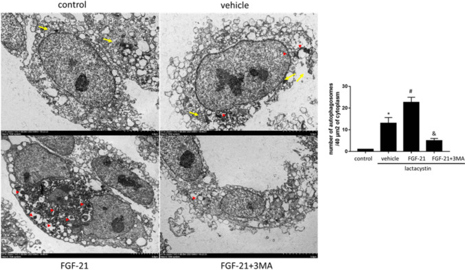
In the electron microscope images, we also observed alteration with autophagy, compared with lactacystin group, in lactacystin + FGF-21 group, more autophagy characterized by double membrane structure devoured intracellular organelles [p = 0.008 < 0.05], indicating that FGF-21 administration induced enhanced autophagy formation and Autophagosomes (Fig. 4).
Fig. 4.
Changes of organelles and autophagosomes in proteasome-damaged SH-SY5Yassessed by transmission electron microscopy (yellow arrow: mitochondria, red triangle: autophagosomes). Analysis chart: the number of autophagosomes was counted every 40 µm2 (10 fields). *P < 0.05 vs. control group, #P < 0.05 vs. lactacystin group and &P < 0.05 vs. lactacystin + FGF-21 group. Values are presented as mean ± standard deviation (n = 3)
Discussion
UPS impairment models are designed to evaluate the role of autophagy in the etiopathogenesis of PD. At present, many studies have used this cell model to study the mechanism of autophagy in the pathogenesis of PD (Shen et al. 2013; Xie et al. 2010; Zhang et al. 2005). In this study, compared with the control group, the survival rate of neurons in lactacystin group decreased significantly, indicating that UPS injury model was successfully established. Many studies have demonstrated that autophagy level in PD model increases. At present, it is still unclear whether autophagy has protective or deleterious role in PD neuropathology. Low levels of autophagy induced by moderate hypoxia or ischemia have protective effects and may prevent activation of apoptosis by degrading and removing damaged Mitochondria (Decker et al. 1980). It has also been suggested that autophagy may be a compensation mechanism and play a protective role by degrading potentially toxic protein.
In this study, we determined whether the protective effect of FGF21 on dopaminergic neurons is related to autophagy. We found that the number of lysosomes increased, the autophagy flux increased and the mitochondrial damage decreased significantly in FGF21-treated cells, which indicated that FGF21 might mediate lysosome-induced autophagy, reduce mitochondrial damage and reduce the incidence of apoptosis.
Studies have shown that autophagy may be regulated by several signal pathways, such as Beclin-11Vps34 complex (Yang et al. 2019), AMPKK–mTOR and PI3KK–AKTT–mTOR (Ling et al. 2016; Zhao et al. 2013). Studies have proved that FGF-21 may play a neuroprotective role by activating AMPKK–mTOR signal transduction pathway in ApoE-KO mice with long-term heat limitation (Rühlmann et al. 2016). However, in the experimental results of this study, p-mTOR and mTOR protein did not change significantly after treated with FGF-21 after proteasome injury. It shows that FGF-21 may enhance autophagy through other pathways than mTOR pathway and protect nerve cells from damage. Beclin-1 is the earliest mammalian autophagy gene located on human chromosome 17q21(40) (Aita et al. 1999). The expression of Beclin-1 in Golgi apparatus mainly regulates autophagy-related proteins by forming Vps34 complex (Yang et al. 2019), and locates these autophagy-related proteins in the precursor structure. In this study, when 3-MA was used to inhibit the expression of autophagy-related genes Beclin-1 and Vps34, compared with lactacystin + FGF-21 treatment group, the nerve cell damage in lactacystin + FGF-21 + 3-MA group was significantly increased, and similar results were observed in the expression of autophagy-related proteins, which indicated that FGF-21-related neuroprotection was enhanced by Beclin–11Vps34 complex. However, the crosstalk mechanism between autophagy and Vps34 is still unclear, which should be discussed in future research.
The purpose of this study is to study the protective effect of exogenous FGF21 on dopaminergic neurons and explore its possible mechanism. There are still some shortcomings in this study: because there is no specific agonist or inhibitor of FGF21 reported at present, the expression levels of FGF21 protein and mRNA in UPS impairment model cells have not been detected. In addition, there is no in vivo experiments to improve the understanding of the role of FGF21 in PD at present, Whether FGF21 can exhibit neuroprotective effect in vivo is worth further study and remains an attractive area of research, with the hope that it could specifically block PD progression.
Supplementary Information
Below is the link to the electronic supplementary material.
Abbreviation
- PD
Parkinson’s disease
- FGF21
Fibroblast growth factor 21
- DA
Dopaminergic
- SN
Substantia nigra
- LNs
Lewis neurites
- UPS
Ubiquitin–proteasome system
- ALS
Autophagosome Degradation System
- FGFR1
FGF21 receptor 1
- BAT
Brown adipose tissue
- ICV
Intracerebroventricular
- AKT
Protein kinase B
- MAPK
Mitogen-activated protein kinase
- FRS2α
FGFR substrate 2α
- rFGF21
Recombinant FGF21
- FBS
Fetal bovine serum
- DMEM
Dulbecco modified Eagle medium
- DMSO
Dimethyl sulfoxide
- OD
Optical density
- LC3B
Light chain 3β
- ANOVA
Analysis of variance
Author contributions
YS and ZZ carried out the studies, participated in collecting data, and drafted the manuscript. YW, SQ and CX performed the statistical analysis and participated in its design. YS and BZ participated in acquisition, analysis, or interpretation of data and draft the manuscript. All authors read and approved the final manuscript.
Data availability
All data generated or analyzed during this study are included in this published article.
Declarations
Conflict of interest
The authors declare that they have no competing interests.
Footnotes
Publisher's Note
Springer Nature remains neutral with regard to jurisdictional claims in published maps and institutional affiliations.
References
- Aita VM, Liang XH, Murty VV, Pincus DL, Yu W, Cayanis E, Kalachikov S, Gilliam TC, Levine B. Cloning and genomic organization of beclin 1, a candidate tumor suppressor gene on chromosome 17q21. Genomics. 1999;59:59–65. doi: 10.1006/geno.1999.5851. [DOI] [PubMed] [Google Scholar]
- Beenken A, Mohammadi M. The FGF family: biology, pathophysiology and therapy. Nat Rev Drug Discov. 2009;8:235–253. doi: 10.1038/nrd2792. [DOI] [PMC free article] [PubMed] [Google Scholar]
- Braak H, Del Tredici K, Rüb U, de Vos RA, Jansen Steur EN, Braak E. Staging of brain pathology related to sporadic Parkinson's disease. Neurobiol Aging. 2003;24:197–211. doi: 10.1016/S0197-4580(02)00065-9. [DOI] [PubMed] [Google Scholar]
- Cantó C, Auwerx J. Cell biology. FGF21 takes a fat bite. Science. 2012;336:675–676. doi: 10.1126/science.1222646. [DOI] [PMC free article] [PubMed] [Google Scholar]
- Chen S, An FM, Yin L, Liu AR, Yin DK, Yao WB, Gao XD. Glucagon-like peptide-1 protects hippocampal neurons against advanced glycation end product-induced tau hyperphosphorylation. Neuroscience. 2014;256:137–146. doi: 10.1016/j.neuroscience.2013.10.038. [DOI] [PubMed] [Google Scholar]
- Chen S, Chen ST, Sun Y, Xu Z, Wang Y, Yao SY, Yao WB, Gao XD. Fibroblast growth factor 21 ameliorates neurodegeneration in rat and cellular models of Alzheimer's disease. Redox Biol. 2019;22:101133. doi: 10.1016/j.redox.2019.101133. [DOI] [PMC free article] [PubMed] [Google Scholar]
- Cuervo AM, Stefanis L, Fredenburg R, Lansbury PT, Sulzer D. Impaired degradation of mutant alpha-synuclein by chaperone-mediated autophagy. Science. 2004;305:1292–1295. doi: 10.1126/science.1101738. [DOI] [PubMed] [Google Scholar]
- Dawson TM, Dawson VL. Molecular pathways of neurodegeneration in Parkinson's disease. Science. 2003;302:819–822. doi: 10.1126/science.1087753. [DOI] [PubMed] [Google Scholar]
- Decker RS, Poole AR, Crie JS, Dingle JT, Wildenthal K. Lysosomal alterations in hypoxic and reoxygenated hearts. II. Immunohistochemical and biochemical changes in cathepsin D. Am J Pathol. 1980;98:445–456. [PMC free article] [PubMed] [Google Scholar]
- Jiang ZS, Srisakuldee W, Soulet F, Bouche G, Kardami E. Non-angiogenic FGF-2 protects the ischemic heart from injury, in the presence or absence of reperfusion. Cardiovasc Res. 2004;62:154–166. doi: 10.1016/j.cardiores.2004.01.009. [DOI] [PubMed] [Google Scholar]
- KakotyS VKC, Tang RD, Yang CH, Dubey SK, Taliyan R. Fibroblast growth factor 21 and autophagy: a complex interplay in Parkinson disease. Biomed Pharmacother. 2020;127:110145. doi: 10.1016/j.biopha.2020.110145. [DOI] [PubMed] [Google Scholar]
- Lansbury PT, Lashuel HA. A century-old debate on protein aggregation and neurodegeneration enters the clinic. Nature. 2006;443:774–779. doi: 10.1038/nature05290. [DOI] [PubMed] [Google Scholar]
- Ling Y, Chen G, Deng Y, Tang H, Ling L, Zhou X, Song X, Yang P, Liu Y, Li Z, Zhao C, Yang Y, Wang X, Kitakaze M, Liao Y, Chen A. Polydatin post-treatment alleviates myocardial ischaemia/reperfusion injury by promoting autophagic flux. Clin Sci (lond) 2016;130:1641–1653. doi: 10.1042/CS20160082. [DOI] [PubMed] [Google Scholar]
- Luo Y, McKeehan WL. Stressed Liver and Muscle Call on Adipocytes with FGF21. Front Endocrinol (lausanne) 2013;4:194. doi: 10.3389/fendo.2013.00194. [DOI] [PMC free article] [PubMed] [Google Scholar]
- Massey AC, Kaushik S, Sovak G, Kiffin R, Cuervo AM. Consequences of the selective blockage of chaperone-mediated autophagy. Proc Natl Acad Sci U S A. 2006;103:5805–5810. doi: 10.1073/pnas.0507436103. [DOI] [PMC free article] [PubMed] [Google Scholar]
- Moran C, Beare R, Wang W, Callisaya M, Srikanth V. Type 2 diabetes mellitus, brain atrophy, and cognitive decline. Neurology. 2019;92:e823–e830. doi: 10.1212/WNL.0000000000006955. [DOI] [PMC free article] [PubMed] [Google Scholar]
- Nelson TJ, Alkon DL. Insulin and cholesterol pathways in neuronal function, memory and neurodegeneration. Biochem Soc Trans. 2005;33:1033–1036. doi: 10.1042/BST0331033. [DOI] [PubMed] [Google Scholar]
- Ohmachi S, Mikami T, Konishi M, Miyake A, Itoh N. Preferential neurotrophic activity of fibroblast growth factor-20 for dopaminergic neurons through fibroblast growth factor receptor-1c. J Neurosci Res. 2003;72:436–443. doi: 10.1002/jnr.10592. [DOI] [PubMed] [Google Scholar]
- Owen BM, Ding X, Morgan DA, Coate KC, Bookout AL, Rahmouni K, Kliewer SA, Mangelsdorf DJ. FGF21 acts centrally to induce sympathetic nerve activity, energy expenditure, and weight loss. Cell Metab. 2014;20:670–677. doi: 10.1016/j.cmet.2014.07.012. [DOI] [PMC free article] [PubMed] [Google Scholar]
- Pankiv S, Clausen TH, Lamark T, Brech A, Bruun JA, Outzen H, Øvervatn A, Bjørkøy G, Johansen T. p62/SQSTM1 binds directly to Atg8/LC3 to facilitate degradation of ubiquitinated protein aggregates by autophagy. J Biol Chem. 2007;282:24131–24145. doi: 10.1074/jbc.M702824200. [DOI] [PubMed] [Google Scholar]
- Ristow M. Neurodegenerative disorders associated with diabetes mellitus. J Mol Med (berl) 2004;82:510–529. doi: 10.1007/s00109-004-0552-1. [DOI] [PubMed] [Google Scholar]
- Rühlmann C, Wölk T, Blümel T, Stahn L, Vollmar B, Kuhla A. Long-term caloric restriction in ApoE-deficient mice results in neuroprotection via Fgf21-induced AMPK/mTOR pathway. Aging (albany NY) 2016;8:2777–2789. doi: 10.18632/aging.101086. [DOI] [PMC free article] [PubMed] [Google Scholar]
- Shen YF, Tang Y, Zhang XJ, Huang KX, Le WD. Adaptive changes in autophagy after UPS impairment in Parkinson's disease. Acta Pharmacol Sin. 2013;34:667–673. doi: 10.1038/aps.2012.203. [DOI] [PMC free article] [PubMed] [Google Scholar]
- Takagi Y, Takahashi J, Saiki H, Morizane A, Hayashi T, Kishi Y, Fukuda H, Okamoto Y, Koyanagi M, Ideguchi M, Hayashi H, Imazato T, Kawasaki H, Suemori H, Omachi S, Iida H, Itoh N, Nakatsuji N, Sasai Y, Hashimoto N. Dopaminergic neurons generated from monkey embryonic stem cells function in a Parkinson primate model. J Clin Invest. 2005;115:102–109. doi: 10.1172/JCI21137. [DOI] [PMC free article] [PubMed] [Google Scholar]
- Taliyan R, Chandran SK, Kakoty V. Therapeutic approaches to Alzheimer's type of dementia: a focus on FGF21 mediated neuroprotection. Curr Pharm Des. 2019;25:2555–2568. doi: 10.2174/1381612825666190716101411. [DOI] [PubMed] [Google Scholar]
- Xie W, Li X, Li C, Zhu W, Jankovic J, Le W. Proteasome inhibition modeling nigral neuron degeneration in Parkinson's disease. J Neurochem. 2010;115:188–199. doi: 10.1111/j.1471-4159.2010.06914.x. [DOI] [PubMed] [Google Scholar]
- Yang S, Guo Y, Zhang W, Zhang J, Zhang Y, Xu P. Effect of FGF-21 on implant bone defects through hepatocyte growth factor (HGF)-mediated PI3K/AKT signaling pathway. Biomed Pharmacother. 2019;109:1259–1267. doi: 10.1016/j.biopha.2018.10.150. [DOI] [PubMed] [Google Scholar]
- Yu P, Zhang C, Gao CY, Ma T, Zhang H, Zhou MM, Yang YW, Yang L, Kong LY. Anti-proliferation of triple-negative breast cancer cells with physagulide P: ROS/JNK signaling pathway induces apoptosis and autophagic cell death. Oncotarget. 2017;8:64032–64049. doi: 10.18632/oncotarget.19299. [DOI] [PMC free article] [PubMed] [Google Scholar]
- Zhang X, Xie W, Qu S, Pan T, Wang X, Le W. Neuroprotection by iron chelator against proteasome inhibitor-induced nigral degeneration. Biochem Biophys Res Commun. 2005;333:544–549. doi: 10.1016/j.bbrc.2005.05.150. [DOI] [PubMed] [Google Scholar]
- Zhao M, Sun L, Yu XJ, Miao Y, Liu JJ, Wang H, Ren J, Zang WJ. Acetylcholine mediates AMPK-dependent autophagic cytoprotection in H9c2 cells during hypoxia/reoxygenation injury. Cell Physiol Biochem. 2013;32:601–613. doi: 10.1159/000354464. [DOI] [PubMed] [Google Scholar]
Associated Data
This section collects any data citations, data availability statements, or supplementary materials included in this article.
Supplementary Materials
Data Availability Statement
All data generated or analyzed during this study are included in this published article.


