Abstract
Background:
Alzheimer’s disease (AD) pathology can be disclosed in vivo using amyloid and tau imaging, unlike non-AD neuropathologies for which no specific markers exist.
Objective:
We aimed to compare brain hypometabolism and tauopathy to unveil non-AD pathologies.
Methods:
Sixty-one patients presenting cognitive complaints (age 48–90), including 32 with positive AD biomarkers (52%), performed [18F]-Fluorodeoxyglucose (FDG)-PET (brain metabolism) and [18F]-MK-6240-PET (tau). We normalized these images using data from clinically normal individuals (n = 30), resulting in comparable FDG and tau z-scores. We computed between-patients correlations to evaluate regional associations. For each patient, a predominant biomarker (i.e., Hypometabolism > Tauopathy or Hypometabolism≤Tauopathy) was determined in the temporal and frontoparietal lobes. We computed within-patient correlations between tau and metabolism and investigated their associations with demographics, cognition, cardiovascular risk factors (CVRF), CSF biomarkers, and white matter hypointensities (WMH).
Results:
We observed negative associations between tau and FDG in 37 of the 68 cortical regions-of-interest (average Pearson’s r = –0.25), mainly in the temporal lobe. Thirteen patients (21%) had Hypometabolism > Tauopathy whereas twenty-five patients (41%) had Hypometabolism≤Tauopathy. Tau-predominant patients were more frequently females and had greater amyloid burden. Twenty-three patients (38%) had Hypometabolism≤Tauopathy in the temporal lobe, but Hypometabolism > Tauopathy in the frontoparietal lobe. This group was older and had higher CVRF than Tau-predominant patients. Patients with more negative associations between tau and metabolism were younger, had worse cognition, and greater amyloid and WMH burdens.
Conclusions:
Tau-FDG comparison can help suspect non-AD pathologies in patients presenting cognitive complaints. Stronger Tau-FDG correlations are associated with younger age, worse cognition, and greater amyloid and WMH burdens.
Keywords: Alzheimer’s disease, mild cognitive impairment, mixed dementias, positron emission tomography
INTRODUCTION
Alzheimer’s disease (AD) is the leading cause of dementia worldwide as it may contribute to 60 to 70% of the cases according to the World Health Organization [1]. AD is defined by the presence of two neuropathologies containing aggregated amyloid-β and tau proteins [2]. However, many patients with a clinical diagnosis of AD, or an amnestic dementia syndrome, have additional pathologies, such as cerebrovascular disease [3], TDP-43 proteinopathy [4], alpha-synucleinopathy [5], or prion diseases [6]. The generic term of “non-AD pathologies” is used when the presence of amyloid and tau pathologies has been excluded, whereas the term “mixed pathologies” is preferred when AD and non-AD pathologies are both observed.
To this day, radiotracers to disclose AD pathology in vivo using positron emission tomography (PET) have been developed. However, non-AD or mixed pathologies cannot be disclosed with specific tracers yet. [18F]-Fluorodeoxyglucose (FDG) PET is a non-specific biomarker of AD that was suggested to disclose non-AD pathologies as well [7]. In AD, tau pathology was shown to mirror brain hypometabolism and clinical symptoms [8]. However, posterior cingulate and parietal hypometabolism is often observed in early AD cases [9, 10] whereas tauopathy typically starts in the medial temporal lobe [11]. One hypothesis is the presence of non-AD pathologies in the parietal lobe, in addition to AD pathology in the medial temporal lobe. An alternative explanation is a disconnection between the medial temporal lobe where tau pathology starts and the parietal lobe where metabolism decreases early [12]. Thus far, several studies directly compared FDG and Tau-PET scans in the framework of AD [7, 8, 13–16]. One interesting observation is the important heterogeneity in the Tau-PET [17] and FDG-PET [18] patterns observed in the AD spectrum that are only partially correlating, suggesting that other pathologies than amyloid and tau contribute to brain metabolism [7].
In this study, we sought to evaluate whether the mismatch between FDG and tau-PET images can inform clinicians about the possibility of non-tau neuropathologies (either in isolation or in combination with AD pathology) in patients attending Memory Clinics with cognitive complaints. To this end, we compared Tau-PET and FDG-PET images acquired in patients attending the Memory Clinic to distinguish tau-related from tau-unrelated hypometabolism using a data-driven approach blinded to clinical diagnoses. Specifically, our goal was to identify and characterize subgroups of patients with a predominantly hypometabolic pattern, suggestive of non-tau, or mixed pathologies from a group of patients with predominant tau pathology, suggesting AD. Thereto, we used a second-generation tau PET radiotracer, namely [18F]-MK-6240 Tau-PET, that was not previously compared to FDG-PET. Regional associations between tau burden and cerebral metabolism were subsequently correlated with the patients’ demographics, cognition, cerebrospinal fluid (CSF) biomarkers, cardiovascular risk factors, and white matter hypointensities (WMH) volumes.
METHODS
Participants
Sixty-one patients having consulted the Saint-Luc Memory Clinic in Brussels for cognitive complaints (age 48–90) and 60 clinically normal (CN, age 57–86) volunteers recruited by advertisements were enrolled in a monocentric study including FDG-PET to assess brain metabolism and [18F]-MK-6240 Tau-PET to estimate tau burden. CN volunteers’ data were here mainly used as reference for the regional tau burden and brain metabolism quantifications. Patients were recruited between June 2019 and September 2022. Exclusion criteria were focal brain lesions, major depression or psychiatric diseases, alcohol or drug abuse, and autosomal-dominant mutations (PS1, PS2, and APP), which were systematically searched for in patients younger than 65 years. The study was approved by the local ethics committee (Eudra-CT number: 2018-003473-94). Written informed consent was obtained from each participant.
Neuropsychological assessment
Each participant underwent a neuropsychological assessment evaluating four cognitive domains: memory (Free and Cued Selective Reminding Test, French version [19]), language (Lexis Naming Test, the Category Fluency Test for animals, and the Letter Fluency Test for the letter ‘P’ [20]), executive functions (Trail Making Test, Luria’s Graphic Sequences (adaptations in French, unpublished)), and the visuospatial functions (Clock Drawing Test and the Praxis part of the CERAD battery [21, 22]). Each domain was assessed based on three measures (for further details about cognitive testing, see [23]). A cognitive domain was considered impaired if performance felt below the 10th percentile of an independent sample (composed of 32 clinically normal individuals who remained cognitively stable over an eight-year period) on at least two out of the three measures. Z-score were also computed for each cognitive domain by referring to the same independent sample of 32 clinically normal individuals and averaged to create a global cognitive z-score [23]. On average, neuropsychological assessments were realized 0.39 year (±0.69) after the FDG-PET acquisition, 0.22 year (±0.44) before Tau-PET acquisition, and 0.08 year (±0.52) before the mean acquisition date between the two PET modalities.
Patients were classified by two experienced neurologists as having: 1) dementia according to the DSM-V criteria for major neurocognitive disorder [24], 2) mild cognitive impairment as defined by a Mini-Mental State Examination (MMSE) score≥24/30 and at least one impaired cognitive domain (MCI), or 3) subjective cognitive impairment (SCI) when performance on all four domains was in the normal range. According to these criteria, the patients group was composed of 17 demented individuals (28%), 39 MCI patients (64%) and 5 SCI patients (8%).
CN volunteers (mean age: 69.2±6.6, 53% female) had MMSE≥27/30 (26/30 if education < 12 years), normal neuropsychological testing according to the abovementioned criteria, and global clinical dementia rating = 0.
Brain MRI
3D T1-weighted images were recorded at 3T (Signa Premier®, GE Healthcare) with a 48-channel phased-array head coil. One hundred and ninety-six sagittal slices were acquired using the following parameters: TR/TE/FA 7.2 ms/2.9 ms/11°; slice thickness 1.2 mm; FOV 270×270 mm2; acquisition matrix 256×256; resolution 1.055×1.055 mm2 (acquisition); ASSET factor 1.75 (parallel imaging). All images were processed through FreeSurfer Software v.6 [25]. WMH lesion load (mm3) was extracted from T1-weighted MRIs. WMH is identified by using spatial intensity gradients across tissue classes [26, 27]. WMH values were log-transformed to account for a positive skew.
Tau [18F]-MK-6240 PET
[18F]-MK-6240 (Cerveau Technologies, Knoxville TN) is an investigational drug studied as second-generation cerebral tau tangles imaging agent. Radiosynthesis was performed at KULeuven (Prof. Van Laere) and shipped to our clinic in less than an hour. Ninety minutes after intravenous administration of [18F]-MK-6240 (target activity 185±5 MBq) a 30-min dynamic acquisition (6x5-min frames) was performed on a Philips Vereos digital PET-CT (Philips Healthcare). Images were reconstructed using manufacturer’s standard reconstruction algorithm (including attenuation, scatter, decay correction, and time-of-flight information). We used averaged-6x5 min standardized uptake value ratios (SUVr) measures with partial volume correction using a Point Spread Function (PSF) reconstruction with a FHWM of 2–6 mm and the cerebral white matter as reference region.
[18F]-Fluorodeoxyglucose PET
In addition to [18F]-MK-6240, all patients underwent a 7-min list-mode FDG-PET according to published guidelines [28] after intravenous injection of FDG (target activity 146–185 MBq) as previously published [23]. Standardized uptake value ratio (SUVr) was uncorrected for partial volume effects and pons was used as reference region. FDG-PET scans were acquired prior [18F]-MK-6240 PET scans, with a mean gap difference of 0.62±0.47 year.
PET-MRI coregistration
For all participants, MK-6240 and FDG-PET were coregistrated using PetSurfer pipeline, a set of tools within FreeSurfer for end-to-end integrated MRI-PET analysis [29, 30]. We extracted data for the 68 cortical regions of interest (ROI) of the Desikan-Killiany Atlas [26].
Cardiovascular risk factor index
A risk factor index for cardiovascular disease was computed for each patient based on the SCORE2 algorithm (or SCORE2-OP for patients older than 70), recommended by the European Society of Cardiology [31, 32].
Statistics
To compare regional FDG and Tau-PET signals, we computed FDG and tau z-scores in each ROI by referring to the CN volunteers’ data and using the following equation: Zpatient = (SUVrpatient – average SUVr CN)/SD SUVr CN. Several analyses were then performed based on these z-scores.
First, we computed Pearson’s correlation coefficients between the FDG z-scores and the tau z-scores across all patients for each of the 68 ROIs, to assess the regional associations between hypometabolism and tau pathology. In some analyses, we distinguished the 18 regions of the temporal lobe from the 42 regions of the frontoparietal lobe, including the cingulate gyrus and the insula, as we assumed that the association between tau burden and brain metabolism may be different in the temporal lobe, where AD tau pathology is known to start, in comparison to the frontoparietal lobe where cerebrovascular pathology or other neuropathologies may be more frequent.
Second, we compared patients’ z-scores to CN volunteers’ z-scores in the temporal and frontoparietal lobes for each PET modality to assess whether significant tau burden or hypometabolism could be detected in each lobe at the group level in patients.
Third, paired t-tests were implemented to compare, within the patient group, FDG z-scores and tau z-scores in the temporal and frontoparietal lobes to test for predominance of tauopathy or hypometabolism in each lobe at the group level.
Fourth, paired t-tests were applied at the individual level to compare FDG and tau z-scores and determine which of these biomarkers was predominant in the temporal and frontoparietal lobes in each patient. Patients were then classified into the four following possible groups: 1) Hypometabolism > Tauopathy in both lobes,2) Hypometabolism≤Tauopathy in both lobes, 3) Hypometabolism > Tauopathy in the temporal but Hypometabolism≤Tauopathy in the frontoparietal lobe, and 4) Hypometabolism≤Tauopathy in the temporal but Hypometabolism > Tauopathy in the frontoparietal lobe. Thereafter, these groups were compared for age, sex, education, apolipoprotein ɛ4 carriage, CSF results (t-tau, Aβ42), MMSE, global cognitive performance, WMH volumes and cardiovascular risk factor scores to determine whether groups with suspected non-tau pathology (Hypometabolism > Tauopathy) had different characteristics than groups with suspected AD tau pathology (Hypometabolism≤Tauopathy).
Fifth, we computed within-patient correlation coefficients to quantify for each patient the association between the FDG z-scores and the tau z-scores in the temporal and frontoparietal lobes. We then tested whether these coefficients were significantly negative (thereby suggesting tau-related hypometabolism) or not significantly different from 0 (thereby suggesting tau-unrelated hypometabolism) across all patients as well as in each patient group. Finally, we assessed the associations between these within-patient Tau-FDG correlation coefficients and patients’ demographics, cognition, cardiovascular risk factors, CSF biomarkers, and WMH volumes.
Statistical analyses were computed with the Stats package available in R software v4.2.2 [33]. Group differences were assessed using the following R functions: shapiro.test to evaluate sample for normality and var.test for homogeneity of variances. Based on the results, two-sample t-tests or wilcoxon tests were computed using the t.test and wilcox.test functions. Proportion differences and correlations were respectively computed using the prop.test and cor.test functions. We did not include covariates in our analyses. Adjusting for education, age, and sex in the analyses implying cognition and WMH did not change our results.
RESULTS
Regional correlations between metabolism and tau burden in patients
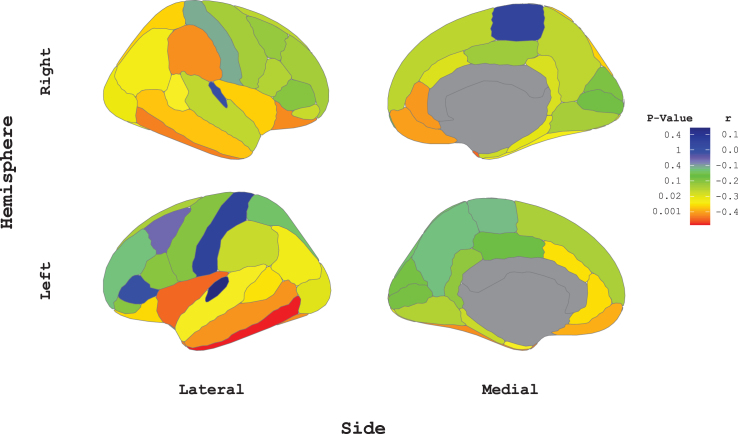
We first correlated tau z-scores with FDG z-scores, in each region of the 68 cortical ROIs, to evaluate the regional associations between tau burden and cerebral metabolism in the entire sample of patients (n = 61). The average Pearson’s correlation across all regions was close to significance (–0.25, p = 0.052). More precisely, 37 out of the 68 cortical ROIs (54%) demonstrated significant negative Tau-FDG associations (Pearson’s R < –0.25, p < 0.05, Fig. 1, yellow), including 16 of the 18 temporal regions (89%), and 19 of the 42 frontoparietal regions (45%). After Bonferroni correction for multiple comparisons (R < –0.42, uncorrected p < 0.0007, corrected p < 0.05, Fig. 1, dark orange and red), the FDG z-scores were still negatively associated with tau z-scores in five regions: the bilateral inferior temporal neocortex, the bilateral temporal pole, and the left insula. No significant positive association between FDG and tau was found in any ROI (max. r = 0.14, positive R in the left post-central, right paracentral gyri, and bilateral transverse, Fig. 1, blue). After adjusting analyses for CSF Aβ1-42, education, age, and sex, the FDG and tau PET signals were significantly associated in the temporal lobe (p = 0.001), and in the frontoparietal lobe (p = 0.002). CSF Aβ1-42 and education were non-significant predictors of FDG data, men had lower FDG only in the temporal whereas FDG decreased with age in both lobes.
Fig. 1.
Between-Patients local Tau-FDG correlations in the whole cortex. The Pearson correlation coefficient between tau burden and glucose metabolism across the 61 patients are plotted in the 68 regions-of-interest. Associated p-values are provided for information. Red and dark orange regions withstand Bonferroni correction, (r < –0.42, p < 0.001). Yellow regions are statistically significant at p = 0.05. Green regions demonstrated marginally significant correlations between tau burden and glucose. Blue regions present an absence of association. This illustration was realized with the ggseg package in R [49].
Regional tau and metabolism differences between patients and CN volunteers
Within the temporal lobe, both FDG and tau z-scores were significantly different between patients and CN volunteers (Table 1, left column titled “All patients”). In the frontoparietal lobe, the metabolism was lower in patients than in CN volunteers, while tau burden did not significantly differ between groups.
Table 1.
Comparison of FDG-predominant, Inconsistent pattern, and Tau-predominant groups
| Mean±SD (min/max) | All patients | Group 1 FDG-predominant | Group 2 Inconsistent pattern | Group 3 Tau-predominant | p (Group 1 versus 2) | p (Group 2 versus 3) | p (Group 1 versus 3) |
| N | 61 | 13 | 23 | 25 | |||
| Frontoparietal Tau (z-score) | 0.78 (±2.04) (–2.53/6.18) | –0.64 (±0.64) (–1.53/0.78) | –0.26 (±0.82) (–2.53/1.11) | 2.48 (±2.10) AB (–0.57/6.18) | 0.162 | <0.001 | <0.001 |
| Temporal Tau (z-score) | 2.81 (±2.83) AB (–1.78/13.34) | –0.32 (±0.71) (–1.78/0.77) | 3.06 (±2.05) AB (–0.01/7.39) | 4.21 (±2.90) AB (0.39/13.34) | <0.001 | 0.150 | <0.001 |
| Frontoparietal FDG (z-score) | –1.52 (±0.85) AB (–3.07/0.54) | –1.75 (±0.79) AB (–2.8/–0.68) | –1.66 (±0.71) AB (–3.07/0.05) | –1.27 (±0.96) A (–3.06/0.54) | 0.732 | 0.123 | 0.136 |
| Temporal FDG (z-score) | –1.23 (±0.92) A (–3.29/1.07) | –1.20 (±0.70) AB (–2.95/–0.26) | –1.40 (±0.82) A (–3.29/0.36) | –1.08 (±1.09) A (–3.08/1.07) | 0.454 | 0.248 | 0.716 |
| Age (y) | 71.42 (±8.42) (47.81–90.37) | 71.37 (±8.69) (57.44–90.37) | 76.45 (±6.4) (56.22/87.25) | 66.82 (±7.45) (47.81/82.76) | 0.031 | <0.001 | 0.100 |
| Male Sex: N (%) | 33 (54%) | 11 (85%) | 10 (43%) | 12 (48%) | 0.016 | 0.753 | 0.028 |
| Education (y) | 15.54 (±4.01) (6–18) | 17.08 (±2.25) (12–18) | 14.09 (±4.65) (6/18) | 16.08 (±3.76) (6/18) | 0.044 | 0.092 | 0.502 |
| ɛ4 carriers: N (%) | 25 (56%) | 5 (45%) | 8 (53%) | 12 (63%) | 0.691 | 0.563 | 0.346 |
| MMSE (/30) | 24.92 (±4.38) (12–30) | 26.85 (±3.93) (15/30) | 24.87 (±3.84) (16/30) | 23.96 (±4.86) (12/30) | 0.034 | 0.748 | 0.054 |
| Global Cognitive (z-score) | –1.53 (±1.07) (–4.22/0.39) | –1.11 (±0.98) (–2.66/0.39) | –1.54 (±0.97) (–3.61/–0.2) | –1.75 (±1.19) (–4.22/0.21) | 0.228 | 0.521 | 0.120 |
| Cardiovascular Risk Factor (% risk over 10 years) | 9.27 (±5.34) (1/32) | 10.15 (±7.79) (2/32) | 11.00 (±4.68) (1/19) | 7.12 (±3.53) (2/16) | 0.255 | 0.002 | 0.285 |
| Log-transformed WMH (mm3) | 8.09 (±0.80) (6.61/10.52) | 8.26 (±0.86) (7.34/10.31) | 8.08 (±0.81) (6.61/10.52) | 8.01 (±0.77) (6.85/10.08) | 0.555 | 0.769 | 0.390 |
| Total Tau (pg/ml) | 571.25 (±363.78) (93/1952) | 479.38 (±408.17) (134/1590) | 579.00 (±406.16) (137/1952) | 614.54 (±303.71) (93/1288) | 0.250 | 0.465 | 0.089 |
| CSF Aβ42 (pg/ml) | 532.42 (±271.02) (115–1369) | 711.85 (±340.82) (299–1369) | 491.25 (±228.34) (226/1174) | 469.54 (±226.65) (115/1029) | 0.045 | 0.637 | 0.014 |
| CSF positivity for AD: N (%) | 32 (52%) | 2 (15%) | 12 (52%) | 18 (72%) | 0.030 | 0.156 | <0.001 |
| Frontoparietal Tau-FDG Correlation | –0.10 (±0.35) C (–0.79/0.60) | 0.00 (±0.25) (–0.41/0.47) | 0.01 (±0.33) (–0.65/0.6) | –0.25 (±0.37) C (–0.79/0.56) | 0.896 | 0.012 | 0.035 |
| Temporal Tau-FDG Correlation | –0.27 (±0.35) CD (–0.80/0.45) | –0.13 (±0.34) (–0.79/0.42) | –0.13 (±0.35) (–0.76/0.45) | –0.47 (±0.26) CD (–0.8/0.03) | 0.966 | <0.001 | 0.003 |
ASignificant difference between and CN (p < 0.05). BSignificant difference between PET z-scores within the same region (p < 0.05). CZ-score is significantly different from 0 (p < 0.05). DTau-FDG correlation is significantly different between regions (p < 0.05). NA, not available. E4 carriers (16 NA), Global Cognitive z-score (4 NA), Cardiovascular Risk Factor scores (1 NA), Log-transformed WMH (1 NA), CSF quantifications (4 NA). CSF positivity for AD is defined by a Total Tau/Aβ42 ratio > 1.1 [47]. For the missing CSF quantifications, CSF positivity was defined based on the [18F] flutemetamol Aβ-PET Centiloid Score (cut-off > 25) [48].
Group-level differences between regional tau burden and metabolism in patients
Among patients, tau z-scores were significantly greater than FDG z-scores (expressed in absolute values) in the temporal lobe, demonstrating greater tauopathy than hypometabolism. In contrast, in the frontoparietal cortex, FDG z-scores were greater than tau z-scores.
Patient classification based on the predominant PET-biomarker
We used paired t-tests to evaluate whether each patient had greater hypometabolism than tauopathy or greater tauopathy than hypometabolism, in the temporal and frontoparietal lobes. Thirteen patients of 61 (21%, Table 1) had significantly greater hypometabolism than tau pathology in both lobes (group subsequently referred to as “FDG-predominant”). Twenty-five patients (41%) had either greater tau z-scores than FDG z-scores in both lobes or tau z-scores that did not statistically differ from FDG z-scores. This group is subsequently referred to as “Tau-predominant” (while also including patients with tau-dependent hypometabolism). Finally, twenty-three patients (38%) had tau burden greater than hypometabolism in the temporal lobe, but greater hypometabolism than tau pathology in the frontoparietal lobe (group subsequently referred to as “Inconsistent pattern”). The opposite pattern was not observed, i.e., individuals who had tau pathology greater thanhypometabolism in the frontoparietal lobe always had tau pathology greater than hypometabolism in the temporal lobe. Of note, the Tau-predominant group had significant hypometabolism compared to CN volunteers in both lobes, whereas the FDG-predominant group did not demonstrate significantly elevated tau pathology compared to CN volunteers in any lobe (tau z-scores not significantly different than zero; Table 1, top lines).
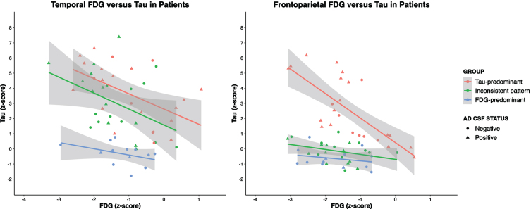
When plotting tau versus FDG z-scores, an association between PET-modalities was observed in the Tau-predominant group whereas it was not observed in the FDG-predominant group. Individuals with an Inconsistent pattern demonstrated a Tau-FDG association in the temporal, but not in the frontoparietal lobe (Fig. 2). Grouping patients based on frontal and parietal lobes separately did not change overall results. This sensitivity analysis is provided in the Supplementary Material.
Fig. 2.
Temporal and Frontoparietal Tau versus FDG in Patients. CSF positivity is defined by a Total Tau/Aβ42 ratio > 1.1 [47]. Group legend: Tau-predominant corresponds to Tau≥FDG both in the temporal and in the frontoparietal. Inconsistent corresponds to Tau≥FDG in the temporal and Tau < FDG in the frontoparietal. FDG-predominant corresponds to Tau < FDG both in the temporal and in the frontoparietal lobe.
Comparison of FDG-predominant, Tau-predominant, and Inconsistent pattern patient groups
Tau-predominant patients were younger and more frequently females than those with an FDG-predominant pattern (Table 1). They also had more abnormal AD CSF biomarkers (significantly lower Aβ42, marginally higher total tau CSF concentrations). In contrast, the Inconsistent pattern group was older and presented higher cardiovascular risk factor scores compared to the Tau-predominant group. This result was also observed when restricting the sample to patients with positive AD CSF biomarkers (AD-CSF+): AD-CSF+ patients with hypometabolism exceeding tau in the frontoparietal lobe (Inconsistent pattern) were older and had a higher cardiovascular risk score than AD-CSF+ Tau-predominant patients. The comparison with the FDG-predominant group was not possible in AD-CSF+ patients because only two AD-CSF+ patients had greater hypometabolism than tau pathology in the temporal lobe, indicating that this group mostly included patients with negative CSF. All groups presented similar WMH volumes. Across all patients, the cardiovascular risk factor was positively correlated with the whole brain WMH (r = 0.32, p = 0.020). However, while higher cardiovascular risk factor scores were not significantly associated with the Tau-FDG association (r = 0.09, p = 0.484), higher WMH were significantly associated with a greater Tau-FDG association (r = –0.30, p = 0.022).
Overall, these results indicate that FDG-predominant patients were less likely to have AD pathology whereas co-existence of frontoparietal hypometabolism and temporal lobe tau deposition was observed in older individuals with higher cardiovascular risk factor scores.
Within-patient correlations between tau burden and metabolism
To quantify the associations between tau burden and metabolism at the individual level, we correlated, for each patient separately, the tau z-scores with the FDG z-scores obtained in each lobe. Across all patients, the Tau-FDG correlation was significantly negative in the temporal lobe (r = –0.27 [CI95 = –0.36, –0.18], one sample t-test = –5.9, p < 0.001), and to a lesser extent, in the frontoparietal lobe (r = –0.10, [CI95 = –0.19, –0.01], one sample t-test = –2.2, p = 0.035).
Patients from the Tau-predominant group had a significant negative correlation coefficient between tau burden and metabolism in both lobes (Table 1, bottom lines), indicating that they had tau-dependent hypometabolism in both these regions. The strength of this correlation was higher for the temporal lobe than for the frontoparietal lobe. In contrast, the correlation between tau burden and cerebral metabolism was not significantly different from 0 for patients in the FDG-predominant nor the Inconsistent pattern groups, suggesting that these patients had tau-unrelated hypometabolism.
Associations between the Tau-FDG within-patient correlations and patients’ characteristics
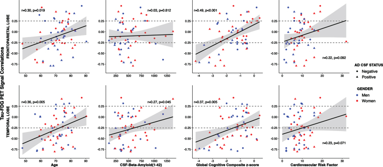
In the temporal lobe, the strength of the Tau-FDG correlation was associated with younger ages (r = 0.36, Fig. 3), lower amyloid concentration in the CSF (r = 0.27), and worse cognition (r = 0.37). A negative Tau-FDG correlation index in the frontoparietal lobe was associated with younger ages (r = 0.30) and worse cognition (r = 0.49), but the association did not reach significance for amyloid levels inthe CSF.
Fig. 3.
Associations between Tau-FDG PET Signal Correlations and Covariables. Dashed lines represent the threshold for significant correlation. AD CSF positivity is defined by a Total Tau/Aβ42 ratio > 1.1 [47].
DISCUSSION
This study aimed to compare FDG- and Tau-PET data to provide information about the presence of non-AD pathologies in patients with variable levels (or absence) of AD pathology. Among all patients, we observed that tauopathy was mainly associated with brain hypometabolism in the temporal lobe, as hypothesized based on the sequence of tau progression in AD [34]. At the individual level, we observed different patterns of tau and FDG biomarkers that could classify patients in three groups with different demographics and CSF AD biomarkers positivity, but similar levels of global cognitive impairment.
First, patients with a tauopathy-predominant pattern (of biomarker) demonstrated a negative association between tau burden and cerebral metabolism in the temporal and frontoparietal lobe. The strength of this correlation was associated with younger ages, worse cognition, and lower levels of amyloid in the CSF, suggesting that the tauopathy-predominant group mainly suffers from AD pathology (72% based on CSF data only).
Second, patients with a hypometabolism-predominant pattern did not significantly from the CN volunteers for the temporal and frontoparietal tau burden. Moreover, in this group, there was no significant association between tau burden and hypometabolism in any cortical region. This group was more frequently males, had normal CSF Aβ42 concentration at the group level, and low prevalence of AD positivity in the CSF (15%). These results suggest that patients with hypometabolism-predominant probably have non-tau pathologies that could explain brain hypometabolism and cognitive impairment.
Third, we observed a group of patients with an Inconsistent pattern of biomarkers depending on the brain regions analyzed. These patients had a Tau-predominant pattern in the temporal lobes, but with a hypometabolism-predominant pattern in the frontoparietal lobe. The opposite pattern (frontoparietal tau predominance with temporal hypometabolism predominance) was however never observed. This Inconsistent pattern may suggest a functional disconnection between the temporal lobe (with tau pathology) and the frontoparietal lobe (with hypometabolism). The brain operates as a highly interconnected network of regions, thus hypometabolism might result from disruptions in functional connectivity or network integrity. If specific brain regions are affected due to underlying pathologies or neurodegenerative processes, brain connectivity between those regions and distal regions might be compromised, leading to decreased metabolic activity in the distal regions. Brain disconnection, as measured using functional MRI or diffusion imaging, was shown to decrease brain metabolism [12, 35], specifically in the posterior cingulate [36], a region that was included in the frontoparietal lobe in the present work. While we did not use connectivity metrics in the present study, it represents a plausible factor contributing to frontoparietal decreased metabolism.
We cannot exclude however the contribution of non-AD pathologies in addition to the presence of AD pathology in the temporal lobe to explain the frontoparietal hypometabolism. Of note, this group of patients (i.e. one displaying an Inconsistent pattern) was older and had higher cardiovascular risk factor scores than patients with higher tau in the frontoparietal lobe, increasing the likelihood of mixed pathologies, as suggested by the neuropathological literature [37]. We observed that with older ages the strength of the association between tau and metabolism decreased, also suggesting the onset of non-tau or mixed pathologies to explain brain hypometabolism. Previous work showed that cardiovascular risk factor scores is associated with cerebrovascular pathologies [38] and a greater risk of cognitive decline in older adults [39].
However, there is evidence in the literature of associations between amyloid and white matter hyperintensities [40]. Furthermore, the association observed between WMH volumes and the Tau-FDG correlation is in line with a post-mortem study reporting association between cortical tau load and white matter lesions [41]. The link between AD pathologies and vascular pathology are thus complex, and they are not two independent pathologies. As WMH represents only one type of co-pathology, its exact contribution among all co-pathologies present in our sample is difficult to analyze given that we have no biomarkers for the other pathologies. The overall negative association between age and Tau-FDG might thus result from a higher prevalence of other types of co-pathologies than WMH, and it is not incompatible with a positive association between WMH and Tau-FDG.
In this study, we specifically focused on the temporal sequence of events in typical AD cases in which tau pathology is likely to accumulate before metabolism starts declining. However, as phenotypes determine the hypothesized sequence of progression, their heterogeneity is worth noting. For example, patients displaying greater brain hypometabolism than tauopathy in the temporal lobe may reflect an underlying Limbic-predominant Aged-related TDP-43 Encephalopathy (LATE) [4]. Furthermore, different regional variants of AD may be present in our cohort. Among others, the dysexecutive variant of AD is known for significant extra-temporal tau accumulation, particularly in frontoparietal regions. However, in some cases, the hypometabolism may be less pronounced in both magnitude and spatial extent [42, 43]. Therefore, dysexecutive cases might exhibit a stronger frontoparietal Tau-FDG relationship compared to typical cases. Similarly, non-amnestic syndromes are likely to have their own distinctive Tau-FDG relationships. Beyond clinical phenotypes, additional factors such as regional PET tracer saturation capacity, patients’ resilience, or the disease’s phase could also impact the Tau-FDG association. Investigating Tau-FDG associations in the frontal and parietal lobes separately did not modify our results (See Supplementary Material 2, Supplementary Figure 1).
A better understanding of the contribution of clinical and pathological heterogeneity in neurodegenerative diseases is needed to include more homogeneous population in clinical trials [44]. Our results advocate for the inclusion of younger participants in trials targeting amyloid or tau, as older age appears to be the main driver of non-AD pathologies. This would decrease the likelihood and severity of mixed pathology in screened individuals, awaiting the development of new biomarkers allowing the implementation of new exclusion criteria.
Limitations and future directions
We cannot ascertain the presence of non-AD or mixed pathologies given the absence of biomarkers for several neuropathologies, such as TDP-43, alpha-synuclein, or several non-AD tauopathies as MK-6240 seems to mostly bind certain tau isoforms (AD 3 R/4 R) [45]. Future work could use MRI to investigate the associations of cerebrovascular disease and brain metabolism, although cardiovascular risk factor scores was shown to be more strongly associated with subsequent cognitive decline than white matter hyperintensities on T1 images [39]. The development of better cerebrovascular biomarkers would facilitate the study of these pathologies and their contribution to cognitive decline in patients attending the Memory Clinic.
Besides non-AD pathologies, several non-AD neurodegenerative diseases involve tau pathology, such as frontotemporal lobar degeneration, corticobasal degeneration, and progressive supranuclear palsy. The affinity of our tau tracer, [F18]-MK-6240, for non-AD tau, is currently being investigated. If the tracer does not bind a specific type of non-AD tau aggregate, the patients are probably considered as FDG-predominant. On the contrary, if the tracer binds to these non-AD tau aggregates, these patients may have been included in the Tau-predominant group and explain why a minority of patients in this group did not demonstrate amyloid pathology in the CSF. Furthermore, off-target binding in the skull and meninges could have been more prominent in the control group than within patients, explaining the higher frequency of patients with lower tau z-scores than controls in the temporal lobe compared to the frontoparietal lobe.
A limitation of our approach is that we grouped patients with tau pathology exceeding or equal to brain hypometabolism altogether. This means that patients with little abnormalities on both FDG and tau biomarkers were included in the Tau-predominant group and/or the group displaying an Inconsistent pattern. This could have decreased the differences between these two groups. We preferred this approach to provide more guarantees that patients with an FDG-predominant pattern had a (non-tau) pathology. Yet, excluding patients with equal abnormalities on both biomarkers did not modify any of our conclusions, although it reduced statistical power.
Due to our limited sample size, we decided not to compare PET modalities on more than two regions of interest. Future studies benefiting from a greater number of patients could base their analyses on a higher number of parcellated brain regions. For example, a recent study showed that an autopsy-derived temporo-limbic FDG-PET signature was able to identify older amnestic patients whose clinical, genetic, and molecular biomarker features are consistent with underlying LATE [46]. The addition of functional MRI or DTI connectivity metrics to FDG and Tau-PET could help distinguish between the effects of disconnection and the contribution of mixed pathologies to explain frontoparietal hypometabolism in the presence of temporal tauopathy.
Conclusions
To date, identifying mixed pathologies in patients within the AD continuum remains a challenge, caused by the increasingly recognized heterogeneity of the disease [17, 18] and the insufficient number of reliable biomarkers to ascertain the existence of non-AD pathologies in-vivo. To address this question, we grouped patients based on their predominant abnormality on one of two biomarkers (i.e., Tau-PET and FDG-PET) both in the temporal and the frontoparietal lobe. This classification allowed to suspect the presence of non-AD pathologies in patients with greater hypometabolism than tau pathology in the temporal lobe; and the presence of mixed-pathologies or a disconnection syndrome in patients with frontoparietal hypometabolism. Stronger Tau-FDG correlations were associated with younger age, worse cognition, greater amyloid burden, and higher WMH volumes. Our results suggest including younger participants in trials targeting amyloid or tau, as older age appears the main driver of non-AD pathologies. Meanwhile, distinguishing disconnection effects from the presence of co-pathologies require additional work with connectivity analyses and the development of biomarkers specific for non-AD pathologies.
Supplementary Material
ACKNOWLEDGMENTS
The authors thank the firm Cerveau Inc. for supplying the [18F]-MK-6240 precursor for PET scan imaging according to a convention with our Clinic. We also thank the Jolly family for their generous donation allowing to fund this project.
SUPPLEMENTARY MATERIAL
The supplementary material is available in the electronic version of this article: https://dx.doi.org/10.3233/JAD-230696.
FUNDING
The Belgian Fund for Scientific Research (FNRS) provided grants for the personnel conducting this research (V.M.: No. ASP029789, B.H.: No. SPD4000041). This work was also supported by the Fonds de la Recherche Scientifique – FNRS for the FRFS-WELBIO under Grant No. 40010035, the Queen Elizabeth Medical Foundation, and the Belgian Alzheimer Research Foundation.
CONFLICT OF INTEREST
The firm Cerveau Inc. supplied the [18F]-MK-6240 precursor for acquiring the PET images analyzed in this article. No other conflicts of interest are reported.
DATA AVAILABILITY
The data supporting the findings of this study are available on request from the corresponding author (Vincent Malotaux, vincent.malotaux@uclouvain.be, Institute of Neuroscience– Université Catholique de Louvain, Brussels, Belgium).
REFERENCES
- [1].World Health Organization, Dementia Fact Sheet No 362, www.who.int/mediacentre/factsheets/fs362/en/.
- [2]. Hyman BT, Phelps CH, Beach TG, Bigio EH, Cairns NJ, Carrillo MC, Dickson DW, Duyckaerts C, Frosch MP, Masliah E, Mirra SS, Nelson PT, Schneider JA, Thal DR, Thies B, Trojanowski JQ, Vinters HV, Montine TJ (2012) National Institute on Aging-Alzheimer’s Association guidelines for the neuropathologic assessment of Alzheimer’s disease. Alzheimers Dement 8, 1–13. [DOI] [PMC free article] [PubMed] [Google Scholar]
- [3]. Attems J, Jellinger KA (2014) The overlap between vascular disease and Alzheimer’s disease–lessons from pathology. BMC Med 12, 206. [DOI] [PMC free article] [PubMed] [Google Scholar]
- [4]. Nelson PT, Dickson DW, Trojanowski JQ, Jack CR, Boyle PA, Arfanakis K, Rademakers R, Alafuzoff I, Attems J, Brayne C, Coyle-Gilchrist ITS, Chui HC, Fardo DW, Flanagan ME, Halliday G, Hokkanen SRK, Hunter S, Jicha GA, Katsumata Y, Kawas CH, Keene CD, Kovacs GG, Kukull WA, Levey AI, Makkinejad N, Montine TJ, Murayama S, Murray ME, Nag S, Rissman RA, Seeley WW, Sperling RA, White CL 3rd, Yu L, Schneider JA (2019) Limbic-predominant age-related TDP-43 encephalopathy (LATE): Consensus working group report. Brain 142, 1503–1527. [DOI] [PMC free article] [PubMed] [Google Scholar]
- [5]. Yamada M, Komatsu J, Nakamura K, Sakai K, Samuraki-Yokohama M, Nakajima K, Yoshita M (2020) Diagnostic criteria for dementia with Lewy bodies: Updates and future directions. J Mov Disord 13, 1–10. [DOI] [PMC free article] [PubMed] [Google Scholar]
- [6]. Zhu C, Aguzzi A (2021) Prion protein and prion disease at a glance. J Cell Sci 134, jcs245605. [DOI] [PubMed] [Google Scholar]
- [7]. Botha H, Mantyh WG, Murray ME, Knopman DS, Przybelski SA, Wiste HJ, Graff-Radford J, Josephs KA, Schwarz CG, Kremers WK, Boeve BF, Petersen RC, Machulda MM, Parisi JE, Dickson DW, Lowe V, Jack CR Jr., Jones DT (2018) FDG-PET in tau-negative amnestic dementia resembles that of autopsy-proven hippocampal sclerosis. Brain 141, 1201–1217. [DOI] [PMC free article] [PubMed] [Google Scholar]
- [8]. Ossenkoppele R, Schonhaut DR, Scholl M, Lockhart SN, Ayakta N, Baker SL, O’Neil JP, Janabi M, Lazaris A, Cantwell A, Vogel J, Santos M, Miller ZA, Bettcher BM, Vossel KA, Kramer JH, Gorno-Tempini ML, Miller BL, Jagust WJ, Rabinovici GD (2016) Tau PET patterns mirror clinical and neuroanatomical variability in Alzheimer’s disease. Brain 139, 1551–1567. [DOI] [PMC free article] [PubMed] [Google Scholar]
- [9]. Landau SM, Harvey D, Madison CM, Koeppe RA, Reiman EM, Foster NL, Weiner MW, Jagust WJ, Alzheimer’s Disease Neuroimaging Initiative (2011) Associations between cognitive, functional, and FDG-PET measures of decline in AD and MCI. Neurobiol Aging 32, 1207–1218. [DOI] [PMC free article] [PubMed] [Google Scholar]
- [10]. Herholz K (2011) Perfusion SPECT and FDG-PET. Int Psychogeriatr 23(Suppl 2), 25–S31. [DOI] [PubMed] [Google Scholar]
- [11]. Braak H, Braak E (1991) Neuropathological stageing of Alzheimer-related changes. Acta Neuropathol 82, 239–259. [DOI] [PubMed] [Google Scholar]
- [12]. Teipel S, Grothe MJ, Alzheimer s Disease Neuroimaging Initiative (2016) Does posterior cingulate hypometabolism result from disconnection or local pathology across preclinical and clinical stages of Alzheimer’s disease? Eur J Nucl Med Mol Imaging 43, 526–536. [DOI] [PMC free article] [PubMed] [Google Scholar]
- [13]. Mayblyum DV, Becker JA, Jacobs HIL, Buckley RF, Schultz AP, Sepulcre J, Sanchez JS, Rubinstein ZB, Katz SR, Moody KA, Vannini P, Papp KV, Rentz DM, Price JC, Sperling RA, Johnson KA, Hanseeuw BJ (2021) Comparing PET and MRI biomarkers predicting cognitive decline in preclinical Alzheimer disease. Neurology 96, e2933–2943. [DOI] [PMC free article] [PubMed] [Google Scholar]
- [14]. Rubinski A, Franzmeier N, Neitzel J, Ewers M, Alzheimer’s Disease Neuroimaging Initiative (2020) FDG-PET hypermetabolism is associated with higher tau-PET in mild cognitive impairment at low amyloid-PET levels. Alzheimers Res Ther 12, 133. [DOI] [PMC free article] [PubMed] [Google Scholar]
- [15]. Lu J, Bao W, Li M, Li L, Zhang Z, Alberts I, Brendel M, Cumming P, Lu H, Xiao Z, Zuo C, Guan Y, Zhao Q, Rominger A (2020) Associations of [(18)F]-APN-1607 Tau PET binding in the brain of Alzheimer’s disease patients with cognition and glucose metabolism. Front Neurosci 14, 604. [DOI] [PMC free article] [PubMed] [Google Scholar]
- [16]. Strom A, Iaccarino L, Edwards L, Lesman-Segev OH, Soleimani-Meigooni DN, Pham J, Baker SL, Landau SM, Jagust WJ, Miller BL, Rosen HJ, Gorno-Tempini ML, Rabinovici GD, La Joie R, Alzheimer’s Disease Neuroimaging Initiative (2022) Cortical hypometabolism reflects local atrophy and tau pathology in symptomatic Alzheimer’s disease. Brain 145, 713–728. [DOI] [PMC free article] [PubMed] [Google Scholar]
- [17]. Vogel JW, Young AL, Oxtoby NP, Smith R, Ossenkoppele R, Strandberg OT, La Joie R, Aksman LM, Grothe MJ, Iturria-Medina Y, Alzheimer’s Disease Neuroimaging Initiative, Pontecorvo MJ, Devous MD, Rabinovici GD, Alexander DC, Lyoo CH, Evans AC, Hansson O (2021) Four distinct trajectories of tau deposition identified in Alzheimer’s disease. Nat Med 27, 871–881. [DOI] [PMC free article] [PubMed] [Google Scholar]
- [18]. Levin F, Ferreira D, Lange C, Dyrba M, Westman E, Buchert R, Teipel SJ, Grothe MJ, Alzheimer’s Disease Neuroimaging Initiative (2021) Data-driven FDG-PET subtypes of Alzheimer’s disease-related neurodegeneration. Alzheimers Res Ther 13, 49. [DOI] [PMC free article] [PubMed] [Google Scholar]
- [19]. Van der Linden M CF, Poitrenaud J, Kalafat M, Calicis, F WC, Adam S, et les membres du GREMEM (2004) L’épreuve de rappel libre/rappel indicé à 16 items (RL/RI16). In L’évaluation des troubles de la mémoire: Présentation de quatre tests de mémoire épisodique (avec leur étalonnage), Solal, ed., Marseille. [Google Scholar]
- [20]. de Partz MP BV, De Wilde V, Seron X, Pillon A (2001) LEXIS: Tests pour l’évaluation des troubles lexicaux chez la personne aphasique, Marseille [Google Scholar]
- [21]. Rouleau I, Salmon DP, Butters N, Kennedy C, McGuire K (1992) Quantitative and qualitative analyses of clock drawings in Alzheimer’s and Huntington’s disease. Brain Cogn 18, 70–87. [DOI] [PubMed] [Google Scholar]
- [22]. Morris JC, Mohs RC, Rogers H, Fillenbaum G, Heyman A (1988) Consortium to establish a registry for Alzheimer’s disease (CERAD) clinical and neuropsychological assessment of Alzheimer’s disease. Psychopharmacol Bull 24, 641–652. [PubMed] [Google Scholar]
- [23]. Ivanoiu A, Dricot L, Gilis N, Grandin C, Lhommel R, Quenon L, Hanseeuw B (2015) Classification of non-demented patients attending a memory clinic using the new diagnostic criteria for Alzheimer’s disease with disease-related biomarkers. J Alzheimers Dis 43, 835–847. [DOI] [PubMed] [Google Scholar]
- [24]. American Psychiatric Association (2013) Diagnostic and Statistical Manual of Mental Disorders (5th Edition). Washington, DC. [Google Scholar]
- [25]. Fischl B (2012) FreeSurfer. Neuroimage 62, 774–781. [DOI] [PMC free article] [PubMed] [Google Scholar]
- [26]. Desikan RS, Segonne F, Fischl B, Quinn BT, Dickerson BC, Blacker D, Buckner RL, Dale AM, Maguire RP, Hyman BT, Albert MS, Killiany RJ (2006) An automated labeling system for subdividing the human cerebral cortex on MRI scans into gyral based regions of interest. Neuroimage 31, 968–980. [DOI] [PubMed] [Google Scholar]
- [27]. Fischl B, Salat DH, van der Kouwe AJ, Makris N, Segonne F, Quinn BT, Dale AM (2004) Sequence-independent segmentation of magnetic resonance images. Neuroimage 23(Suppl 1), S69–84. [DOI] [PubMed] [Google Scholar]
- [28]. Varrone A, Asenbaum S, Vander Borght T, Booij J, Nobili F, Nagren K, Darcourt J, Kapucu OL, Tatsch K, Bartenstein P, Van Laere K, European Association of Nuclear Medicine Neuroimaging Committee (2009) EANM procedure guidelines for PET brain imaging using [18F]FDG, version 2. Eur J Nucl Med Mol Imaging 36, 2103–2110. [DOI] [PubMed] [Google Scholar]
- [29]. Greve DN, Svarer C, Fisher PM, Feng L, Hansen AE, Baare W, Rosen B, Fischl B, Knudsen GM (2014) Cortical surface-based analysis reduces bias and variance in kinetic modeling of brain PET data. Neuroimage 92, 225–236. [DOI] [PMC free article] [PubMed] [Google Scholar]
- [30]. Greve DN, Salat DH, Bowen SL, Izquierdo-Garcia D, Schultz AP, Catana C, Becker JA, Svarer C, Knudsen GM, Sperling RA, Johnson KA (2016) Different partial volume correction methods lead to different conclusions: An (18)F-FDG-PET study of aging. Neuroimage 132, 334–343. [DOI] [PMC free article] [PubMed] [Google Scholar]
- [31]. SCORE2-OP working group and ESC Cardiovascular risk collaboration (2021) SCORE2-OP risk prediction algorithms: Estimating incident cardiovascular event risk in older persons in four geographical risk regions. Eur Heart J 42, 2455–2467. [DOI] [PMC free article] [PubMed] [Google Scholar]
- [32]. SCORE2 working group and ESC Cardiovascular risk collaboration (2021) SCORE2 risk prediction algorithms: New models to estimate 10-year risk of cardiovascular disease in Europe. Eur Heart J 42, 2439–2454. [DOI] [PMC free article] [PubMed] [Google Scholar]
- [33]. R Core Team (2022) R: A language and environment for statistical computing. R Foundation for Statistical Computing, Austria. Vienna. [Google Scholar]
- [34]. Braak H, Alafuzoff I, Arzberger T, Kretzschmar H, Del Tredici K (2006) Staging of Alzheimer disease-associated neurofibrillary pathology using paraffin sections and immunocytochemistry. Acta Neuropathol 112, 389–404. [DOI] [PMC free article] [PubMed] [Google Scholar]
- [35]. Palesi F, Vitali P, Chiarati P, Castellazzi G, Caverzasi E, Pichiecchio A, Colli-Tibaldi E, D’Amore F, D’Errico I, Sinforiani E, Bastianello S (2012) DTI and MR volumetry of hippocampus-PC/PCC circuit: In search of early micro- and macrostructural signs of Alzheimer’s disease. Neurol Res Int 2012, 517876. [DOI] [PMC free article] [PubMed] [Google Scholar]
- [36]. Bozoki AC, Korolev IO, Davis NC, Hoisington LA, Berger KL (2012) Disruption of limbic white matter pathways in mild cognitive impairment and Alzheimer’s disease: A DTI/FDG-PET study. Hum Brain Mapp 33, 1792–1802. [DOI] [PMC free article] [PubMed] [Google Scholar]
- [37]. Tome SO, Thal DR (2021) Co-pathologies in Alzheimer’s disease: Just multiple pathologies or partners in crime? Brain 144, 706–708. [DOI] [PubMed] [Google Scholar]
- [38]. Williamson W, Lewandowski AJ, Forkert ND, Griffanti L, Okell TW, Betts J, Boardman H, Siepmann T, McKean D, Huckstep O, Francis JM, Neubauer S, Phellan R, Jenkinson M, Doherty A, Dawes H, Frangou E, Malamateniou C, Foster C, Leeson P (2018) Association of cardiovascular risk factors with MRI indices of cerebrovascular structure and function and white matter hyperintensities in young adults. JAMA 320, 665–673. [DOI] [PMC free article] [PubMed] [Google Scholar]
- [39]. Rabin JS, Schultz AP, Hedden T, Viswanathan A, Marshall GA, Kilpatrick E, Klein H, Buckley RF, Yang HS, Properzi M, Rao V, Kirn DR, Papp KV, Rentz DM, Johnson KA, Sperling RA, Chhatwal JP (2018) Interactive associations of vascular risk and beta-amyloid burden with cognitive decline in clinically normal elderly individuals: Findings from the Harvard Aging Brain Study. JAMA Neurol 75, 1124–1131. [DOI] [PMC free article] [PubMed] [Google Scholar]
- [40]. Alban SL, Lynch KM, Ringman JM, Toga AW, Chui HC, Sepehrband F, Choupan J, Alzheimer’s Disease Neuroimaging Initiative (2023) The association between white matter hyperintensities and amyloid and tau deposition. Neuroimage Clin 38, 103383. [DOI] [PMC free article] [PubMed] [Google Scholar]
- [41]. McAleese KE, Firbank M, Dey M, Colloby SJ, Walker L, Johnson M, Beverley JR, Taylor JP, Thomas AJ, O’Brien JT, Attems J (2015) Cortical tau load is associated with white matter hyperintensities. Acta Neuropathol Commun 3, 60. [DOI] [PMC free article] [PubMed] [Google Scholar]
- [42]. Corriveau-Lecavalier N, Barnard LR, Lee J, Dicks E, Botha H, Graff-Radford J, Machulda MM, Boeve BF, Knopman DS, Lowe VJ, Petersen RC, Jack CR Jr., Jones DT (2023) Deciphering the clinico-radiological heterogeneity of dysexecutive Alzheimer’s disease. Cereb Cortex 33, 7026–7043. [DOI] [PMC free article] [PubMed] [Google Scholar]
- [43]. Townley RA, Graff-Radford J, Mantyh WG, Botha H, Polsinelli AJ, Przybelski SA, Machulda MM, Makhlouf AT, Senjem ML, Murray ME, Reichard RR, Savica R, Boeve BF, Drubach DA, Josephs KA, Knopman DS, Lowe VJ, Jack CR Jr., Petersen RC, Jones DT (2020) Progressive dysexecutive syndrome due to Alzheimer’s disease: A description of 55 cases and comparison to other phenotypes. Brain Commun 2, fcaa068. [DOI] [PMC free article] [PubMed] [Google Scholar]
- [44]. Younes K, Sha SJ (2023) The most valuable player or the tombstone: Is tau the correct target to treat Alzheimer’s disease? Brain 146, 2211–2213. [DOI] [PubMed] [Google Scholar]
- [45]. Aguero C, Dhaynaut M, Normandin MD, Amaral AC, Guehl NJ, Neelamegam R, Marquie M, Johnson KA, El Fakhri G, Frosch MP, Gomez-Isla T (2019) Autoradiography validation of novel tau PET tracer [F-18]-MK-6240 on human postmortem brain tissue. Acta Neuropathol Commun 7, 37. [DOI] [PMC free article] [PubMed] [Google Scholar]
- [46]. Grothe MJ, Moscoso A, Silva-Rodriguez J, Lange C, Nho K, Saykin AJ, Nelson PT, Scholl M, Buchert R, Teipel S, Alzheimer’s Disease Neuroimaging Initiative (2023) Differential diagnosis of amnestic dementia patients based on an FDG-PET signature of autopsy-confirmed LATE-NC. Alzheimers Dement 19, 1234–1244. [DOI] [PMC free article] [PubMed] [Google Scholar]
- [47]. Bayart JL, Hanseeuw B, Ivanoiu A, van Pesch V (2019) Analytical and clinical performances of the automated Lumipulse cerebrospinal fluid Abeta(42) and T-Tau assays for Alzheimer’s disease diagnosis. J Neurol 266, 2304–2311. [DOI] [PubMed] [Google Scholar]
- [48]. Hanseeuw BJ, Malotaux V, Dricot L, Quenon L, Sznajer Y, Cerman J, Woodard JL, Buckley C, Farrar G, Ivanoiu A, Lhommel R (2021) Defining a Centiloid scale threshold predicting long-term progression to dementia in patients attending the memory clinic: An [(18)F] flutemetamol amyloid PET study. Eur J Nucl Med Mol Imaging 48, 302–310. [DOI] [PMC free article] [PubMed] [Google Scholar]
- [49]. Mowinckel AM, Vidal-Piñeiro D (2020) Visualization of Brain Statistics With R Packages ggseg and ggseg3d. Adv Methods Pract Psychol Sci 3, 466–483. [Google Scholar]
Associated Data
This section collects any data citations, data availability statements, or supplementary materials included in this article.
Supplementary Materials
Data Availability Statement
The data supporting the findings of this study are available on request from the corresponding author (Vincent Malotaux, vincent.malotaux@uclouvain.be, Institute of Neuroscience– Université Catholique de Louvain, Brussels, Belgium).


