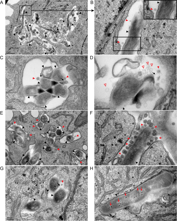
Fig 6.
Transmission electron microscopy of phage infection of intracellular M. abscessus. GD20-infected THP-1 cells were incubated with BPsΔ for 24 h (A–D). THP-1 phagosome with BPsΔ phage adsorbed to intracellular GD20 (A and B). BPsΔ phage tails adsorb to intracellular GD20 (C). BPsΔ phage, with empty and intact capsids, adsorbed to intracellular GD20 (D). GD82-infected THP-1 cells were incubated with BPsΔ for 24 h. Colocalized phage and bacteria are observed in multiple phagosomes in the same cell (E). GD82-infected THP-1 cells incubated with BPsΔ for 48 h. Multiple phages adsorbed to the bacterial pole are observed (F). GD82-infected A549 cells incubated with BPsΔ for 24 h and a phage tail adsorbed to intracellular bacteria (G). GD82-infected A549 cells were incubated with BPsΔ for 48 h, and intracellular phage progeny was observed (H). Black arrowheads indicate intracellular M. abscessus, red arrowheads indicate adsorbed phage, black arrows indicate phage tails, red outlined arrowheads indicate empty phage capsid, and red arrows indicate phage progeny. Only a subset of phage particles and bacteria are indicated. Black boxes indicate areas of higher magnification (A and B). For additional TEM images, see Fig. S5.

