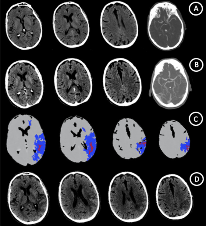
An 82 yo man with aphasia and right-side weakness performed NCCT and single-phase CTA from 2.5 h from symptoms onset in spoke center that showed a large parenchymal hypodensity referring to acute ischemic stroke with ASPECT score of 6 (Insula, M2, M3, M6 segments) due to distal M1 cerebral artery occlusion. 3.5 h later, neuroimaging study protocol with NCCT, multi-phase CT-Angiography and CT-Perfusion in hub center was repeated. CT-Perfusion was realized with OLEA Perfusion Software that define total hypoperfused tissue with Tmax > 6 s and infarct core with rCBF < 40%. A spontaneous partial vascular recanalization with leptomeningeal vasodilatation occurred and CTP showed a little ischemic core (3,8 ml) and large penumbra (108,5 ml). 3 days after, NCCT uphold the ischemic core size. This case shows a typical Perfusion Scotoma with underestimation of ischemic core volume > 6 h from symptom onset due to early reperfusion from partial recanalization as well as luxury perfusion [1, 2] (Fig. 1).
Fig. 1.
Neuroimaging study protocol in spoke and hub center: NCCT with ASPECT score of 6 and distal M1 cerebral artery occlusion. (A) NCCT, mCTA and CTP > 6 h from symptom onset with spontaneous recanalization and ischemic core underestimation due to luxury perfusion (B,C). NCCT at 3 days uphold a large ischemic core size (D)
Funding
Open access funding provided by Università degli Studi di Firenze within the CRUI-CARE Agreement. No targeted funding reported.
Declarations
Ethical approval and Informed consent statement
Full consent was obtained from the patient’s parents for the case report and the study was approved by the authors’ institutional ethics committee.
Conflict of interest
None.
Footnotes
Publisher's Note
Springer Nature remains neutral with regard to jurisdictional claims in published maps and institutional affiliations.
References
- 1.Abrams K, Dabus G. Perfusion Scotoma: A Potential Core Underestimation in CT Perfusion in the Delayed time window in patients with acute ischemic stroke. AJNR Am J Neuroradiol. 2022;43(6):813–816. doi: 10.3174/ajnr.A7524. [DOI] [PMC free article] [PubMed] [Google Scholar]
- 2.Nagar VA, McKinney AM, Karagulle AT, et al. Reperfusion phenomenon masking acute and subacute infarcts at dynamic perfusion CT: confirmation by fusion of CT and diffusion-weighted MR images. AJNR Am J Neuroradiol. 2009;193:1629–38. doi: 10.2214/AJR.09.2664. [DOI] [PubMed] [Google Scholar]


