Persoonia : Molecular Phylogeny and Evolution of Fungi logoLink to Persoonia : Molecular Phylogeny and Evolution of Fungi
. 2022 Dec 20;49:261–350. doi: 10.3767/persoonia.2022.49.08

Fungal Planet description sheets: 1436–1477

YP Tan 1,2, SL Bishop-Hurley 1, RG Shivas 2, DA Cowan 3, G Maggs-Kölling 4, SSN Maharachchikumbura 5, U Pinruan 6, KL Bransgrove 7, S De la Peña-Lastra 8, E Larsson 9, T Lebel 10, S Mahadevakumar 11, A Mateos 12, ER Osieck 13, A Rigueiro-Rodríguez 8, S Sommai 6, K Ajithkumar 14, A Akulov 15, FE Anderson 16, F Arenas 17, S Balashov 18, Á Bañares 19, DK Berger 20, MV Bianchinotti 16,21, S Bien 22, P Bilański 23, A-G Boxshall 24, M Bradshaw 25, J Broadbridge 26, FJS Calaça 27, C Campos-Quiroz 28, J Carrasco-Fernández 28, JF Castro 28, S Chaimongkol 6,29, S Chandranayaka 30, Y Chen 5, D Comben 31, JDW Dearnaley 32, AS Ferreira-Sá 27, K Dhileepan 31, ML Díaz 16,21, PK Divakar 33, S Xavier-Santos 27, A Fernández-Bravo 34, J Gené 34, FE Guard 35, M Guerra 28, S Gunaseelan 36, J Houbraken 37, K Janik-Superson 38, R Jankowiak 23, M Jeppson 9, Ž Jurjević 18, M Kaliyaperumal 36, LA Kelly 7, K Kezo 36, AN Khalid 39, P Khamsuntorn 6, D Kidanemariam 20, M Kiran 40, E Lacey 41, GJ Langer 22, LV López-Llorca 42,43, JJ Luangsa-ard 6, P Lueangjaroenkit 44,45, HT Lumbsch 46, JG Maciá-Vicente 47,48, LS Mamatha Bhanu 49, TS Marney 1, JE Marqués-Gálvez 17, A Morte 17, A Naseer 39, A Navarro-Ródenas 17, O Oyedele 50, S Peters 22, S Piskorski 51, L Quijada 52, GH Ramírez 16,53, K Raja 36, A Razzaq 39, VJ Rico 33, A Rodríguez 17, M Ruszkiewicz-Michalska 51, RM Sánchez 16,21, C Santelices 28, AS Savitha 54, M Serrano 8, L Leonardo-Silva 27, H Solheim 55, S Somrithipol 6, MY Sreenivasa 56, H Stępniewska 23, D Strapagiel 57, T Taylor 31, D Torres-Garcia 34, J Vauras 58, M Villarreal 59, CM Visagie 60, M Wołkowycki 61, W Yingkunchao 6,29, E Zapora 61, JZ Groenewald 37, PW Crous 37,60
PMCID: PMC10792226  PMID: 38234383

Abstract

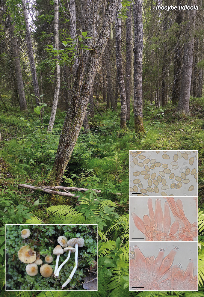
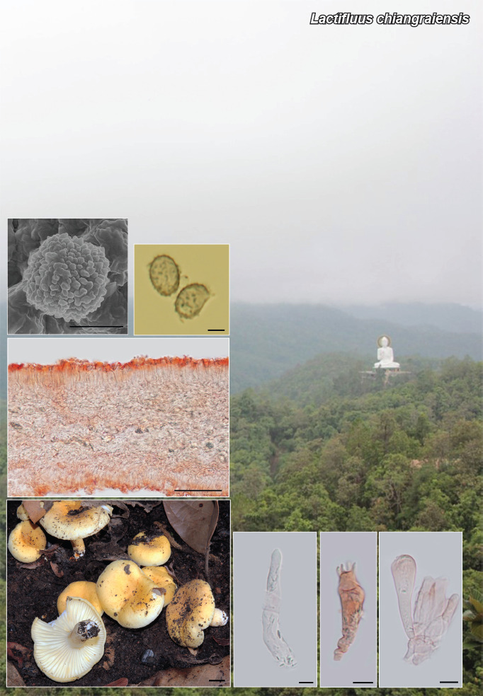
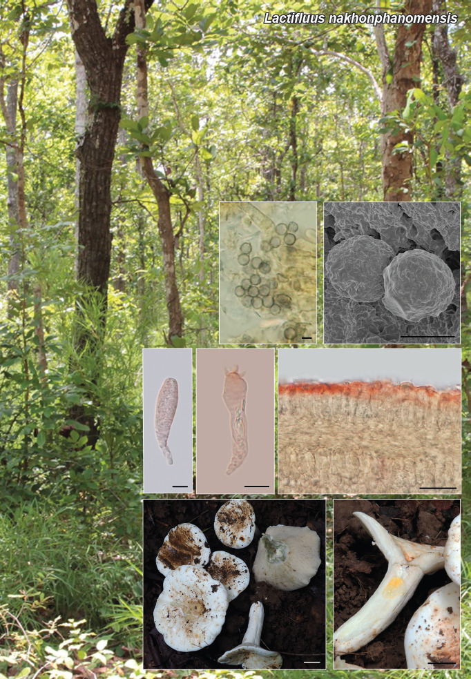
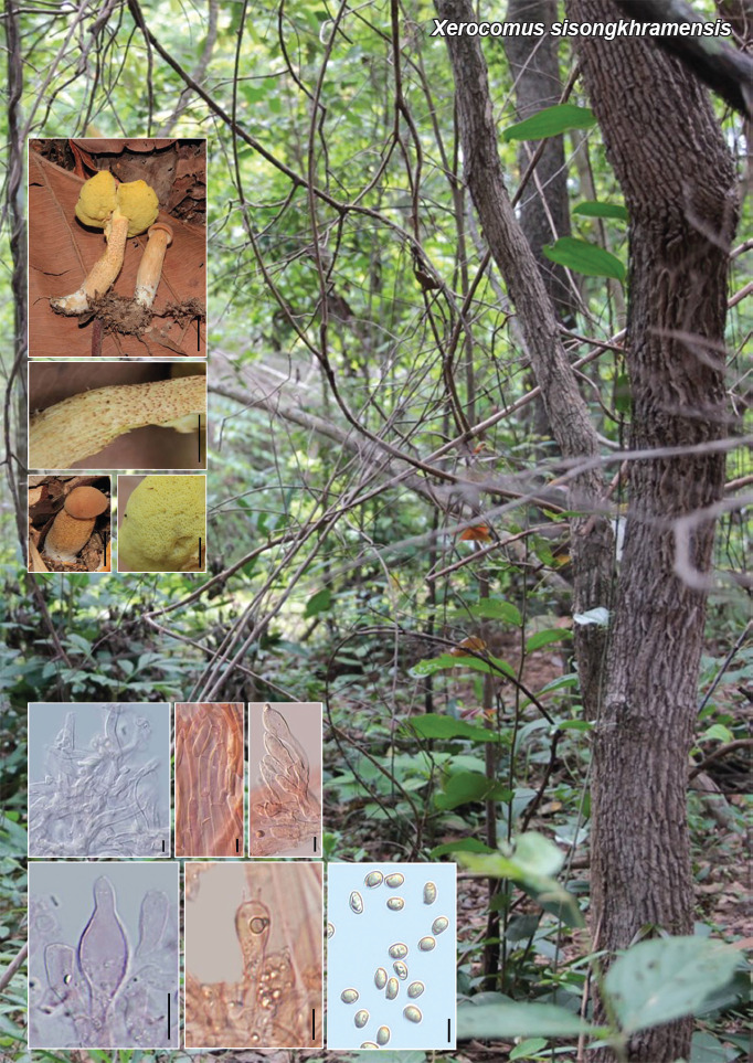
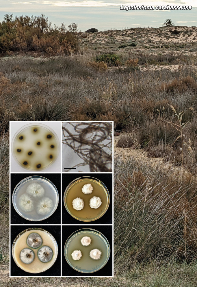
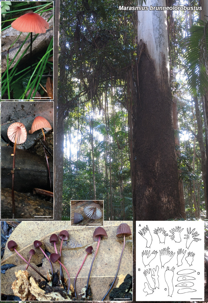
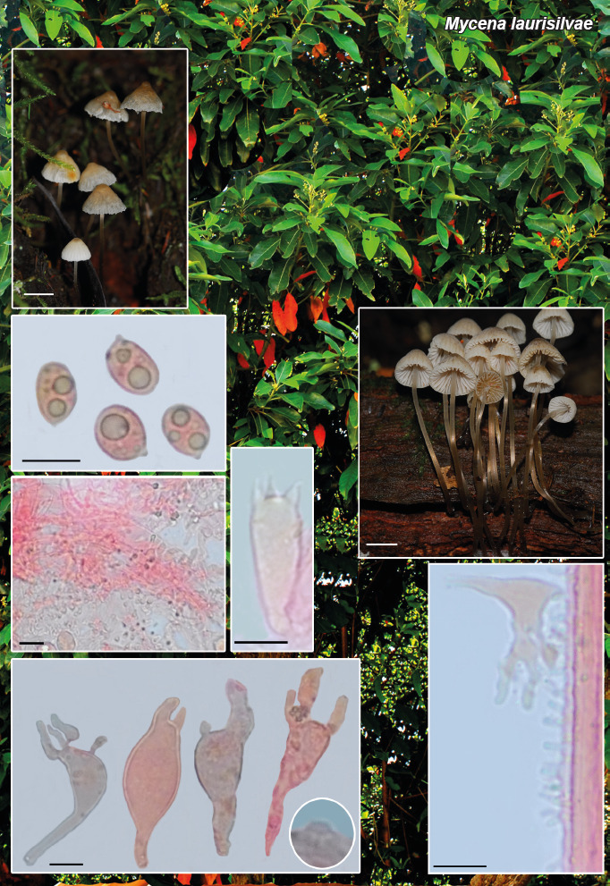
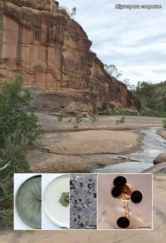
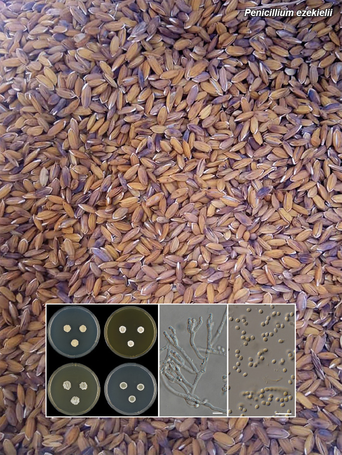
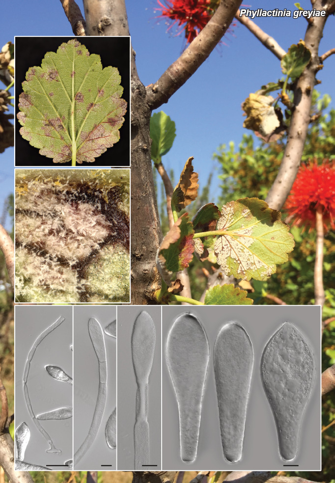
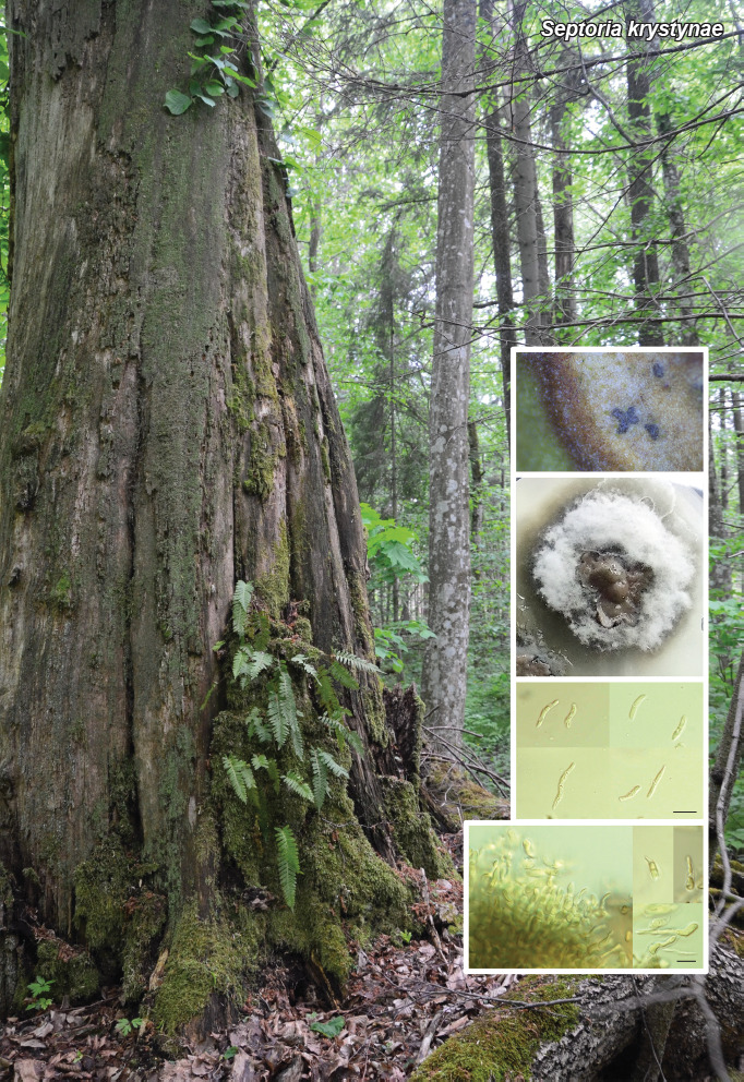
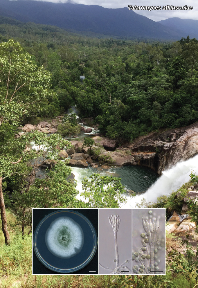
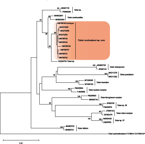
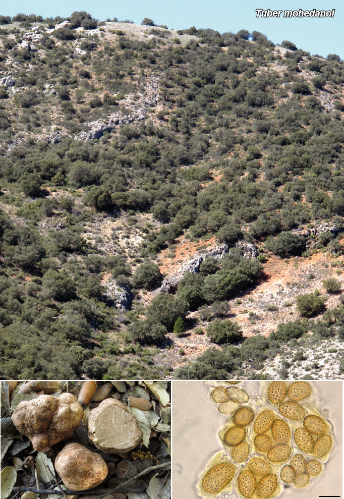
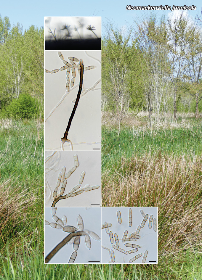
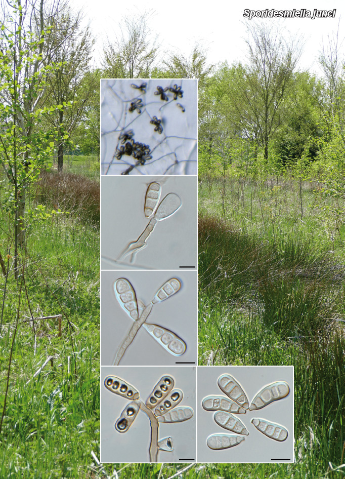
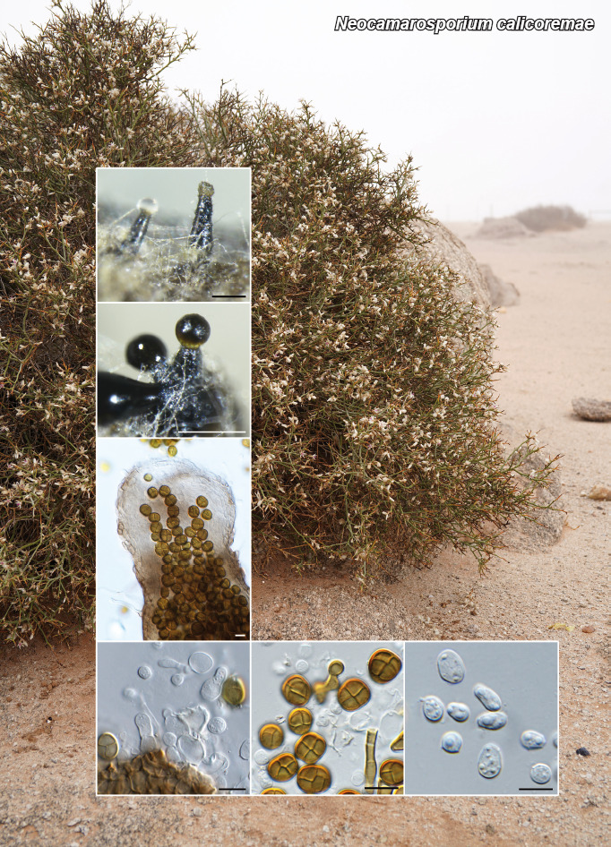
Novel species of fungi described in this study include those from various countries as follows: Argentina, Colletotrichum araujiae on leaves, stems and fruits of Araujia hortorum. Australia, Agaricus pateritonsus on soil, Curvularia fraserae on dying leaf of Bothriochloa insculpta, Curvularia millisiae from yellowing leaf tips of Cyperus aromaticus, Marasmius brunneolorobustus on well-rotted wood, Nigrospora cooperae from necrotic leaf of Heteropogon contortus, Penicillium tealii from the body of a dead spider, Pseudocercospora robertsiorum from leaf spots of Senna tora, Talaromyces atkinsoniae from gills of Marasmius crinis-equi and Zasmidium pearceae from leaf spots of Smilaxglyciphylla. Brazil, Preussia bezerrensis from air. Chile, Paraconiothyrium kelleni from the rhizosphere of Fragaria chiloensis subsp. chiloensis f. chiloensis. Finland, Inocybe udicola on soil in mixed forest with Betula pendula, Populus tremula, Picea abies and Alnus incana. France, Myrmecridium normannianum on dead culm of unidentified Poaceae. Germany, Vexillomyces fraxinicola from symptomless stem wood of Fraxinus excelsior. India, Diaporthe limoniae on infected fruit of Limonia acidissima, Didymella naikii on leaves of Cajanus cajan, and Fulvifomes mangroviensis on basal trunk of Aegiceras corniculatum. Indonesia, Penicillium ezekielii from Zea mays kernels. Namibia, Neocamarosporium calicoremae and Neocladosporium calicoremae on stems of Calicorema capitata, and Pleiochaeta adenolobi on symptomatic leaves of Adenolobus pechuelii. Netherlands, Chalara pteridii on stems of Pteridium aquilinum, Neomackenziella juncicola (incl. Neomackenziella gen. nov.) and Sporidesmiella junci from dead culms of Juncus effusus. Pakistan, Inocybe longistipitata on soil in a Quercus forest. Poland, Phytophthora viadrina from rhizosphere soil of Quercus robur, and Septoria krystynae on leaf spots of Viscum album. Portugal (Azores), Acrogenospora stellata on dead wood or bark. South Africa, Phyllactinia greyiae on leaves of Greyia sutherlandii and Punctelia anae on bark of Vachellia karroo. Spain, Anteaglonium lusitanicum on decaying wood of Prunus lusitanica subsp. lusitanica, Hawksworthiomyces riparius from fluvial sediments, Lophiostoma carabassense endophytic in roots of Limbarda crithmoides, and Tuber mohedanoi from calcareus soils. Spain (Canary Islands), Mycena laurisilvae on stumps and woody debris. Sweden, Elaphomyces geminus from soil under Quercus robur. Thailand, Lactifluus chiangraiensis on soil under Pinus merkusii, Lactifluus nakhonphanomensis and Xerocomus sisongkhramensis on soil under Dipterocarpus trees. Ukraine, Valsonectria robiniae on dead twigs of Robinia hispida. USA, Spiralomyces americanus (incl. Spiralomyces gen. nov.) from office air. Morphological and culture characteristics are supported by DNA barcodes.

Citation: Tan YP, Bishop-Hurley SL, Shivas RG, et al. 2022. Fungal Planet description sheets: 1436–1477. Persoonia 49: 261–350. https://doi.org/10.3767/persoonia.2022.49.08

Keywords: ITS nrDNA barcodes, LSU, new taxa, systematics

Acknowledgements

The work of Pedro Crous and colleagues benefitted from funding by the European Union’s Horizon 2020 research and innovation program (RISE) under the Marie Sklodowska-Curie grant agreement No. 101008129, project acronym ‘Mycobiomics’, and the Dutch NWO Roadmap grant agreement No. 2020/ENW/00901156, project ‘Netherlands Infrastructure for Ecosystem and Biodiversity Analysis – Authoritative and Rapid Identification System for Essential biodiversity information’ (acronym NIEBA-ARISE). Don Cowan gratefully acknowledges funding support from the National Research Foundation (grant number 137954) and from the University of Pretoria. Jose G. Maciá-Vicente acknowledges support from the German Research Foundation under grant MA7171/1-1, and from the Landes-Offensive zur Entwicklung Wissenschaftlich-ökonomischer Exzellenz (LOEWE) of the state of Hesse within the framework of the Cluster for Integrative Fungal Research (IPF). Cecilia Santelices and colleagues acknowledge financial support from the FONDECYT INICIACIÓN project No. 11191074 from the Chilean National Agency for Research and Development (ANID) and to the FONDEQUIP Program from ANID (grant: EQM200205) for funding a platform of equipment for preservation of microbial genetic resources. Hanna Stepniewska and colleagues were financed by the Ministry of Science and Higher Education of the Republic of Poland (SUB/040013/D019). Saúl De la Peña-Lastra and colleagues would like to thank the Secretaria Regional do Ambientee Alterações Climáticas Açores for the permission granted for sampling (Licença n° 16/2021/DRAAC). The research of P.K. Divakar and colleagues was supported by the Spanish Ministry of Science and Innovation (PID2019-105312GB-I00/AEI/10.13039/501100011033). Furthermore, Luis Alberto Parra is acknowledged for his help on nomenclature, and Donald H. Pfister for his revision of the text. Antonio Mateos and colleagues would like to thank the great naturalist Cristóbal Burgos Morillo for his help in processing the images and Amalio Gutiérrez for his collaboration, company and cooperation on surveys. Gustavo Hernán Ramírez and colleagues acknowledge German Barros and Martín Bonacci for their help with molecular studies. Frances E. Guard and colleagues thank the curation staff at RBGV, PERTH and BRI for their help with loans and processing of specimens. Ángel Bañares and colleagues thank Pablo Alvarado ALVALAB and Fernanda Rodríguez Fariña Unidade de Bioloxía Molecular (SAI) Universidade da Coruña who assisted with the generation of the ITS sequence data. The research of D. Kidanemariam and D.K. Berger was supported by the National Research Foundation of South Africa (grant nr 120398) and the University of Pretoria. Cobus M. Visagie and co-authors are grateful for the advice received from Uwe Braun and thank Konstanze Bensch (MycoBank) for Latin assistance. Steffen Bien and colleagues thank Ulrike Damm from the Senckenberg Natural History Museum Görlitz for technical support. Teresa Lebel and colleagues acknowledge the Royal Botanic Gardens Victoria and the Friends of the Royal Botanic Gardens Victoria for funding and support for the Northern Territory field and lab work; Northern Territory Herbarium staff for space and lab access and help with collection curation; State Herbarium of South Australia for support of the undergraduate intern J. Broadbridge; their research was also supported through funding from Australian Biological Resources Study grant (4-EHOJ161) to the State Herbarium of South Australia. Asunción Morte is grateful to the Fundación Séneca - Agencia de Ciencia y Tecnología de la Región de Murcia (20866/PI/18) for financial support. Malarvizhi Kaliyaperumal and Sugantha Gunaseelan would like to acknowledge DST, SERB-EMR (EMR/2016/003078) for the financial assistance. Malarvizhi Kaliyaperumal and Kanaga valli Raja are grateful to TANSCST, DOTE, Chennai (Student Scheme Project, 2018 (BS-014)) for providing financial aid. Attihalli S. Savitha and co-authors thank Bettadapura R. Nuthan, Department of Studies in Microbiology, for his kind support in culture maintenance and laboratory assistance. Shivannegowda Mahadevakumar thanks the Director of KSCSTE – Kerala Forest Research Institute for support. Mikael Jeppson, Jukka Vauras and Ellen Larsson thank the Swedish Taxonomy Initiative (SLU Artdatabanken, Uppsala) (grant to E. Larsson dha.2019.4.3-13). The study of Daniel Torres-Garcia and colleagues was partially supported by the Spanish Ministerio de Economía, Industria y Competitividad (grant PID2021-128068NB-100). Shivannegowda Mahadevakumar and co-authors thank Bettadapura R. Nuthan, Department of Studies in Microbiology, University of Mysore and Bettadapura R. Meghavarshinigowda, Department of Studies in Botany, University of Mysore, for their assistance in the laboratory. Umpawa Pinruan and colleagues were financially supported by the Platform Technology Management Section, National Center for Genetic Engineering and Biotechnology (BIOTEC), Project Grant No. P19-50231. Antônio Sérgio Ferreira-Sá and colleagues thank the Coordenação de Aperfeiçoamento de Pessoal de Nível Superior (CAPES) for granting PhD scholarships (Finance code – 001) to ASFS and LLS.

REFERENCES

  1. Akata I, Halici MG, Uzun Y. 2011. Additional macrofungi records from Trabzon province for the mycobiota of Turkey. Turkish Journal of Botany 35: 309–314. [Google Scholar]
  2. Álvarez VI, Raymundo T, Valenzuela R. 2016. Hongos histerioides (Dothideomycetes, Ascomycota) del bosque tropical caducifolio en el Parque Nacional Lagunas de Chacahua, Oaxaca, México. Acta Botánica Mexicana 116: 49–64. [Google Scholar]
  3. An YY, Zeng XY, Geng K, et al. 2019. One new species and one new record of Zasmidium in China. Biodiversity Data Journal 9: e59001. [DOI] [PMC free article] [PubMed] [Google Scholar]
  4. Andreasen M, Skrede I, Jaklitsch WM, et al. 2021. Multi-locus phylogenetic analysis of lophiostomatoid fungi motivates a broad concept of Lophiostoma and reveals nine new species. Persoonia 46: 240–271. [DOI] [PMC free article] [PubMed] [Google Scholar]
  5. Antonin V, Noordeloos M. 2010. A monograph of marasmioid and collybioid fungi in Europe. Eching, IHW-Verlag. [Google Scholar]
  6. Archana S, Kharwar RN, Raghvendra S, et al. 2014. A new species of Zasmidium (Mycosphaerellaceae) from India. Sydowia 66: 309–312. [Google Scholar]
  7. Arenal F, Platas G, Peláez F. 2007. A new endophytic species of Preussia (Sporormiaceae) inferred from morphological observations and molecular phylogenetic analysis. Fungal Diversity 25: 1–17. [Google Scholar]
  8. Ariyawansa HA, Tanaka K, Thambugala KM, et al. 2014. A molecular phylogenetic reappraisal of the Didymosphaeriaceae (= Montagnulaceae). Fungal Diversity 68: 69–104. [Google Scholar]
  9. Arzanlou M, Groenewald JZ, Gams W, et al. 2007. Phylogenetic and morphotaxonomic revision of Ramichloridium and allied genera. Studies in Mycology 58: 57–93. [DOI] [PMC free article] [PubMed] [Google Scholar]
  10. Asgari B, Zare R. 2010. Two new species of Preussia from Iran. Nova Hedwigia 90: 533–548. [Google Scholar]
  11. Bandini D, Oertel B, Eberhardt U. 2021a. Noch mehr Risspilze (3): Einundzwanzig neue Arten der Familie Inocybaceae. Mycologica Bavarica 22: 31–138. [Google Scholar]
  12. Bandini D, Oertel B, Eberhardt U. 2021b. A fresh outlook on the smooth-spored species of Inocybe: type studies and 18 new species. Mycological Progress 20: 1019–1114. [Google Scholar]
  13. Bandini D, Oertel B, Eberhardt U. 2022. Noch mehr Risspilze (3): Einundzwanzig neue Arten der Familie Inocybaceae. Mycologia Bavarica 22: 31–138. [Google Scholar]
  14. Bao D-F, McKenzie EHC, Bhat DJ, et al. 2020. Acrogenospora (Acrogenosporaceae, Minutisphaerales) appears to be a very diverse genus. Frontiers in Microbiology 11: 1606. [DOI] [PMC free article] [PubMed] [Google Scholar]
  15. Baral HO, Quijada L. 2020. Nomenclatural novelties. Index Fungorum 10: 1–3. [Google Scholar]
  16. Bashir H, Chen J, Jabeen S, et al. 2021. An overview of Agaricus section Hondenses and Agaricus section Xanthodermatei with description of eight new species from Pakistan. Scientific Reports 11: 12905. [DOI] [PMC free article] [PubMed] [Google Scholar]
  17. Bell A. 2005. An illustrated guide to the coprophilous Ascomycetes of Australia. CBS Biodiversity Series (Utrecht) 3: 1–173. [Google Scholar]
  18. Bezerra JDP, Sandoval-Denis M, Paiva LM, et al. 2017. New endophytic Toxicocladosporium species from cacti in Brazil, and description of Neocladosporium gen. nov. IMA Fungus 8: 77–97. [DOI] [PMC free article] [PubMed] [Google Scholar]
  19. Bien S, Kraus C, Damm U. 2020. Novel collophorina-like genera and species from Prunus trees and vineyards in Germany. Persoonia 45: 46–67. [DOI] [PMC free article] [PubMed] [Google Scholar]
  20. Boehm EWA, Mugambi GK, Miller AN, et al. 2009a. A molecular phylogenetic reappraisal of the Hysteriaceae, Mytilinidiaceae and Gloniaceae (Pleosporomycetidae, Dothideomycetes) with keys to world species. Studies in Mycology 64: 49–83. [DOI] [PMC free article] [PubMed] [Google Scholar]
  21. Boehm EWA, Schoch CL, Spatafora JW. 2009b. On the evolution of the Hysteriaceae and Mytilinidiaceae (Pleosporomycetidae, Dothideomycetes, Ascomycota) using four nuclear genes. Mycological Research 113: 461–479. [DOI] [PubMed] [Google Scholar]
  22. Bon M. 1998. Novitates, 1, Marasmiaceae et Dermolomataceae comb., st. et sp. nov. / Novitates – Genre Inocybe, sous-genre Clypeus Britz., Documents Mycologiques 28: 1–45. [Google Scholar]
  23. Bonito GM, Gryganskyi AP, Trappe JM, et al. 2010. A global meta-analysis of Tuber ITS rDNA sequences: species diversity, host associations and long-distance dispersal. Molecular Ecology 19: 4994–5008. [DOI] [PubMed] [Google Scholar]
  24. Bradshaw M, Braun U, Pfister D. 2022. Phylogeny and taxonomy of the genera of the Erysiphaceae, part 1, Golovinomyces. Mycologia 114(6): 964–993. [DOI] [PubMed] [Google Scholar]
  25. Braun U, Cook RTA. 2012. Taxonomic manual of the Erysiphales (powdery mildews). CBS Biodiversity Series 11. CBS, Utrecht, The Netherlands. [Google Scholar]
  26. Braun U, Crous PW, Nakashima C. 2014. Cercosporoid fungi (Mycosphaerellaceae) 2. Species on monocots (Acoraceae to Xyridaceae, excluding Poaceae). IMA Fungus 5: 203–390. [DOI] [PMC free article] [PubMed] [Google Scholar]
  27. Burgess TI, Simamora AV, White D, et al. 2018. New species from Phytophthora Clade 6a: evidence for recent radiation. Persoonia 41: 1–17. [DOI] [PMC free article] [PubMed] [Google Scholar]
  28. Carneiro de Almeida DA, Pascholati Gusmão LF, Miller AN. 2014. A new genus and three new species of hysteriaceous ascomycetes from the semiarid region of Brazil. Phytotaxa 176: 298–308. [Google Scholar]
  29. Ceruti A, Fontana A, Nosenzo C. 2003. Le specie europee del genere Tuber: una revisione storica. Vol. 37. Italy, Turin, Museo Regionale di Scienze Naturali. [Google Scholar]
  30. Chakraborty D, Parihar A, Mehta N, et al. 2017. A new species of Xerocomus (Boletaceae) from India. Mycosphere 8: 44–50. [Google Scholar]
  31. Chevallier FF. 1826. Flore générale des environs de Paris, Vol I. Ferra Librairie-Editeur, Paris, France. [Google Scholar]
  32. Chupp C. 1954. A monograph of the fungus genus Cercospora. Published by the author, Ithaca, New York, USA. [Google Scholar]
  33. Cock MJW, Evans HC. 1984. Possibilities for biological control of Cassia tora and C. obtusifolia. Tropical Pest Management 30: 339–350. [Google Scholar]
  34. Crespo A, Ferencova Z, Pérez-Ortega S, et al. 2010. Austroparmelina, a new Australasian lineage in parmelioid lichens (Parmeliaceae, Ascomycota): a multigene and morphological approach. Systematics and Biodiversity 8: 209–221. [Google Scholar]
  35. Crossland FLS. 1900. New and critical British Fungi found in West Yorkshire. The Naturalist. 1900: 5–10. [Google Scholar]
  36. Crous PW, Boers J, Holdom D, et al. 2022. Fungal Planet description sheets: 1383–1435. Persoonia 48: 261–371. [DOI] [PMC free article] [PubMed] [Google Scholar]
  37. Crous PW, Cowan DA, Maggs-Kölling G, et al. 2020a. Fungal Planet description sheets: 1112–1181. Persoonia 45: 251–409. [DOI] [PMC free article] [PubMed] [Google Scholar]
  38. Crous PW, Cowan DA, Maggs-Kölling G, et al. 2021a. Fungal Planet description sheets: 1182–1283. Persoonia 46: 313–528. [DOI] [PMC free article] [PubMed] [Google Scholar]
  39. Crous PW, Osieck ER, Jurjević Ž, et al. 2021b. Fungal Planet description sheets: 1284–1382. Persoonia 47: 178–374. [DOI] [PubMed] [Google Scholar]
  40. Crous PW, Schumacher RK, Wingfield MJ, et al. 2018. New and interesting fungi. 1. Fungal Systematics and Evolution 1: 169–215. [DOI] [PMC free article] [PubMed] [Google Scholar]
  41. Crous PW, Shivas RG, Quaedvlieg W, et al. 2014. Fungal Planet description sheets: 214–280. Persoonia 32: 184–306. [DOI] [PMC free article] [PubMed] [Google Scholar]
  42. Crous PW, Wingfield MJ, Chooi YH, et al. 2020b. Fungal Planet description sheets: 1042–1111. Persoonia 44: 301–459. [DOI] [PMC free article] [PubMed] [Google Scholar]
  43. Crous PW, Wingfield MJ, Schumacher RK, et al. 2020c. New and interesting fungi. 3. Fungal Systematics and Evolution 6: 157–231. [DOI] [PMC free article] [PubMed] [Google Scholar]
  44. Damm U, Cannon PF, Liu F, et al. 2013. The Colletotrichum orbiculare species complex: Important pathogens of field crops and weeds. Fungal Diversity 61: 29–59. [Google Scholar]
  45. Damm U, Cannon PF, Woudenberg JHC, et al. 2012. The Colletotrichum boninense species complex. Studies in Mycology 73: 1–36. [DOI] [PMC free article] [PubMed] [Google Scholar]
  46. Damm U, Verkley GJ, Crous PW, et al. 2008. Novel Paraconiothyrium species on stone fruit trees and other woody hosts. Persoonia 20: 9–17. [DOI] [PMC free article] [PubMed] [Google Scholar]
  47. De Beer ZW, Marincowitz S, Duong TA, et al. 2016. Hawksworthiomyces gen. nov. (Ophiostomatales), illustrates the urgency for a decision on how to name novel taxa known only from environmental nucleic acid sequences (ENAS). Fungal Biology 120: 1323–1340. [DOI] [PubMed] [Google Scholar]
  48. De Leo F, Urzì C, De Hoog GS. 1999. Two Coniosporium species from rock surfaces. Studies in Mycology 43: 70–79. [Google Scholar]
  49. Dennis RWG. 1955. Ascomycetes from Tristan da Cunha. Results of the Norwegian Scientific Expedition to Tristan da Cunha (1937–1938) 36–38: 1–10. [Google Scholar]
  50. Dennis RWG. 1981. British Ascomycetes. Vaduz, Cramer. [Google Scholar]
  51. Divakar PK, Crespo A, Kraichak E, et al. 2017. Using a temporal phylogenetic method to harmonize family- and genus-level classification in the largest clade of lichen-forming fungi. Fungal Diversity 84: 101–117. [Google Scholar]
  52. Divakar PK, Kauff F, Crespo A, et al. 2013. Understanding phenotypical character evolution in parmelioid lichenized fungi (Parmeliaceae, Ascomycota). PLoS ONE 8: e83115. [DOI] [PMC free article] [PubMed] [Google Scholar]
  53. Doveri F. 2004. Fungi Fimicoli Italici – a guide to the recognition of Basidiomycetes and Ascomycetes living on faecal material. Associazione Micologica Bresadola, 1st ed. [Google Scholar]
  54. Ellis MB. 1972. Dematiaceous Hyphomycetes. XI. Mycological Papers 131: 1–25. [Google Scholar]
  55. Ellis MB, Ellis JP. 1985. Microfungi on land plants. An identification handbook. Macmillan Publishing Company. [Google Scholar]
  56. Fan YG, Wu RH, Bau T. 2018. Two new species and eight newly recorded species of subg. Inocybe from China. Journal of Fungal Research 16: 70–83. [Google Scholar]
  57. Farr DF, Rossman AY. 2022. Fungal Databases, U.S. National Fungus Collections, ARS, USDA. https://nt.ars-grin.gov/fungaldatabases/ [Retrieved 25 Sept. 2022]. [Google Scholar]
  58. Fisher PJ. 1985. The anamorph of Claussenomyces atrovirens. Transactions of the British Mycological Society 85: 759–760. [Google Scholar]
  59. Gamundi IJ, Giaiotti AL. 1995. A new species of Claussenomyces (Helotiales) from southern South America. New Zealand Journal of Botany 33: 513–517. [Google Scholar]
  60. Gilliam MS. 1976. The genus Marasmius in the northeastern United States and adjacent Canada. Mycotaxon 4: 1–144. [Google Scholar]
  61. Goh TK, Hyde KD, Tsui KM. 1998. The hyphomycete genus Acrogenospora, with two new species and two new combinations. Mycological Research 102: 1309–1315. [Google Scholar]
  62. Gonçalves MFM, Aleixo A, Vicente TFL, et al. 2019. Three new species of Neocamarosporium isolated from saline environments: N. aestuarinum sp. nov., N. endophyticum sp. nov. and N. halimiones sp. nov. Mycosphere 10: 608–621. [Google Scholar]
  63. Guindon S, Dufayard JF, Lefort V, et al. 2010. New algorithms and methods to estimate maximum-likelihood phylogenies: assessing the performance of PhyML 3.0. Systematic Biology 59: 307–321. [DOI] [PubMed] [Google Scholar]
  64. Hale ME, Jr. 1973. Fine structure of the cortex in the lichen family Parmeliaceae viewed with the scanning-electron microscope. Smithsonian Contributions to Botany 10: 1–92. [Google Scholar]
  65. Hale ME, Jr. 1976. A monograph of the lichen genus Pseudoparmelia Lynge (Parmeliaceae). Smithsonian Contributions to Botany 31: 1–62. [Google Scholar]
  66. Hale ME, Jr. 1980. Taxonomy and distribution of the Parmelia flaventior group (Lichens: Parmeliaceae). Journal of the Hattori Botanical Laboratory 47: 75–84. [Google Scholar]
  67. Hansen EM, Wilcox WF, Reeser PW, et al. 2009. Phytophthora rosacearum and P. sansomeana, new species segregated from the Phytophthora megasperma “complex”. Mycologia 101: 129–135. [DOI] [PubMed] [Google Scholar]
  68. Hattori T, Sakayaroj J, Jones EBG, et al. 2014. Three species of Fulvifomes (Basidiomycota, Hymenochaetales) associated with rots on mangrove tree Xylocarpus granatum in Thailand. Mycoscience 55: 344–354. [Google Scholar]
  69. He MQ, Chuankid B, Hyde KD, et al. 2018. A new section and species of Agaricus subgenus Pseudochitonia from Thailand. MycoKeys 40: 53–67. [DOI] [PMC free article] [PubMed] [Google Scholar]
  70. Heinemann P. 1978. Essai d’une clé de determination des genres Agaricus et Micropsalliota. Sydowia 30: 6–37. [Google Scholar]
  71. Houbraken J, Kocsubé S, Visagie CM, et al. 2020. Classification of Aspergillus, Penicillium, Talaromyces and related genera (Eurotiales): An overview of families, genera, subgenera, sections, series and species. Studies in Mycology 95: 5–169. [DOI] [PMC free article] [PubMed] [Google Scholar]
  72. Huelsenbeck JP, Ronquist F. 2001. MrBayes: Bayesian inference of phylogenetic trees. Bioinformatics 17: 754–755. [DOI] [PubMed] [Google Scholar]
  73. Husbands DR, Henkel TW, Bonito G, et al. 2013. New species of Xerocomus (Boletales) from the Guiana Shield, with notes on their mycorrhizal status and fruiting occurrence. Mycologia 105: 422–435. [DOI] [PubMed] [Google Scholar]
  74. Hyde KD, Jones EBG, Liu J-K, et al. 2013. Families of Dothideomycetes. Fungal Diversity 63: 1–313. [Google Scholar]
  75. Hyde KD, Tennakoon DS, Jeewon R, et al. 2019. Fungal diversity notes 1036–1150: taxonomic and phylogenetic contributions on genera and species of fungal taxa. Fungal Diversity 96: 1–242. [Google Scholar]
  76. Jaklitsch W, Fournier J, Voglmayr H. 2018. Two unusual new species of Pleosporales: Anteaglonium rubescens and Atrocalyx asturiensis. Sydowia 70: 129–140. [DOI] [PMC free article] [PubMed] [Google Scholar]
  77. Jankowiak R, Stępniewska H, Bilański P. et al. 2014. Occurrence of Phytophthora plurivora and other Phytophthora species in oak forests of southern Poland and their association with site conditions and the health status of trees. Folia Microbiologica 59: 531–542. [DOI] [PubMed] [Google Scholar]
  78. Jayasiri SC, Hyde KD, Jones EBG, et al. 2018. Taxonomic novelties of hysteriform Dothideomycetes. Mycosphere 9: 803–837. [Google Scholar]
  79. Jayasiri SC, Hyde KD, Jones EBG, et al. 2019. Diversity, morphology and molecular phylogeny of Dothideomycetes on decaying wild seed pods and fruits. Mycosphere 10: 1–186. [Google Scholar]
  80. Jayasiri SC, Jones EBG, Kang J-C, et al. 2016. A new species of genus Anteaglonium (Anteagloniaceae, Pleosporales) with its asexual morph. Phytotaxa 263: 233–244. [Google Scholar]
  81. Kalyaanamoorthy S, Minh BQ, Wong TKF, et al. 2017. ModelFinder: Fast model selection for accurate phylogenetic estimates. Nature Methods 14: 587–589. [DOI] [PMC free article] [PubMed] [Google Scholar]
  82. Katoh K, Standley DM. 2013. MAFFT multiple sequence alignment software version 7: improvements in performance and usability. Molecular Biology and Evolution 30: 772–780. [DOI] [PMC free article] [PubMed] [Google Scholar]
  83. Katoh K, Toh H. 2008. Recent developments in the MAFFT multiple sequence alignment program. Briefings in Bioinformatics 9: 286–298. [DOI] [PubMed] [Google Scholar]
  84. Kearse M, Moir R, Wilson A, et al. 2012. Geneious Basic: an integrated and extendable desktop software platform for the organization and analysis of sequence data. Bioinformatics 28: 1647–1649. [DOI] [PMC free article] [PubMed] [Google Scholar]
  85. Kennedy AH, Schoch CL, Marrero G, et al. 2022. Publicly available and validated DNA reference sequences are critical to fungal identification and global plant protection efforts: A use-case in Colletotrichum. Plant Disease 106: 1573–1596. [DOI] [PMC free article] [PubMed] [Google Scholar]
  86. Khan MB, Naseer A, Aqdus F, et al. 2022. Inocybe quercicola sp. nov. (Agaricales, Inocybaceae), from Pakistan. Microbial Biosystems 6: 22–29. [Google Scholar]
  87. Kimura M. 1980. A simple method for estimating evolutionary rate of base substitutions through comparative studies of nucleotide sequences. Journal of Molecular Evolution 16: 111–120. [DOI] [PubMed] [Google Scholar]
  88. Kong HZ, Liang ZQ. 2003. A new Penicillium species isolated from Jiangxi, China. Mycosystema 22: 4–5. [Google Scholar]
  89. Kornerup A, Wanscher JH. 1978. Methuen handbook of colour, 3rd ed. Eyre Methuen, London. [Google Scholar]
  90. Kornerup A, Wanscher JH. 1981. Metheun’s handbook of colours, 3rd edn. Metheun & Co. Ltd., London. [Google Scholar]
  91. Kruys Å. 2015. New species of Preussia with 8-celled ascospores (Sporormiaceae, Pleosporales, Ascomycota). Phytotaxa 234: 143–150. [Google Scholar]
  92. Kumar S, Stecher G, Li M, et al. 2018. MEGA X: Molecular Evolutionary Genetics Analysis across computing platforms. Molecular Biology and Evolution 35: 1547–1549. [DOI] [PMC free article] [PubMed] [Google Scholar]
  93. Kumar S, Stecher G, Tamura K. 2016. MEGA7: Molecular Evolutionary Genetics Analysis version 7.0 for bigger datasets. Molecular Biology and Evolution 33: 1870–1874. [DOI] [PMC free article] [PubMed] [Google Scholar]
  94. Kuyper TW. 1986. A revision of the genus Inocybe in Europe. I. Subgenus Inosperma and the smooth-spored species of subgenus Inocybe. Persoonia Supplement 3: 1–247. [Google Scholar]
  95. Lanfear R, Frandsen PB, Wright AM, et al. 2017. PartitionFinder 2: new methods for selecting partitioned models of evolution for molecular and morphological phylogenetic analyses. Molecular Biology and Evolution 34: 772–773. [DOI] [PubMed] [Google Scholar]
  96. Lebel T, Cooper JA, Castellano MA, et al. 2021. Three independent evolutionary events of sequestrate Lactifluus species in Australasia. Fungal Systematics and Evolution 8: 9–25. [DOI] [PMC free article] [PubMed] [Google Scholar]
  97. Luo ZL, Hyde KD, Liu JK, et al. 2019. Freshwater Sordariomycetes. Fungal Diversity 99: 451–660. [Google Scholar]
  98. Lygis V, Vasiliauskas R, Larsson KH, et al. 2005. Wood-inhabiting fungi in stems of Fraxinus excelsior in declining ash stands of northern Lithuania, with particular reference to Armillaria cepistipes. Scandinavian Journal of Forest Research 20: 337–346. [Google Scholar]
  99. Maciá-Vicente JG, Ferraro V, Burruano S, et al. 2012. Fungal assemblages associated with roots of halophytic and non-halophytic plant species vary differentially along a salinity gradient. Microbial Ecology 64: 668–679. [DOI] [PubMed] [Google Scholar]
  100. Mackey AP, Miller EN, Palmer WA. 1997. Sicklepod (Senna obtusifolia) in Queensland. Queensland. Department of Natural Resources, Dept. of Natural Resources, Woolloongabba, Qld. [Google Scholar]
  101. Man in ‘t Veld WA, Rosendahl K, Van Rijswick PCJ, et al. 2019. Multiple Halophytophthora spp. and Phytophthora spp. including P. gemini, P. inundata, and P. chesapeakensis sp. nov. isolated from the seagrass Zostera marina in the Northern Hemisphere. European Journal of Phytopathology 153: 341–357. [Google Scholar]
  102. Manamgoda DS, Cai L, McKenzie EHC, et al. 2012. Two new Curvularia species from northern Thailand. Sydowia 64: 255–266. [Google Scholar]
  103. Marin-Felix Y, Groenewald JZ, Cai L, et al. 2017. Genera of phytopathogenic fungi: GOPHY 1. Studies in Mycology 86: 99–216. [DOI] [PMC free article] [PubMed] [Google Scholar]
  104. Matheny BP, Swenie RA. 2018. The Inocybe geophylla group in North America: a revision of the lilac species surrounding I. lilacina. Mycologia 110: 618–634. [DOI] [PubMed] [Google Scholar]
  105. McNeill J, Barrie FF, Buck WR, et al. (eds). 2012. International Code of Nomenclature for algae, fungi, and plants (Melbourne Code). Regnum Vegetabile no. 154.
  106. Miller MA, Pfeiffer W, Schwartz T. 2010. Creating the CIPRES Science Gateway for inference of large phylogenetic trees. In: Proceedings of the Gateway Computing Environments Workshop (GCE), 14 Nov. 2010, New Orleans, LA: 1–8. [Google Scholar]
  107. Minh BQ, Schmidt HA, Chernomor O, et al. 2020. IQ-TREE 2: New models and efficient methods for phylogenetic inference in the genomic era. Molecular Biology and Evolution 37: 1530–1534. [DOI] [PMC free article] [PubMed] [Google Scholar]
  108. Miyamoto Y, Maximov TC, Bryanin S, et al. 2022. Host phylogeny is the primary determinant of ectomycorrhizal fungal community composition in the permafrost ecosystem of eastern Siberia at a regional scale. Fungal Ecology 55: 101117. [Google Scholar]
  109. Molia A, Larsson E, Jeppson M, et al. 2020. Elaphomyces section Elaphomyces (Eurotiales, Ascomycota) – taxonomy and phylogeny of North European taxa, with the introduction of three new species. Fungal Systematics and Evolution 5: 283–300. [DOI] [PMC free article] [PubMed] [Google Scholar]
  110. Moral C. 2004. Filogenia y caracterización molecular de hongos basidiomicetos del género Mycena. PhD thesis, Universidad Autónoma de Madrid, Spain. [Google Scholar]
  111. Mugambi GK, Huhndorf SM. 2009. Parallel evolution of hysterothecial ascomata in ascolocularous fungi (Ascomycota, Fungi). Systematics and Biodiversity 7: 453–464. [Google Scholar]
  112. Nag Raj TR, Kendrick WB. 1975. A monograph of Chalara and allied genera. Wilfred Laurier University Press, Waterloo, Ontario, Canada. [Google Scholar]
  113. Nguyen L-T, Schmidt HA, Von Haeseler A, et al. 2015. IQ-TREE: A fast and effective stochastic algorithm for estimating maximum-likelihood phylogenies. Molecular Biology and Evolution 32: 268–274. [DOI] [PMC free article] [PubMed] [Google Scholar]
  114. Ortiz-Santana B, Lodge DJ, Baroni TJ, et al. 2007. Boletes from Belize and the Dominican Republic. Fungal Diversity 27: 247–416. [Google Scholar]
  115. Ouellette GB, Korf RP. 1979. Three new species of Claussenomyces from Macaronesia. Mycotaxon 10: 255–264. [Google Scholar]
  116. Paz A, Bellanger JM, Lavoise C, et al. 2017. The genus Elaphomyces (Ascomycota, Eurotiales): a ribosomal DNA-based phylogeny and revised systematics of European ‘deer truffles’. Persoonia 38: 197–239. [DOI] [PMC free article] [PubMed] [Google Scholar]
  117. Pegler DN, Young TWK. 1972. Studies on African Agaricales II. Kew Bulletin 23: 219–249. [Google Scholar]
  118. Quaedvlieg W, Verkley GJM, Shin H-D, et al. 2013. Sizing up Septoria. Studies in Mycology 75: 307–390. [DOI] [PMC free article] [PubMed] [Google Scholar]
  119. Quijada L, Baral HO, Jaen-Molina R, et al. 2014. Phylogenetic and morphological circumscription of the Orbilia aurantiorubra group. Phytotaxa 175: 001–018. [Google Scholar]
  120. Raper KB, Fennell DI. 1948. New species of Penicillium. Mycologia 40: 507–585. [PubMed] [Google Scholar]
  121. Robich G. 2006. A revised key to the species of Mycena section Fragilipedes of the Northern Hemisphere. Persoonia 19: 1–43. [Google Scholar]
  122. Ronquist F, Huelsenbeck JP. 2003. MrBayes 3: Bayesian phylogenetic inference under mixed models. Bioinformatics 19: 1572–1574. [DOI] [PubMed] [Google Scholar]
  123. Ronquist F, Teslenko M, et al. 2012. MrBayes 3.2: Efficient Bayesian Phylogenetic Inference and Model Choice Across a Larga Model Space. Systematics Biology 61: 539–542. [DOI] [PMC free article] [PubMed] [Google Scholar]
  124. Rossman A, Crous PW, Hyde KD, et al. 2015. Recommended names for pleomorphic genera in Dothideomycetes. IMA Fungus 6: 507–523. [DOI] [PMC free article] [PubMed] [Google Scholar]
  125. Ryberg M, Nilsson RH, Kristiansson E, et al. 2008. Mining metadata from unidentified ITS sequences in GenBank: a case study in Inocybe (Basidiomycota). BMC Evolutionary Biology 8: 50. [DOI] [PMC free article] [PubMed] [Google Scholar]
  126. Salvador-Montoya CA, Popoff OF, Reck M, et al. 2018. Taxonomic delimitation of Fulvifomes robiniae (Hymenochaetales, Basidiomycota) and related species in America: F. squamosus sp. nov. Plant Systematics and Evolution 304: 445–459. [Google Scholar]
  127. Schoch CL, Shoemaker RA, Seifert KA, et al. 2006. A multigene phylogeny of the Dothideomycetes using four nuclear loci. Mycologia 98: 1041–1052. [DOI] [PubMed] [Google Scholar]
  128. Scholler M, Schmidt A, Meeboon J, et al. 2018. Phyllactinia fraxinicola, another Asian fungal pathogen on Fraxinus excelsior (common ash) introduced to Europe? Mycoscience 59: 85–88. [Google Scholar]
  129. Silva M, Barreto RW, Pereira OL, et al. 2016. Exploring fungal mega-diversity: Pseudocercospora from Brazil. Persoonia 37: 142–172. [DOI] [PMC free article] [PubMed] [Google Scholar]
  130. Singer R. 1976. Marasmieae (Basidiomycetes - Tricholomataceae). Flora Neotropica 17: 1–347. [Google Scholar]
  131. Sivanesan A. 1987. Graminicolous species of Bipolaris, Curvularia, Drechslera, Exserohilum and their teleomorphs. Mycological Papers 158: 1–261. [Google Scholar]
  132. Sivanesan A, Alcorn JL, Shivas RG. 2003. Three new graminicolous species of Curvularia (anamorphic fungi) from Queensland, Australia. Australian Systematic Botany 16: 275–278. [Google Scholar]
  133. Smith G. 1939. Some new species of mould fungi. Transactions of the British Mycological Society 22: 252–256. [Google Scholar]
  134. Spegazzini CL. 1880. Fungi argentini Pugillus 2. Anales de la Sociedad Científica Argentina 10: 5–33. [Google Scholar]
  135. Spegazzini CL. 1910. Mycetes Argentinenses. Series V. Anales del Museo Nacional de Buenos Aires, Series 3, 13: 329–467. [Google Scholar]
  136. Stamatakis A. 2014. RAxML version 8: A tool for phylogenetic analysis and post-analysis of large phylogenies. Bioinformatics 30: 1312–1313. [DOI] [PMC free article] [PubMed] [Google Scholar]
  137. Stamatakis A, Hoover P, Rougemont J. 2008. A rapid bootstrap algorithm for the RAxML web servers. Systematic Biology 57: 758–771. [DOI] [PubMed] [Google Scholar]
  138. Stangl J. 1989. Die Gattung Inocybe in Bayern. Hoppea 46: 5–388. [Google Scholar]
  139. Summerbell RC, Gueidan C, Schroers HJ, et al. 2011. Acremonium phylogenetic overview and revision of Gliomastix, Trichothecium and Sarocladium. Studies in Mycology 68: 139–162. [DOI] [PMC free article] [PubMed] [Google Scholar]
  140. Swart L, Crous PW, Denman S, et al. 1998. Fungi occurring on Proteaceae. I. South African Journal of Botany 64: 137–142. [Google Scholar]
  141. Swofford DL. 2002. PAUP*: Phylogenetic Analysis Using Parsimony (*and other methods). Version 4.0b10. Sinauer Associates, Sunderland, Massachusetts. [Google Scholar]
  142. Swofford DL. 2003. PAUP* 4.0: Phylogenetic Analysis Using Parsimony (*and other methods). Sinauer Associates, Sunderland, Massachusetts. [Google Scholar]
  143. Tamura K, Stecher G, Peterson D, et al. 2013. MEGA6: Molecular evolutionary genetics analysis v. 6.0. Molecular Biology and Evolution 30: 2725–2729. [DOI] [PMC free article] [PubMed] [Google Scholar]
  144. Tan YP, Bransgrove KL, Marney TS, et al. 2022. Nomenclatural novelties. Index Fungorum 511: 1–8. [Google Scholar]
  145. Tao G, Liu ZY, Liu F, et al. 2013. Endophytic Colletotrichum species from Bletilla ochracea (Orchidaceae), with descriptions of seven new species. Fungal Diversity 61: 139–164. [Google Scholar]
  146. The Royal Horticultural Society . 2015. RHS colour chart, sixth revised edition. Royal Horticultural Society, London. [Google Scholar]
  147. Thongklang N, Nawaz R, Khalid AN, et al. 2014. Morphological and molecular characterization of three Agaricus species from tropical Asia (Pakistan, Thailand) reveals a new group in section Xanthodermatei. Mycologia 106: 1220–1232. [DOI] [PubMed] [Google Scholar]
  148. Tibpromma S, Hyde KD, McKenzie EHC, et al. 2018. Fungal diversity notes 840–928: micro-fungi associated with Pandanaceae. Fungal Diversity 93: 1–160. [Google Scholar]
  149. Trifinopoulos J, Nguyen L-T, Von Haeseler A, et al. 2016. W-IQ-TREE: a fast online phylogenetic tool for maximum likelihood analysis. Nucleic Acids Research 44: W232–W235. [DOI] [PMC free article] [PubMed] [Google Scholar]
  150. Tulasne LR, Tulasne C. 1851. Fungi Hypogaei, Histoire et Monographie des Champignons Hypogés. Klincksieck F. (ed.), Paris, France. [Google Scholar]
  151. Unterseher M, Peršoh D, Schnittler M. 2013. Leaf-inhabiting endophytic fungi of European Beech (Fagus sylvatica L.) co-occur in leaf litter but are rare on decaying wood of the same host. Fungal Diversity 60: 43–54. [Google Scholar]
  152. Verkley G, Silva M, Wicklow D, et al. 2004. Paraconiothyrium, a new genus to accommodate the mycoparasite Coniothyrium minitans, anamorphs of Paraphaeosphaeria, and four new species. Studies in Mycology 50: 323–335. [Google Scholar]
  153. Verkley GJM, Quaedvlieg W, Shin H-D, et al. 2013. A new approach to species delimitation in Septoria. Studies in Mycology 75: 213–305. [DOI] [PMC free article] [PubMed] [Google Scholar]
  154. Vestergren T. 1896. Bidrag till kännedomen om Götlands svampflora. Bihang till Kungliga Svenska Vetenskaps-Akademiens Handlingar 22: 1–29. [Google Scholar]
  155. Videira SIR, Groenewald JZ, Nakashima C, et al. 2017. Mycosphaerellaceae – chaos or clarity? Studies in Mycology 87: 257–421. [DOI] [PMC free article] [PubMed] [Google Scholar]
  156. Waipara NW, Winks CJ, Gianotti AF, et al. 2006. Surveys for potential biocontrol agents for moth plant in New Zealand and Argentina. New Zealand Plant Protection 59: 18–22. [Google Scholar]
  157. Wang J, Shao S, Liu C, et al. 2021. The genus Paraconiothyrium: species concepts, biological functions, and secondary metabolites. Critical Reviews in Microbiology 47: 781–810. [DOI] [PubMed] [Google Scholar]
  158. Wang M, Liu F, Crous PW, et al. 2017. Phylogenetic reassessment of Nigrospora: Ubiquitous endophytes, plant and human pathogens. Persoonia 39: 118–142. [DOI] [PMC free article] [PubMed] [Google Scholar]
  159. Wijayawardene NN, Crous PW, Kirk PM, et al. 2014. Naming and outline of Dothideomycetes–2014 including proposals for the protection or suppression of generic names. Fungal Diversity 69: 1–55. [DOI] [PMC free article] [PubMed] [Google Scholar]
  160. Winter G. 1883. L. Rabenhorstii fungi Europaei et extraeuropaei exsiccati, Klotzschii herbarii vivi mycologici continuatio. Editio nova. Series secunda. Cent. 30: 2901–3000. [Google Scholar]
  161. Wu G, Li Y-C, Zhu Z-T, et al. 2016. One hundred noteworthy boletes from China. Fungal Diversity 81: 25–188. [Google Scholar]
  162. Yanna Hyde KD. 2002. New saprobic fungi on fronds of palms from northern Queensland, Australia. Australian Systematic Botany 15: 755–764. [Google Scholar]
  163. Yeh Y-H, Kirschner R. 2019. A new species of Alpestrisphaeria (Dothideomycetes) with monodictys-like anamorph and revision of three Monodictys species. Mycological Progress 18: 703–711. [Google Scholar]
  164. Zhou JL, Su SY, Su HY, et al. 2016. A description of eleven new species of Agaricus sections Xanthodermatei and Hondenses collected from Tibet and the surrounding areas. Phytotaxa 257: 99–121. [Google Scholar]
  165. Zhou Y, Gong G, Zhang S, et al. 2014. A new species of the genus Trematosphaeria from China. Mycological Progress 13: 33–43. [Google Scholar]
  166. Zogg H. 1962. Die Hysteriaceae s.str. und Lophiaceae, unter besonderer Berücksichtigung der mitteleuropäischen Formen. Beiträge zur Kryptogamenflora der Schweiz, Band 11: 1–190. [Google Scholar]
Persoonia. 2022 Dec 20;49:264–265.

Fungal Planet 1436 – 20 December 2022

Acrogenospora stellata De la Peña-Lastra, A. Mateos & Quijada, sp. nov.

Saúl De la Peña-Lastra 1, Antonio Mateos 2, Luis Quijada 3, Antonio Rigueiro-Rodríguez 4

Etymology. The specific epithet refers to the star-shaped form of the hysterothecia.

Classification — Acrogenosporaceae, Minutisphaerales, Dothideomycetes.

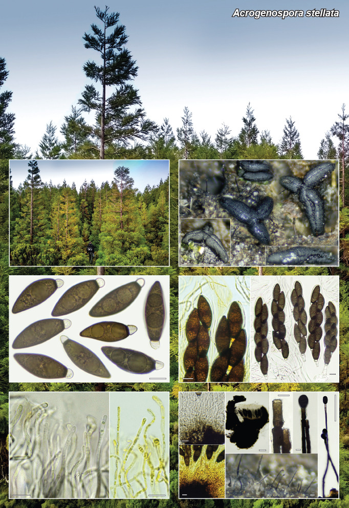
Hysterothecia 0.8–1.5 mm long, 0.2–0.4 mm wide, fusiform, simple or frequently branched in a three-pointed star shape, carbonaceous, straight or somewhat curved, ends obtuse to acuminate, compressed and smooth or somewhat laterally grooved, and with a narrower supporting base, appearing to have a pseudostipitate, longitudinally fissured with invaginated groove or cleft. Peridium thick at base (up to 354 μm thick) and apically thinner (68–100 μm thick), carbonaceous, smooth or slightly rough, consisting of pseudoparenchyma of very dark pigmented cells, irregular to subglobose, 9–12 μm diam. Pseudoparaphyses cylindrical, fragile, narrow, 1.5–2 μm thick, slightly enlarged towards the apical cell, up to 2.7 μm diam, septate, hyaline, simple or bifurcate at lower cells, with sparse yellowish globose guttules, and embedded in a hyaline gelatinous material. Asci bitunicate, cylindrical-clavate, 150–178 × 21–33 μm, 8-spored, spores biseriate, pars spori-fera 110–135 μm, ascus apex inamyloid after IKI treatment, base short pedicellate, without croziers. Hypothecium of textura globulosa-intricata, with brownish pigment and dextrinoid reaction in IKI. Ascospores fusoid-clavate, greenish brown, (34–)34.5–37.9–40.8(–43) × (11–)11.9–12.8–13.7(–15.6) μm; Q = (2.5–)2.7–3–3.3; n = 30, unequally bicellular, separated by a septum; upper coloured cell (30.6–)31.4–33.2–35(–36.3) × (10.8–)12.2–12.8–13.4(–15.6) μm; Q = (2.3–)2.4–2.6–2.8 (–3.1) μm, with one extreme subacute and the other one truncate, usually with one or two large and several smaller guttules, lower hyaline cell, papilla-like, (4.6–)4.9–5.4–5.8(–5.9) × (3.6–)4.2–4.5–4.9 μm. Asexual morph: colonies effuse, black and hairy. Conidiophores blackish brown or black, 200–570 μm long, 6–10 μm wide, with broadened base (12–20 μm), thick-walled, smooth, solitary, septate, unbranched, straight or flexuous. Conidiogenous cells monoblastic, terminal and percurrent. Conidia ellipsoidal or broadly ellipsoidal, dark brown, olive or blackish brown, smooth, 25–30 × 15–20 μm, truncate at base with a hilum 8–10 μm wide; aseptate.

Habitat & Distribution — Gregarious, more or less solitary on dead wood decorticated with algae and bryophytes of residual laurel forests planted with Cryptomeria japonica. Only known so far from Terceira Island (Azores, Portugal).

Typus. Portugal, Azores, Terceira, Angra do Heroísmo, Algar do Carvão, Terra Brava, N38°44'09" W27°12'0.4", 670 m a.s.l., more or less solitary on dead wood or bark of residual laurel forests, 14 Jan. 2022, A. Mateos & S. De la Peña (holotype AMI-SPL1243, ITS and LSU sequences GenBank OP439740 and OP439739, MycoBank MB 845631).

Colour illustrations. Portugal, Azores, Terceira, Algar do Carvão, laurel forests planted with Cryptomeria japonica, where the holotype of Acrogenospora stellata was collected. Right column: ascomata in upper photo correspond with the holotype; middle photo corresponds with: detail of *ascus apex (left, IKI-2), mature asci (right, *H2O) with detail base ascus without crozier; the bottom photo is: *hypothecium (left, H2O, IKI-2); hysterothecia section in middle-upper photo (H2O); asexual morph colony on wood (middle-bottom photo); conidiophores, conidiogenous cells and conidia (right, three pictures, *H2O). Left column: middle photo ascospores (*H2O); and the bottom photo is pseudoparaphyses (H2O, IKI-2). Scale bar = 100 μm (section hysterothecia, asexual morph colony on wood and conidiophores, right), 10 μm (all others).

Notes — The asexual genus Acrogenospora was protected against its sexual genus Farlowiella (Wijayawardene et al. 2014, Rossman et al. 2015) based on the diversity and wider use of Acrogenospora rather than following the priority principle of the International Code of Nomenclature (ICN; McNeill et al. 2012). Farlowiella was historically included in the family Hysteriaceae together with seven other genera (Chevallier 1826, Zogg 1962). Phylogenetic studies pointed out the difficulty in the morphological interpretation of genera in Hysteriaceae (Schoch et al. 2006). Based on morphological and phylogenetic evidence, Boehm et al. (2009a, b) removed Farlowiella from Hysteriaceae and placed the genus among the incertae sedis in Pleosporomycetidae. At the same time, Boehm et al. (2009a, b) showed that the sexual morph Farlowiella was in the same clade as the asexual morph Acrogenospora. After Rossman et al. (2015) concluded that the name Acrogenospora should be protected against Farlowiella, the new family Acrogenosporaceae was erected to include the genus, based on phylogenetic analyses and morphological differences with Minutisphaeraceae (Jayasiri et al. 2018). Recent studies have generated molecular data for supporting the asexual-sexual connection (Jayasiri et al. 2018, Hyde et al. 2019, Bao et al. 2020). Sequence editing, alignment (taxa sampling provided in FP1436-2) and BI analysis were performed using Geneious (v. 6.1.7, https://www.geneious.com). Based on megablast search of NCBIs GenBank nucleotide database, the closest hits using the ITS sequence were Acrogenospora carmichaeliana (HF677172, ident. = 87.5 %) and different strains under the name Acrogenospora sp. such as AH-2022a (ON176303, 87.3 %) or Dothideomycetes sp. like LS-2013g (KF513514, 85.6 %). Our phylogenetic analyses strongly supported the inclusion of the new species Acrogenospora stellata within the genus and in the family Acrogenosporaceae, showing it to be distinct from other species. The new species clusters in an unsupported clade together with A. olivaceospora and A. sphaerocephala. The former has larger conidia than A. stellata (32–36.9 × 28–32.8 μm), and the latter has subsphaerical, wider conidia (15.5–30 μm) (Bao et al. 2020). From those accepted asexual morphs without sequences the most morphologically similar to A. stellata are: A. megalospora (obovoid conidia and narrower conidial attachment (5–8 μm vs 8–10 μm in A. stellata; Goh et al. 1998)); A. ovalia (shorter, multiseptate conidiophores with multiple proliferations at apex; Goh et al. 1998); and A. setiformis (shorter conidia, 16–24 μm vs 25–30 μm in A. stellata; Ellis 1972). All other asexual taxa without sequence data have larger conidia. Only two species in the genus are known to have described sexual morphs, A. altissima (F. australis) from the remote Tristan da Cunha Islands in the South Atlantic and A. carmichaeliana from Europe; both have smaller spores than A. stellata, 13–15 × 6–7.5 μm and 18–27 × 7–12 μm, respectively. Furthermore, only A. stellata has star-shaped hysterothecia (Dennis 1955, 1981, Ellis 1972, Ellis & Ellis 1985).

graphic file with name per-2022-49-8-g001.jpg

Acrogenospora stellata

Supplementary material

FP1436-1

Phylogenetic tree.

FP1436-2

Taxa with the accession numbers used for the phylogenetic tree.

Persoonia. 2022 Dec 20;49:266–267.

Fungal Planet 1437 – 20 December 2022

Agaricus pateritonsus T. Lebel, Boxshall & Broadbridge, sp. nov.

Teresa Lebel 1, Jasmin Broadbridge 2, Amelia-Grace Boxshall 3

Etymology. In reference to the overall shape of younger sporocarps = bowl haircut/pageboy = L. patera (bowl) and tonsus (haircut). Common name: the Pageboy Agaricus.

Classification — Agaricaceae, Agaricales, Agaricomycetes.

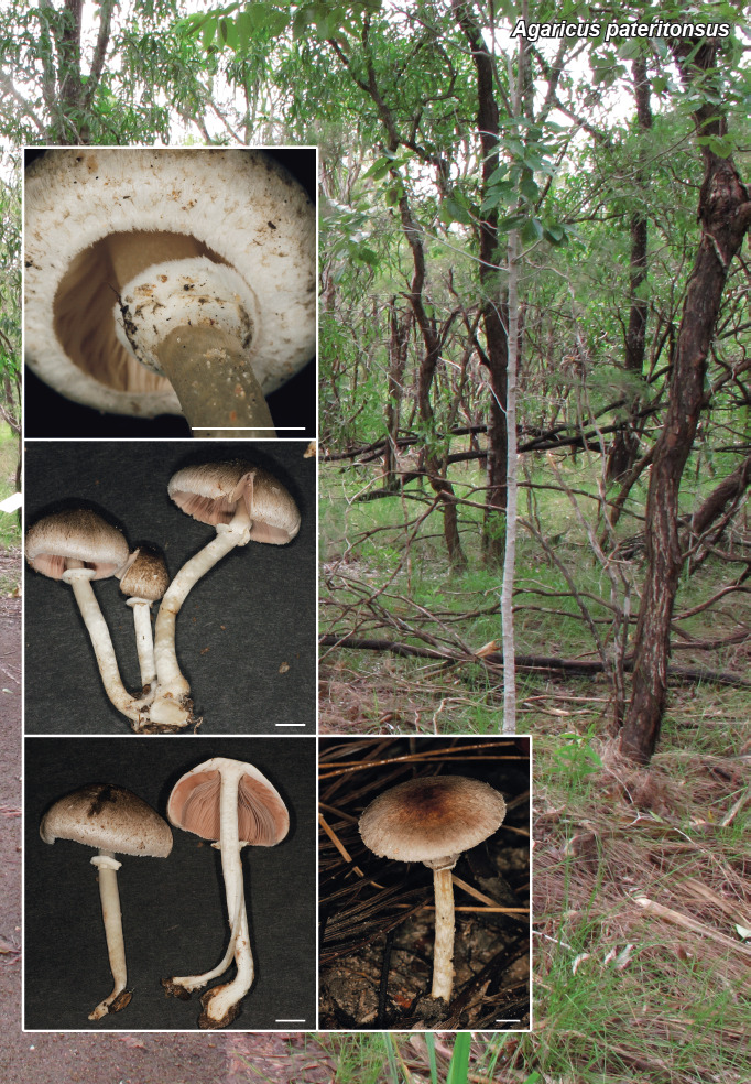
Pileus 9–60(–70) mm diam, up to 28 mm thick at the disk, convex with a blunt to squarish apex when younger, becoming broadly umbonate to planoconvex or occasionally shallowly depressed at maturity; surface dry, pale cream to pale greyish brown with overlying very fine dark brown-purple fibrillose scales that are denser at the apex, becoming pale lilac-pinkish with age, pileus margin finely striate; very slight narrow band of red staining at apex of lamellae and stipe in cross section that fades. Lamellae free, fine, crowded, edges even, initially pale to rich pink, deepening to dark dull brown, lamellulae in 2–3 series. Stipe 25–65 × 3.5–6 mm, central, cylindrical tapering slightly to apex, solid when younger becoming hollow at maturity, up to 7 mm at slightly bulbous base, silky and dry, appearing smooth and shiny to ‘pearlescent’, off-white to very pale grey, context not staining. Annulus in upper quarter of stipe, < 3 mm wider than stipe, ± 3–4 mm high, comprising universal and partial veils, frequently appearing inferous or inverted due to tension caused by stipe elongation and pileus expansion; universal veil pale and smooth, with pigmented fibrillose patches corresponding to the pileus fibrils and darkening with maturity; partial veil thick, white, cottony to floccose, with roughly torn margin and corresponding appendicular fragments on margin of pileus. Odour mostly reported as not distinctive, may be pleasant and slightly sweet. No colour change on touching the stipe or in the context.

Basidiospores (4.6–)5.4–6.9 × (3.1–)3.3–4.3 μm (5.73 ± 0.47 × 3.79 ± 0.26 μm, Q= 1.12–1.80, n = 63), oval to mildly reniform, smooth, thick-walled, lightly pigmented golden brown to dark brown in mature basidiospores, protruding at point of attachment. Immature basidiospores contain little to no pigment. Basidia (13.2–)14.8–19.6(–20.2) × 5–6.8(–7.2) μm (17.02 ± 1.94 × 5.78 ± 0.81 μm, Q = 2.12–4.50, n = 21), mostly 4-spored, clavate, smooth, some sterigmata long and rounded at apex. Cheilocystidia present, 15.3–17.3 × (3.4–)4.6–6.4 μm (16.39 ± 0.75 × 5.32 ± 1.04 μm, Q = 2.64–4.74, n = 7), smooth, clavate, appearing similar in size and shape to mature basidia. Pleurocystidia absent. Hymenophoral trama composed of interwoven septate hyphae 5.2–16.34 μm, mildly to highly inflated, appearing ‘jumbled’, smooth, no pigment. Subhymenium hyphae 4.98–6.37 μm, parenchymous. Pileipellis made up of thin but occasionally inflated, septate, interwoven hyphae 1.96–5.5 μm, not unidirectional, smooth, faintly pigmented in areas, terminal hyphae blunt, protruding slightly from pileus surface. Clamp connections observed in one out of four specimens within the hymenophoral trama.

Habit, Habitat & Distribution — Caespitose in groups or solitary (with slightly bulbous base if solitary). Subtropical mixed forest of Eucalyptus, Acacia, Callitris with open understory of herbs and grasses.

Typus. Australia, Northern Territory, Palmerston, Territory Wildlife Park, walking track to Goose Lagoon, 21 Jan. 2014, G.M. Bonito, M.D. Barrett, T. Lebel & C.N. Barrett GB486 (holotype MEL2382833, ITS-LSU sequence GenBank KP012708, MycoBank MB 845486).

Additional materials examined. Australia, Northern Territory, Darwin, Glen Holmes Reserve, 1st small creek line past locked gate, 20 Jan. 2014, G.M. Bonito, M.D. Barrett, T. Lebel & C.N. Barrett GB460 (MEL2382806, ITS-LSU GenBank sequence KP012687); Palmerston, Howard Springs Reserve, 23 Jan. 2014, G.M. Bonito, M.D. Barrett & T. Lebel GB512 (MEL2382859, ITS-LSU GenBank sequence KP012733); Palmerston, Howard Springs Reserve, ± 500 m from entrance, 23 Jan. 2014, G.M. Bonito, M.D. Barrett & T. Lebel GB510 (MEL2382857, ITS-LSU GenBank sequence KP012731).

Notes — Agaricus pateritonsus sp. nov. is a member of section Xanthodermatei subsect. Paradoxi and is phylogenetically sister to A. brunneogracilis (ML 96; PP 0.94) from Thailand according to Maximum likelihood and Bayesian analyses (see FP1437; Stamatakis 2014, He et al. 2018, Bashir et al. 2021). Support values for deeper nodes, including support for subsect. Paradoxi, are weak in our analyses of this single gene, however, our results are similar to those found previously by He et al. (2018) and Bashir et al. (2021). Both species have fine fibrillose scales that completely cover the pileus (greyish brown in brunneogracilis, purplish brown in pateritonsus), crowded lamellae, silky stipe, little colour change or bruising reactions, and a mild but pleasant odour (Zhou et al 2016). However, A. pateritonsus has much larger sporocarps, that are sometimes caespitose rather than solitary, lamellae that are darker at maturity, the stipe does not have a hollow central strand, and the annulus is more robust. Of other similar taxa, A. microvolvatulus has a volva-like or abrupt bulbous stipe base and much smaller spores (Heinemann 1978, Thongklang et al. 2014) and A. murinocephalus has a pileus with a central disc of greyish brown to black scales and a truncated umbo. Some variation exists between collections, in a cross section of GB510, an older specimen, the context immediately oxidised dark greyish brown (but it was a little waterlogged when collected). This collection also exhibits a much darker pileus cap surface than the other specimens collected, which vary from dark fibrils denser at the apex and scattering towards the margin to mid to dark purplish brown fibrils covering the surface of the cap.

Colour illustrations. Australia, Northern Territory, Palmerston, Territory Wildlife Park, subtropical mixed forest, holotype site. Basidiocarp showing inverted annulus and veil remnants on pileus margin; umbonate cap, pileus pigmentation and striate margin giving A. pateritonsus its name; cross section of pileus showing young lamellae; and mature basidiome in subtropical mixed forest. Scale bars = 10 mm. (Photo credits C.N. Barrett & T. Lebel).

graphic file with name per-2022-49-8-g002.jpg

Agaricus pateritonsus

Supplementary material

FP1437

Phylogenetic tree.

Persoonia. 2022 Dec 20;49:268–269.

Fungal Planet 1438 – 20 December 2022

Anteaglonium lusitanicum A. Mateos, De la Peña-Lastra & M. Serrano, sp. nov.

Antonio Mateos Izquierdo 1, Saúl De la Peña-Lastra 2, Miguel Serrano 2, Antonio Rigueiro-Rodríguez 2

Etymology. The epithet lusitanicum refers to the fact that it was collected on wood of Prunus lusitanica subsp. lusitanica and, moreover, has been collected in the ancient Roman region of Lusitania.

Classification — Anteagloniaceae, Pleosporales, Dothideomycetes.

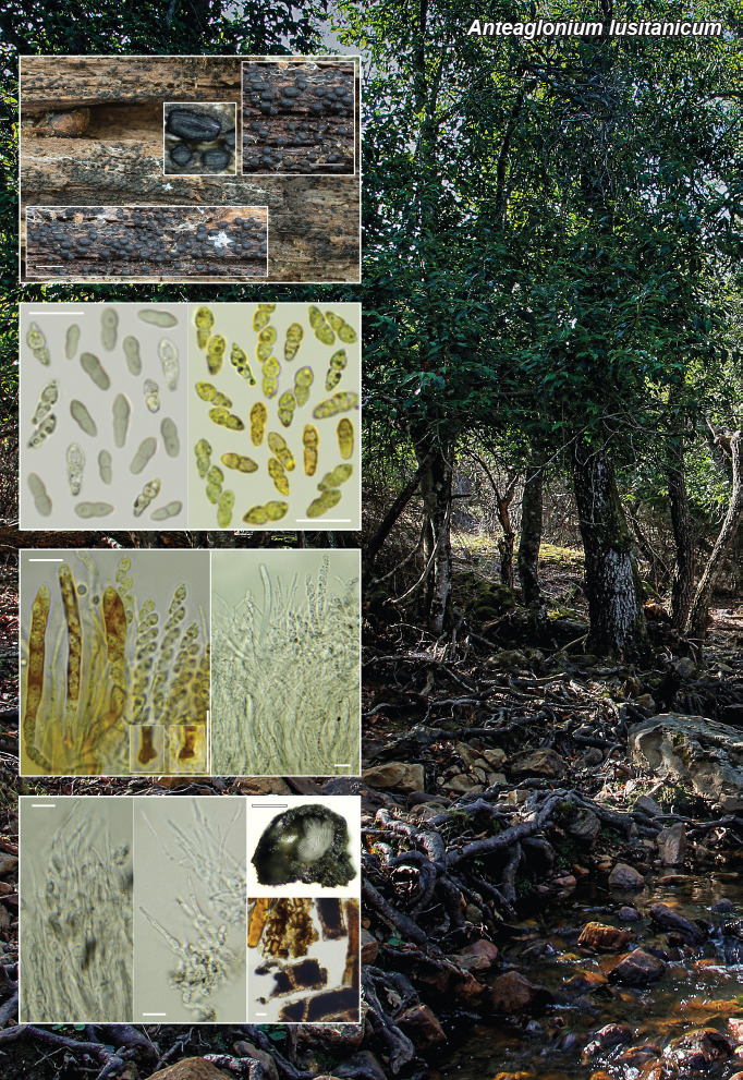
Hysterothecia *150–500 μm long, 150–300 μm wide, 150–250 μm high, ovoid, subglobose, oblong or ellipsoid, simple, not branched, parallel oriented, carbonaceous consistency, straight, obtuse ends, shallow, tending to darken the surrounding substrate forming a subiculum, with extractable pigments in 10 % KOH greenish brown not intense, gregarious. Peridium 26–65 μm thick, wider in the middle and at the base, and at the apical end thinner, carbonaceous, slightly rugose, with many striae on both sides, with shallow longitudinal cleavage furrow in the centre, inner walls composed of small pseudoparenchymatous cells, irregularly shaped, pigmented, 4–8 μm diam and 1–2 μm wide wall, and other larger cells in the remaining wall dark brown or orange pigmented, septate, 18–21 μm diam, with thick walls 2–3 μm. Hamathecium trabeculate, with numerous filamentous pseudoparaphyses, very thin, *0.8–1.8 μm wide, cylindrical, but sometimes with pointed apex, hyaline, simple or branched at various levels, but generally protruding above the asci, septate, with vacuolar guttules (VBs) inside. Asci *(51.7–)53.4–66.8–86.1(–90) × 4–4.7–5.6(–5.7) μm, 8-spored, bitunicate, cylindrical, hyaline, obliquely to irregularly uniseriate, base provided with croziers; ascus apex inamyloid after IKI treatment. Ascospores *(6.8–)7.4–8.5–10(–10.8) × (2.8–)2.9–3.1–3.4(–3.5) μm; Q = (2.2–)2.4–2.7–3.1(–3.6); n = 40; biconical or dimorphic, fusoid, with ovoid or obtuse apices in general but sometimes somewhat acuminate, hyaline, bicellular, with 1–2 guttules in each cell, thin-walled, smooth, distal cells broader than lower, wedge-shaped, proximal cell wedge-shaped or oblong, strongly constricted at septum. Asexual morph not known.

Habitat & Distribution — Gregarious, in more or less numerous groups in laurel forest areas, on decaying wood of Prunus lusitanica subsp. lusitanica. Currently known only from the type location in western Spain (Cáceres, Spain).

Typus. Spain, Cáceres, Alía, Lorera de la Trucha, N39°32'50.57" W5°14'55.08", 640 m a.s.l., gregarious growth on decaying wood of Prunus lusitanica subsp. lusitanica (Rosaceae), 20 Feb. 2021, A. Mateos, S. De la Peña & A. Gutiérrez (holotype AMI-SPL647, ITS and LSU sequences GenBank OP441407 and OP441665, MycoBank MB 845630).

Notes — After their transfer from the Hysteriaceae, based on molecular studies, of the genera Hysterographium, Farlowiella and Glonium (see FP1436 of this paper), the genus Glonium was divided into Psiloglonium in the Hysteriaceae and Glonium in the Gloniaceae (Boehm et al. 2009a) and shortly thereafter the new genus, Anteaglonium, was proposed in the Pleosporales (Mugambi & Huhndorf 2009). The genus was subsequently placed in a new family, the Anteagloniaceae by Hyde et al. (2013). Anteaglonium lusitanicum has the typical characters of the genus, but differs well from the other species of the genus; the most morphologically dissimilar are A. latirostrum and A. brasiliense which have erumpent and irregular hysterothecia, with very fusoid and much larger asco-spores (Mugambi & Huhndorf 2009, Carneiro de Almeida et al. 2014); also A. gordoniae, recently described (Jayasiri et al. 2019), has fusoid ascospores with several septa, being larger (20–22 × 1.5–3 μm) than A. lusitanicum; A. rubescens differs from all other species in the genus by brown didymospores, which disarticulate within asci, and by the production of a redorange to pink pigment (Jaklitsch et al. 2018); more similar to our species is A. thailandicum, but it differs by having smaller spores (6.4–7.8 × 2.4–3.1 μm), shorter asci, aseptate pseudoparaphyses, on a black thin crust, without KOH extractable pigments (Jayasiri et al. 2016); A. subglobosum has shorter subglobose ascomata, covered with tomentum and with reddish wall, smaller spores (6–7 × 2–3 μm) (Mugambi & Huhndorf 2009); A. parvulum is different in that the hysterothecae are partly sunken into the substrate, with acuminate apices, without crust or dark subiculum, without KOH-extractable pigments and with smaller spores (5.2–8 × 2–3.2 μm) (Jayasiri et al. 2016); with a similar morphology to A. lusitanicum, A. abbreviatum, differs by its larger, sometimes erumpent hysterothecae, thicker peridium, with a disordered arrangement on the substrate, shorter asci and smaller spores (6.4–7.2 × 2.4–3.2) (Álvarez et al. 2016).

Phylogeny — Maximum Likelihood (ML) and Bayesian analyses from a combined dataset of ITS and 28S LSU nrDNA markers produced a tree with similar topologies, recovering A. lusitanicum in a nested position within the fully supported clade (Bayesian posterior probability (BPP) = 1 / ML bootstrap = 100 %) of genus Anteaglonium. The new species grouped in a clade (BPP = 0.90 / ML bootstrap = 73 %) with Anteaglonium parvulum as sister species. BLAST searches found no identical sequences to A. lusitanicum in the GenBank nucleotide database; the ITS and 28S LSU nrDNA accessions from A. parvulum being the most similar, with percentage of identity ranging from 94.6 % to 96.5 % in the ITS and from 98 % to 99 % in the LSU.

Colour illustrations. Spain, Alía, Lorera de la Trucha, laurissilva of Prunus lusitanica subsp. lusitanica, where the holotype of Anteaglonium lusitanicum was collected. View of hysterothecia holotype on host surface; ascospores (*H2O, *IKI 2); ascus apex, detail base ascus with crozier (*IKI 2) and mature asci (*H2O); pseudoparaphyses (*H2O), section through hysterothecium (H2O), peridial cells in vertical section (H2O). Scale bar = 1 mm (hysterothecia), 100 μm (section hysterothecia), 10 μm (all others).

graphic file with name per-2022-49-8-g003.jpg

Anteaglonium lusitanicum

Supplementary material

FP1438-1

Phylogenetic tree.

FP1438-2

Table. Taxa with ITS, LSU and GenBank accession numbers included in the phylogenetic tree.

Persoonia. 2022 Dec 20;49:270–271.

Fungal Planet 1439 – 20 December 2022

Colletotrichum araujiae G.H. Ramirez, F. Anderson & Bianchin., sp. nov.

Gustavo Hernán Ramírez 1, Freda Elizabeth Anderson 2, Romina Magalí Sánchez 3, Marina Lucía Díaz 3, María Virginia Bianchinotti 3

Etymology. Name refers to the host genus, Araujia, from which it was isolated.

Classification — Glomerellaceae, Glomerellales, Sordariomycetes.

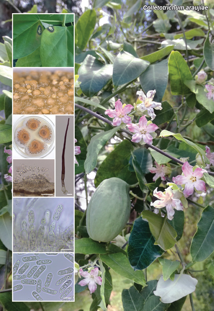
Sexual morph not observed. Asexual morph on sterile host stems. Conidiomata acervular, conidiophores and setae developing from pale brown, angular to rounded cells. Setae rarely produced. Conidiophores hyaline, smooth-walled, aseptate or septate, up to 40 μm long. Conidiogenous cells hyaline, smooth-walled, cylindrical to ampulliform, sometimes extending to form new conidiogenous loci, 11.5–22 × 4–7.5 μm, opening 1–2 μm diam, collarette ≤ 0.5 μm, periclinal thickening distinct. Conidia hyaline, smooth-walled, aseptate, straight, cylindrical, apex and base rounded, hilum visible, granular to guttulate content, often with two polar guttules, (16.5–)18–22(–24.5) × 6.5–7(–8) μm, av. ± SD = 20.1 ± 1.8 × 6.8 ± 0.4 μm, L/W ratio = 2.9.

Asexual morph on PDA. Vegetative hyphae 1–6 μm diam, hyaline, smooth-walled, septate, branched. Conidiomata acervular, consisting of conidiophores and setae formed directly on hyphae. Setae dark brown, paler towards the base, smooth and thick-walled, often verruculose towards the tip, 3–6-septate, 90–200 μm long, cylindrical or inflated basally, 3.5–6.5 μm diam, tips rounded. Conidiophores hyaline, smooth-walled. Conidiogenous cells hyaline, smooth-walled, cylindrical to ampulliform, often extending to form new conidiogenous loci, 11–25 × 3.5–7 μm, opening 1–1.5 μm diam, periclinal thickening conspicuous. Conidia hyaline, smooth-walled, aseptate, straight, cylindrical, rounded at the ends, sometimes with a short prominent hilum, contents granular or guttulate, 18–21 × 6–8.5(–10) μm, av. ± SD = 19.4 ± 1 × 7.3 ± 0.9 μm, L/W ratio = 2.7. Appressoria single or in small groups of 2–3, medium to dark brown, smooth-walled, ovoid, navicular or irregular in shape, margins entire or undulate, 9–15 × 6.5–11 μm, av. ± SD = 11.1 ± 1.4 × 8.8 ± 1.1 μm, L/W ratio = 1.3.

Culture characteristics — (PDA at 25 °C, 12 h photoperiod): Colonies flat with entire margin, centre of the colonies covered with conidial masses with no aerial mycelium, moderate white aerial mycelium towards the margins, 25.5–27 mm in 7 d (40–42 mm in 10 d). Conidial masses salmon to orange.

Typus. Argentina, Buenos Aires Province, -35.584°S -62.997°W, on leaves, stems and fruits of Araujia hortorum (Apocynaceae), 13 July 2015, G.H. Ramírez (holotype BBB:Ah35–04, culture ex-type BBB:GR3504, ITS, gapdh and tub2 sequences GenBank OP035058, OP067659 and OP067660, MycoBank MB 845057).

Notes — There are only a few records of Colletotrichum and Gloeosporium species occurring on Araujia hortorum. Colletotrichum cf. orbiculare was isolated from purple spots on the leaves of A. hortorum (Waipara et al. 2006) although the authors did not provide a description of their isolate. Colletotrichum araujiae has wider conidia than any of the species known in the C. orbiculare species complex (Damm et al. 2013). Gloeosporium americanum was described on leaves of A. hortorum, and has smaller conidia (10–12 × 4–5 μm) than C. araujiae (Spegazzini 1880).

Based on a megablast search in GenBank nucleotide database, the closest hits using the ITS sequence had highest similarity to Colletotrichum brasiliense (strain PaL-3, GenBank MN646264; Identities = 549/554 (99 %), no gaps), C. hippeastri (strain F269, GenBank MW995568; Identities = 546/554 (99 %), no gaps) and C. parsonsiae (strain CBS 128525, GenBank MH865006.1; Identities = 545/555 (98 %), one gap); closest hits using the gapdh sequence had highest similarity to Colletotrichum brasiliense (strain PaL-3, GenBank MN657408; Identities = 216/223 (97 %), no gaps), C. parsonsiae (strain CBS 128525, GenBank JQ005320; Identities = 216/223 (97 %), no gaps) and C. hippeastri (strain CBS 241.78, GenBank JQ005319; Identities = 213/223 (96 %), no gaps); closest hits using the tub2 sequence had highest similarity to Colletotrichum brasiliense (strain CBS 128528, GenBank JQ005668; Identities = 496/496 (100 %, no gaps), C. parsonsiae (strain CBS 128525, GenBank JQ005667; Identities = 490/498 (98 %), two gaps) and C. condaoense (strain CBS 134299, GenBank MH229923; Identities = 484/493 (98 %), two gaps).

Phylogenies inferred from concatenated ITS, tub2 and gapdh sequences place the fungus in the C. boninense species complex (Damm et al. 2012), where it forms a distinct single-strain lineage. Although it is phylogenetically closely related to C. brasiliense, a pathogen of Passiflora (Passifloraceae), there are differences in the size of its conidia (larger in C. araujiae) and in the shape of its appressoria (deeply lobed in C. brasiliense) (Damm et al. 2012). Colletotrichum araujiae can be distinguished from C. parsonsiae (a pathogen of Parsonsia, Apocynaceae) by having wider conidia, and from C. hippeastri (a pathogen of Hippeastrum, Amaryllidaceae) by having shorter conidia and differently shaped appressoria.

Colour illustrations. Araujia hortorum growing in Argentina. Foliar symptoms; acervuli on inoculated Araujia stem; colonies on PDA; acervulum; setae; conidiogenous cells; conidia. Scale bars = 50 μm (acervulum), 10 μm (all others).

graphic file with name per-2022-49-8-g004.jpg

Colletotrichum araujiae

Supplementary material

FP1439-1

Phylogenetic tree.

FP1439-2

Table. Strains of Colletotrichum spp. studied with GenBank accession numbers.

Persoonia. 2022 Dec 20;49:272–273.

Fungal Planet 1440 – 20 December 2022

Curvularia fraserae Y.P. Tan, Bishop-Hurley, L. Kelly & R.G. Shivas, sp. nov.

Yu Pei Tan 1, Sharon L Bishop-Hurley 1, Lisa Kelly 2, Roger G Shivas 3

Etymology. Named after Lillian Ross Fraser (1908–1987), an Australian botanist, mycologist and plant pathologist. In 1937, Lilian Fraser became the first Australian woman to earn a Doctor of Science in New South Wales. Lilian Fraser studied the taxonomy of sooty moulds; citrus diseases, including Phytophthora root rot; and collected many rare and unusual smut fungi. One of her biographers describes her as independent and forthright, with ideas ahead of her time, who dedicated her life to the pursuit of knowledge. Lillian Fraser has been commemorated in the names of several species of Australian fungi, including Balladyna fraserae, Euantennaria fraserae, Hadrosporium fraserianum, Hendersonia fraserae, Langdonia fraseriana, Meliola fraserae, and Metacapnodium fraserae.

Classification — Pleosporaceae, Pleosporales, Dothideomycetes.

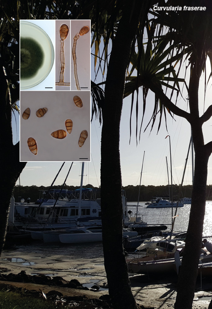
Asexual morph on maize leaf agar (MLA): Hyphae pale brown, smooth, branched, septate, 3–5 μm wide. Conidiophores single or grouped, unbranched, straight to flexuous, septate, sub-hyaline, 50–300 × 3–5 μm, lateral or terminal, unbranched or sparingly branched. Conidiogenous cells intercalary and terminal, geniculate, swollen, subhyaline, smooth, polytretic. Conidia ellipsoidal to ovoid, 15–23 × 8–11 μm, 1–3-septate, apical cell rounded, basal cell obconical, pale brown, basal cell often subhyaline; hila thickened and protuberant, 1.5–2.5 μm wide, darkened.

Culture characteristics (25 °C, 7 d, in darkness) — Colony on potato dextrose agar (PDA) 6 cm diam, surface velutinous with some aerial mycelium, olivaceous black, margin fimbriate.

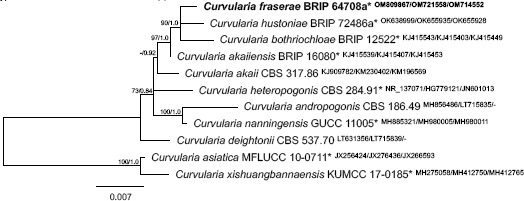
Phylogenetic tree of selected Curvularia species based on maximum likelihood analysis of the ITS, gapdh and tef1α gene regions. Analyses were performed on the Geneious Prime ® 2022 platform (Biomatters Ltd.) using RAxML v. 8.2.11 (Stamatakis 2014) and MrBayes v. 3.2.6 (Huelsenbeck & Ronquist 2001), both based on the GTR substitution model with gamma-distribution rate variation. The scale bar represents substitutions per nucleotide position. RAxML bootstrap (bs) values greater than 70 % and Bayesian posterior probabilities (pp) greater than 0.8 are given at the nodes (bs/pp). GenBank accession numbers are indicated (superscript ITS/gapdh/tef1α). Curvularia sporobolicola ex-type strain BRIP 23040b was used as outgroup. Novel taxon is indicated in bold. Ex-type strains indicated with an asterisk (*).

Typus. Australia, Queensland, Gin Gin, from dying leaf of Bothriochloa insculpta (Poaceae), 24 May 2016, L. Kelly (holotype BRIP 64708a preserved as metabolically inactive culture, ITS, gapdh and tef1a sequences GenBank OM809867, OM721558 and OM714552, MycoBank MB 845307).

Notes — Curvularia fraserae is phylogenetically related to three other graminicolous species, C. akaiiensis, C. bothriochloae and C. hustoniae. Curvularia fraserae is distinguished from C. hustoniae (ex-type strain BRIP 72486a) by sequence comparison of the ITS region (GenBank OK638999; Identities 674/677 (99 %), one gap; unique nucleotide at positions 165(C), 263(C)), gapdh (GenBank OK655935; Identities 529/535 (99 %), no gap; unique nucleotide at positions 72(A), 239(T), 284(C), 338(A), 359(C), 437(G)), and tef1a (GenBank OK655928; Identities 892/899 (99 %), three gaps; unique nucleotide at positions 97(C), 166(C), 619(C), 676(G)). Curvularia fraserae has shorter conidia than C. akaiiensis (cf. 22–28 × 7–16) and C. bothriochloae (cf. 30–47 × 15–25 μm) (Sivanesan et al. 2003). The morphology of C. hustoniae is not known (Tan et al. 2022).

Based on a megablast search of NCBIs GenBank nucleotide database, the closest relevant hits using the ITS sequence are C. akaiiensis (culture CBS 127726, GenBank LT631342; Identities 674/676 (99 %), no gaps), C. hustoniae (culture BRIP 72486a, GenBank OK638999; Identities 674/677 (99 %), one gap) and C. akaii (culture CBS 127728, GenBank LT631341; Identities 674/678 (99 %), two gaps). The closest relevant hits using the gapdh sequence are C. akaiiensis (culture CBS 127726, GenBank LT715799; Identities 538/542 (99 %), no gaps), C. hustoniae (culture BRIP 72486a, GenBank OK655935; Identities 529/535 (99 %), no gaps) and C. deightonii (culture CBS 537.70, GenBank LT715839; Identities 533/542 (98 %), no gaps). The closest relevant hits using the tef1a sequence are C. hustoniae (culture BRIP 72486a, GenBank OK655928; Identities 892/899 (99 %), three gaps), C. akaii (culture CBS 317.86, GenBank KM196569; Identities 891/899 (99 %), three gaps) and C. lolii (culture LIPP:CU1, GenBank MT881706; Identities 887/899 (99 %), three gaps).

graphic file with name per-2022-49-8-i001.jpg

Colour illustrations. Australia, Queensland, Tin Can Bay islet. Colonies on PDA, conidiophores, and conidia. Scale bars = 1 cm (colony), 10 μm (all others).

graphic file with name per-2022-49-8-g005.jpg

Curvularia fraserae

Persoonia. 2022 Dec 20;49:274–275.

Fungal Planet 1441 – 20 December 2022

Curvularia millisiae Y.P. Tan, Bishop-Hurley, E. Lacey, Dhileepan & R.G. Shivas, sp. nov.

Yu Pei Tan 1, Sharon L Bishop-Hurley 1, Kunjithapatham Dhileepan 2, Ernest Lacey 3, Roger G Shivas 4

Etymology. Named after Nancy Fannie Millis (1922–2012), an Australian microbiologist who made many outstanding contributions in agriculture, environmental protection, medicine, and engineering. Nancy Millis overcame gender discrimination in the 1950s when women were not employed in many workplaces to establish the first applied microbiology course taught at an Australian university. One of her biographers noted that on her professorial appointment at The University of Melbourne in 1982, she remarked that universities could be a ‘bit of a club where blokes tend to appoint blokes’. In 2002, Nancy Millis was featured on the 45c Australian postage stamp that honoured prominent medical scientists who had become ‘living legends’.

Classification — Pleosporaceae, Pleosporales, Dothideomycetes.

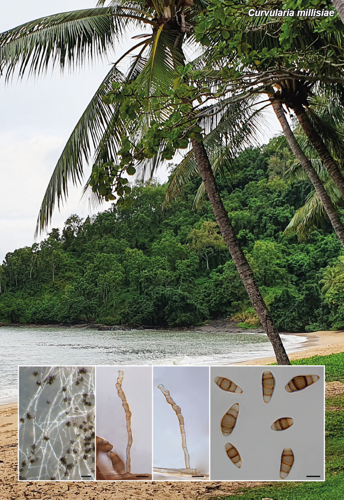
Asexual morph on maize leaf agar (MLA): Conidiophores erect, straight to flexuous, geniculate towards apex, brown, cylindrical, smooth, 50–400 μm long, septate, lateral or terminal, unbranched or sparingly branched. Conidiogenous cells intercalary and terminal, brown, smooth, polytretic with darkened scars. Conidia cylindrical to ellipsoidal, straight or slightly curved, 18–37 × 8.5–11 μm, (2–)4–5-distoseptate, pale brown to brown, rounded at the apex, basal cell obconical, the third cell from base often swollen and darker than the others, end cells pale brown; wall smooth, basal cell sometimes minutely roughened; hila conspicuous, slightly protuberant, thickened.

Culture characteristics (25 °C, 10 d, in darkness) — Colony on potato dextrose agar (PDA) 4 cm diam, surface velutinous with scant white aerial mycelium, olivaceous grey, margin irregular.

Typus. Australia, Queensland, Daintree, from yellowing leaf tips of Cyperus aromaticus (Cyperaceae), 3 Sept. 2020, K. Dhileepan, M.D.E. Shivas & R.G. Shivas (holotype BRIP 71718a preserved as metabolically inactive culture, ITS, LSU, gapdh and tef1a sequences GenBank OK661031, OK661032, OK636415 and OK636413, MycoBank MB 845324).

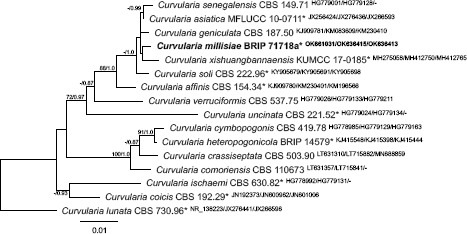
Notes — Curvularia millisiae is phylogenetically related to C. asiatica, C. geniculata, C. senegalensis, C. soli and C. xishuangbannaensis. All species share similar morphological characters with 3–5 distoseptate conidia that fall within the range 15–48 × 8–20 μm (Sivanesan 1987, Manamgoda et al. 2012, Marin-Felix et al. 2017, Tibpromma et al. 2018).

Curvularia millisiae is distinguished from C. xishuangbannensis (ex-type strain KMUCC 17-0185) by sequence comparison of the ITS region (GenBank MH275058; Identities 514/516 (99 %), one gap; unique nucleotide at position 486(G)), gapdh (GenBank MH412750; Identities 523/532 (98 %), one gap; unique nucleotide at positions 74(T), 108(T), 202(A), 212(A), 340(C), 367(C), 454(C), 508(T)) and tef1a (GenBank MH412765; Identities 890/892 (99 %), two gaps).

Based on a megablast search of NCBIs GenBank nucleotide database, the closest relevant hits using the ITS region are C. senegalensis (strain 15-838, GenBank MT476857, Identities 593/593 (100 %)), C. trifolii (isolate RM35, GenBank MG664781, Identities 593/593 (100 %)) and C. clavata (strain M5, GenBank KX610320, Identities 593/593 (100 %)). The closest relevant hits using gapdh sequence are C. asiatica (as C. asianensis; strain LC11959, GenBank MN264083; Identities 567/576 (98 %), no gaps), C. geniculata (culture CBS 351.65, GenBank LT715833; Identities 567/576 (98 %), no gaps) and C. senegalensis (culture CBS 149.71, GenBank LT715787; Identities 565/576 (98 %), no gaps). The closest relevant hits using the tef1α sequence are C. asiatica (as C. asianensis; strain LC11959, GenBank MN263941; Identities 926/926 (100 %), no gaps), C. soli (culture CBS 222.96, GenBank KY905698; Identities 921/ 921 (100 %), no gaps) and C. geniculata (strain LC11961, GenBank MN263960; Identities 925/926 (99 %), no gaps).

graphic file with name per-2022-49-8-i002.jpg

Colour illustrations. Australia, Queensland, Trinity Beach. Conidiophores on PDA; conidiophores and conidia. Scale bars = 100 μm (conidiophores on PDA), 10 μm (conidia).

Phylogenetic tree of selected Curvularia species based on maximum likelihood analysis of the ITS, gapdh and tef1a gene regions. Analyses were performed on the Geneious Prime ® 2022 platform (Biomatters Ltd.) using RAxML v. 8.2.11 (Stamatakis 2014) and MrBayes v. 3.2.6 (Huelsenbeck & Ronquist 2001), both based on the GTR substitution model with gamma-distribution rate variation. The scale bar represents substitutions per nucleotide position. RAxML bootstrap (bs) values greater than 70 % and Bayesian posterior probabilities (pp) greater than 0.8 are given at the nodes (bs/pp). GenBank accession numbers are indicated (superscript ITS/gapdh/tef1a). Curvularia lunata ex-type strain CBS 730.96 was used as outgroup. Novel taxon is indicated in bold. Ex-type strains indicated with an asterisk (*).

graphic file with name per-2022-49-8-g006.jpg

Curvularia millisiae

Persoonia. 2022 Dec 20;49:276–277.

Fungal Planet 1442 – 20 December 2022

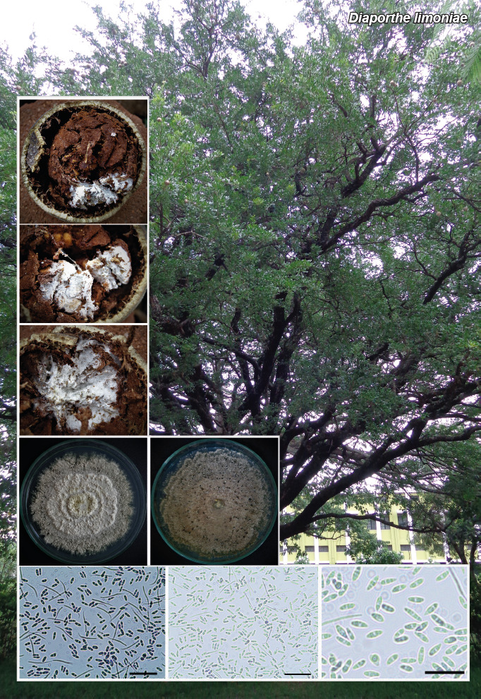
Diaporthe limoniae Mahadevak., Y. Chen, Maharachch., L.S.M. Bhanu & Chandran., sp. nov.

Shivannegowda Mahadevakumar 1, Yanpeng Chen 2, Sajeewa S N Maharachchikumbura 2, Lakkur Sannaiah Mamatha Bhanu 3, Siddaiah Chandranayaka 4

Etymology. Named after the host genus from which it was isolated, Limonia.

Classification — Diaporthaceae, Diaporthales, Sordariomycetes.

Conidiomata pycnidial, solitary, rarely aggregated, subglobose, dark brown to black, thick-walled up to 5 mm diam, covered with hyphal outgrowths, superficial, no ostiole observed, exuding a creamy mucoid conidial mass; wall is pseudoparenchymatous of dark brown textura angularis, outer layer cells are thicker than the inner layers. Paraphyses straight, flexuous, hyaline, smooth- and thin-walled, cylindrical, tapering towards the apex with 1–2 basal septa, unbranched. Conidiogenous cells lining the cavity, straight, hyaline, smooth- and thin-walled, cylindrical, tapering towards the apex, aseptate and rarely 1–2 septa observed, unbranched, collarette up to 1 μm long and variable in length, dimorphic, short conidiogenous cells (6.8–)10.6–12.8(–14.62) × (0.9–)1.52–2.1(–2.4) μm, long conidiogenous cells (12.8–)24.4–28.32(–38.2) × (1.02–)2.2–2.8(–3.4) μm, mostly phialidic, often intermingled with paraphyses. Alpha conidia cylindrical to ellipsoidal, with rounded apex, and obtuse to truncate base, hyaline, smooth, thin-walled, aseptate, bi-guttulate, with conspicuous guttule at each end, straight to slightly curved, (4.8–)5.6–8.2(–9.8) × (1.02–)1.7–2.31(–2.62) μm. Beta conidia hyaline, filiform, hamate, eguttulate, aseptate, (14–)18.72–22.3(–25.6) × (1.6–)1.7–1.9(–2.1) μm.

Culture characteristics — Colonies on potato dextrose agar (PDA) reaching 48 mm diam after 7 d at 26 °C. Surface flat, velvety, with filiform margin, circular, whitish to pale. Reverse yellowish to pale and become buff towards the centre. Conidiomata pycnidial, solitary or aggregated, half-immersed, pale brown to dark brown or black.

Typus. India, Karnataka, Mysuru, Botanical Garden, Department of Studies in Botany, University of Mysore, on infected fruit of Limonia acidissima (Rutaceae), 2 Oct. 2016, S. Mahadevakumar (holotype UOM-IOE-12/22; living ex-type culture DIA-12, ITS, LSU and tub2 sequences GenBank MZ573108, MZ573109 and MZ615698, MycoBank MB 843159).

Notes — Based on a megablast search of NCBIs GenBank nucleotide database, the closest hits using the ITS sequence of D. limoniae had highest similarity to Diaporthe sp. SM-2017a (isolate MKS2, GenBank MF085558; Identities = 472/474 (99.58 %), no gaps), D. vaccinii (isolate ALE-98, GenBank MF380717; Identities = 466/470 (99.15 %), three gaps), Diaporthe sp. CS-2021a (isolate MFLUCC17-0329, GenBank OL780507; Identities 465/470 (98.94 %) three gaps). Similarly, the closest hits using the LSU region sequence had highest similarity to Diaporthe cotoneastri (culture CBS 439.82, GenBank MH873257; Identities = 566/568 (99.65 %), no gaps), Diaporthe ambigua (culture CBS 134.42, GenBank MH867598; Identities = 566/568 (99.65 %), no gaps) and D. eres (culture CBS 186.37, GenBank MH867392; Identities = 566/568 (100 %), no gaps). Further, the closest hit using tub2 gene sequence had highest similarity to Diaporthe citrichinensis (as Diaporthe sp. FH-2013b; isolate cgyg3, GenBank KC357461; Identities = 470/487 (96.51 %), one gap (0 %)), D. citriasiana (isolate ZJUD034B, GenBank KJ420829; Identities = 466/483 (96.48 %), one gap (0 %)), D. citrichinensis (isolate PSCG462, GenBank MK691286; Identities = 470/487 (96.45 %), one gap (0 %)) and D. citrichinensis (isolate CNUCC201909, GenBank MH104872; Identities = 465/484 (96.07 %), one gap (0 %)). Based on the phylogeny, D. limoniae is closely related to D. sennicola, but morphologically they differ with respect to the shape of alpha conidia and length of conidiogenous cells. Morphologically, D. limoniae has similarity to Phomopsis azadirachtae viz., alpha conidia, beta conidia, and colony morphology; however, except for ITS nrDNA, the LSU and tub2 gene sequence data did not share any similarity with P. azadirachtae.

Colour illustrations. Limonia acidissima found growing in Manasagangotri Campus, University of Mysore from where the fruit sample associated with D. limoniae was collected. Fruit of L. acidissima associated with D. limoniae; pure cultures of D. limoniae on PDA medium (19-d-old); microscopic view of alpha conidia and beta conidia of D. limoniae. Scale bars = 20 μm.

graphic file with name per-2022-49-8-g007.jpg

Diaporthe limoniae

Supplementary material

FP1442-1

Table. Details of reference sequences retrieved and used in phylogenetic tree construction and analysis.

FP1442-2

Phylogenetic tree.

Persoonia. 2022 Dec 20;49:278–279.

Fungal Planet 1443 – 20 December 2022

Didymella naikii Savitha, Ajithk., Mahadevak., Maharachch. & Sreenivasa, sp. nov.

Attihalli S Savitha 1, Kadaiah Ajithkumar 2, Shivannegowda Mahadevakumar 3, Sajeewa SN Maharachchikumbura 4, Marikunte Y Sreenivasa 5

Etymology. Named after Prof. M.K. Naik for his outstanding contribution to the field of plant pathology in India.

Classification — Didymellaceae, Pleosporales, Dothideomycetes.

Mycelium composed of septate, branched, subhyaline to pale brown, smooth, 1–8 μm wide hyphae, sometimes aggregated into closely appressed strands. Conidiomata pycnidial, produced abundantly, partly immersed in the agar; solitary, flask-shaped, pale to blackish brown, often partly immersed, pseudoparenchymatous, with an ostiole that, when immature, appeared to contain isodiametric multicellular occlusions, bearing necks ranging 80–100 μm long and 30–40 μm wide at the extreme tip, (48-)60–100(–112) × (152–)180–210(–227) μm; pycnidial wall composed of isodiametric to ellipsoidal cells, 3–6 layers, (5.2–)7.1–9.8(–10.4) μm thick. Conidiogenous cells phialidic, hyaline, simple, smooth-walled, borne on the innermost cells of the pycnidial wall, subglobose to broadly flask-shaped, isodiametric in size, 3–5 μm diam. Conidia hyaline, simple, eguttulate, cylindrical, obtuse at each end, smooth, aseptate, (8.9–)10.7–12.4(–14.6) × (2.1–)2.7–3.8(–4.2) μm. Chlamydospores observed on potato dextrose agar (PDA) along with some simple hyphal swellings.

Culture characteristics — Colonies on PDA within 2 wk in a 12/12 dark/light regime at 28 °C, dark olivaceous, lanose aerial mycelium, black and stromatic beneath the surface, margin thin and cream-coloured, reverse black; zonate and similar to above but obscured by medium.

Typus. India, Karnataka, Raichur, on leaves of Cajanus cajan (Fabaceae), 12 May 2018, A.S. Savitha (holotype NFCCI 5104, culture ex-type PLS3, ITS, LSU and tub2 sequences GenBank OM952211, OM830704, OM858681, MycoBank MB 842072).

Additional materials examined. India, Karnataka, Mysore, leaf spot of C. cajan, 10 Jan. 2020, S. Mahadevakumar, UOM-IOE 2020/11, culture MYS1, ITS, LSU and tub2 sequences GenBank OM948732, OM830702 and OM858679; Karnataka, Doddamaragowdanahally, leaf spot of C. cajan, 19 Dec. 2021, S. Mahadevakumar, UOM-IOE 2021/02, culture MYS2, ITS, LSU and tub2 sequences GenBank OM952210, OM830703 and OM858680.

Notes — Two Phoma species are well-known to cause a leaf spot disease on pigeon pea plants, namely Phoma cajanicola and Didymella herbarum (= Phoma herbarum). However, the new species described is distinct from P. cajanicola and D. herbarum in colony characters as well as in conidial shape and size.

Based on a megaBLAST search of NCBIs GenBank nucleotide database, the closets hit using the ITS sequence of PLS3 had highest similarity to Didymella sp. (isolate L29, GenBank MG198901; Identities = 707/761 (92.9 %), 20 gaps (2 %)), Didymella sp. (isolate 63JAN, GenBank MW723759; Identities = 691/740 (93.38 %), 20 gaps (2 %)). Similarly, the closest hit using the LSU region sequence of PLS3 had highest similarity to Didymella microchlamydospora (culture CBS 105.95, GenBank NG_069838; Identities = 559/559 (100 %), no gaps), Aschochyta neopisi (specimen MFLU 16-0291, GenBank MT177945; Identities = 559/559 (100 %), no gaps) and Didymella anserina (isolate 18S0023, GenBank MN368300; Identities = 559/559 (100 %), no gaps). Further, the closest hit using the tub2 gene sequence of PLS3 had highest similarity to Didymella curtsii (isolate Z3, GenBank KP185125; Identities = 285/299 (95 %), no gaps), Didymella pomorum (isolate PP4, GenBank MN473122; Identities = 286/298 (96 %), two gaps (0 %)) and Didymella sp. (isolate MFLUCC 16-0489, GenBank MH104872; Identities = 286/300 (95 %), two gaps (0 %)). Didymella naikii is phylogenetically related to D. gardeniae (CBS 302.79, CBS 626.68) and D. prolaticolla (CBS 126182)

Colour illustrations. Cajanus cajan in agricultural fields in Raichur, Karnataka, India. Symptomatic leaf of Cajanus cajan; colony on PDA; chlamydospores and hyphae; pycnidia; conidia. Scale bars = 20 mm (culture plate); 20 μm (chlamydospores and pycnidium); 10 μm (conidia).

graphic file with name per-2022-49-8-g008.jpg

Didymella naikii

Supplementary material

FP1443-1

Phylogenetic tree.

FP1443-2

Table. List of reference sequences used in phylogenetic tree construction and analysis of Didymella naikii.

Persoonia. 2022 Dec 20;49:280–281.

Fungal Planet 1444 – 20 December 2022

Elaphomyces geminus Jeppson & E. Larss., sp. nov.

Mikael Jeppson 1, Ellen Larsson 1

Etymology. The name refers to its similarity with Elaphomyces quercicola.

Classification — Elaphomycetaceae, Eurotiales, Eurotiomycetes.

Ascomata subglobose, 15–25 mm diam. Outer peridium composed of a dense layer of yellowish brown to orange brown, pyramidal warts with acute tips, 1–1.5 per mm. Section of peridium shows warts with yellowish tips. Inner peridium marbled, yellow brown to pinkish ochraceous brown or violaceous brown with small marbles, somewhat irregularly to radially arranged with whitish veins. Mature gleba pulverulent, dark brown to black. The ascomata are not covered by a strongly adhering mycelial coating. Peridial warts composed of interwoven yellowish hyphae, inwards hyaline and more compacted. Bundles of hyaline septate hyphae separate the warts. Inner peridium composed of hyaline hyphae with abundant extracellular reddish brown pigments. Ascospores dark brown, globose, 16–21 μm (Hoyer, ornamentation excluded), 18–26 (KOH 3 %, ornamentation excluded), with a dense echinate ornamentation of rod-shaped spines (2–2.5 μm long), with age more or less curved, coalescing to form meshes and irregular patches on fully developed spores. Asci not observed.

Ecology & Distribution — On its Swedish locality associated with Quercus robur in a warm slope with a dense cover of deciduous trees on sandy - loamy soil (marine deposits), bordering a westerly directed siliceous rocky outcrop, close to seashore. In Sweden to date recorded only from the type locality, but a sequence deposited in GenBank (OM672783) confirms its presence also in North America (USA, North Carolina).

Typus. Sweden, Bohuslän, Resteröd, Ulvesund, Ulvövägen, under Quercus robur (Fabaceae), N58.222894° E11.887675°, 12 Mar. 2022, E. Larsson EL9-22 (holotype GB-0207643, ITS-LSU sequence GenBank OP222195, MycoBank MB 845542).

Additional materials examined. Elaphomyces geminus: Sweden, Bohuslän, Resteröd, Ulvesund, Ulvövägen, under Q. robur, N58.222904° E11.887708°, 4 Apr. 2022, E. Larsson EL17-26 (GB-0207642, ITS sequence GenBank OP222193); ibid., 22 Sept. 2021, E. Larsson EL122-21 (GB-0207644, ITS sequence GenBank OP222194). Elaphomyces quercicola: Sweden, Bohuslän, Resteröd, Ulvesund, Grinddalen 12, under Q. robur, N58.225230° E11.890586°, 25 July 2020, E. Larsson EL79-20 (GB-0207641, ITS-LSU sequence GenBank OP222192).

Notes — Elaphomyces geminus belongs to Elaphomyces sect. Elaphomyces subsect. Muricati, encompassing species with a marbled peridium visible when sectioned. The type species of this subsection, E. muricatus, was recently epitypified by Molia et al. (2020). The proposed new species is closely related to E. quercicola that was described in Paz et al. (2017), also being associated with Quercus spp. The latter is distinguished by its pyramid-like peridial warts alternating with conical and obtuse warts and the whole ascoma is covered by a strongly adhering mycelial coating. In E. geminus the outer peridial layer is almost entirely covered with pyramidal, acute warts and a conspicuous mycelial coating is absent. The ascospores are on average slightly larger in E. geminus and more strongly ornamented as compared with E. quercicola. Despite similarity in morphology and ecology the sequence differences in the ITS region are distinct between E. geminum and E. quercicola and they must be regarded as independent species (differing by 15 substitutions and four insertion/deletion events). Although so far only recorded on few occasions, the occurrence of E. geminus in both Europe and North America suggests that the species has a wide distribution range.

graphic file with name per-2022-49-8-i003.jpg

Colour illustrations. Elaphomyces geminus habitat from the type locality in Bohuslän, Sweden. Ascomata (fresh, holotype); close-up of sectioned peridium; ascospores (holotype). Scale bars = 10 mm (ascomata, section of peridium), 10 μm (ascospores).

Phylogram obtained using PAUP v. 4.0a (Swofford 2003) based on ITS and LSU data showing the position of E. geminus as a sister species to E. quercicola within subsection Muricati. Heuristic searches with 1 000 random-addition sequence replicates and tree bisection-reconnection (TBR) branch swapping were performed. Relative robustness of clades was assessed by the bootstrap method using 1 000 heuristic search replicates with 100 random taxon addition sequence replicates and TBR branch swapping. Bootstrap support values (> 74 %) are indicated on branches. Elaphomyces geminus is marked in bold and the holotype is indicated.

graphic file with name per-2022-49-8-g009.jpg

Elaphomyces geminus

Persoonia. 2022 Dec 20;49:282–283.

Fungal Planet 1445 – 20 December 2022

Fulvifomes mangroviensis S. Gunaseelan, K. Raja, K. Kezo & M. Kaliyaperumal, sp. nov.

Sugantha Gunaseelan 1, Kanaga valli Raja 1, Kezhocuyi Kezo 1, Malarvizhi Kaliyaperumal 1

Etymology. The species epithet ‘mangroviensis’, signifies the habitat of the species (mangrove).

Classification — Hymenochaetaceae, Hymenochaetales, Agaricomycetes.

Basidiomata annual, pileate, appianate, sessile, soft, to light corky when dry. Pilei dimidiate, convex, projecting up to 4.2 cm long, 5.7 cm broad and 1.5 cm thick at the base. Pileal surface brownish yellow (5C8; Kornerup & Wanscher 1981) to tan brown (6E5), tomentose to smooth, azonate, lacking cracks and crust. Margin greyish orange (5B5), acute to obtuse, 1 mm thick. Pore surface pale orange (5A3) to dark brown (6F8). Pores round to angular, smooth, 3–5 per mm. Context homogenous, corky, yellowish brown (5D8), up to 1 cm thick. Tube layer hard corky, yellow (5C6) to light brown (6D8), tubes up to 0.6 cm thick.

Hyphal system dimitic in trama, subdimitic to dimitic in context, tissue darkening in KOH without hyphal swelling. Context hyphae loosely interwoven, generative hyphae thin- to thick-walled, frequently septate, rarely branched, infrequently encrusted, hyaline to brown, 2–7 μm. Skeletal hyphae or Skeletoid hyphae thick-walled with narrow to wide lumen, dominant, occasionally with secondary septa, branched, golden yellow, 2.5–8.2 μm. Trama hyphal system tightly interwoven. Generative hyphae thin- to thick-walled, branched, septate, hyaline to brown, rarely encrusted with simple crystals, 2–4.5 μm. Skeletal hyphae dominant, narrow lumen, aseptate, unbranched, yellow to golden yellow, 2.6–6.2 μm. Setae and chlamydospores absent. Cystidioles yellow, subulate, rare, 5.7–14 × 2–4.7 μm. Basidioles broadly clavate 6–12 × 3.9–5.3 μm size. Basidia broadly clavate, yellow, four sterigmata, 6.2–14 × 3.9–6.2 μm. Basidiospores thick-walled, broadly ellipsoid to subglobose, golden yellow to brown, (4.6–)4.9–5.7(–5.9) × (3–)3.9–4.9(–5) μm, Q = 1.14, (n = 50/2), Q = 1.05–1.3, CB ; IKI 7

Typus. India, Tamil Nadu, Thiruvarur district, Muthupet, N10°46' E79°51', basal trunk of Aegiceras corniculatum (Primulaceae), 18 Mar. 2018, S. Gunaseelan (holotype MUBL4012, ITS and LSU sequences GenBank MW040083 and MW048909, Index Fungorum IF 558180).

Additional material examined. India, Tamil Nadu, Thiruvarur district, Muthupet, N10°48' E79°53', basal trunk of A. corniculatum, 18 Mar. 2018, M. Kaliyaperumal, KSM-MP12a, ITS and LSU sequences GenBank OM897221 and OM897222.

Notes — The phylogeny inferred from the ITS and nLSU sequences demonstrated that the new species F. mangroviensis nested in the Fulvifomes clade and that the two specimens of F. mangroviensis (MUBL4012 and KSM-MP12a) form a lineage with strong support (100 % BS). Fulvifomes mangroviensis shares similar characters with other mangrove inhabiting species reported elsewhere in lacking a crust near the pileus. Besides, in our Indian species, basidiomatal characters are consistent with F. halophilus, F. mangrovicus, F. merrillii, F. siamensis and F. xylocarpicola, in having applanate, sessile, broadly attached pilei and soft corky basidiomata but the former differs by annual, brownish yellow to tan brown pilei, azonate, lacking cracks, smooth to tomentose, larger pores (3–5 per mm), presence of cystidioles and variation in basidiospores size (4.6–5.9 × 3–5 μm). A subdimitic to dimitic hyphal system in context and dimitic in trama has been observed in F. mangrovensis. Similar features have also been described in F. halopilus, F. mangrovicus and F. merrillii, species reported on mangrove trees (Hattori et al. 2014), but F. mangrovensis varies in morphological and microscopical features such as basidiospore size, and the presences of cystidioles. Fulvifomes mangroviensis differs from F. robiniae in pileus character, pores per mm, hyphal system, as well as shape and size of basidiospores (Salvador-Montoya et al. 2018).

Based on a BLAST search of NCBIs GenBank nucleotide database, the closest hits using the ITS sequence had highest similarity with Fulvifomes sp. SP-2012a (strain MU20, GenBank JX104691; Identities = 439/490 (90 %), 20 gaps (4 %)), Fulvifomes sp. (isolate AVM15F2, GenBank MT543106; Identities = 438/490 (89 %), 20 gaps (4 %)) and Fulvifomes halophilus (isolate JV 1502/4, GenBank MH390427; Identities = 438/490 (89 %), 20 gaps (4 %)). Closest hits using the LSU sequence are Fulvifomes siamensis (isolate Dai 18309, GenBank MH390389; Identities = 1010/1064 (95 %), 18 gaps (1 %)), Fulvifomes dracaenicola (voucher Dai 22093, GenBank MW559804; Identities = 1009/1065 (95 %), 18 gaps (1 %)) and Fulvifomes costaricensis (isolate JV 1607/103, GenBank MH390386; Identities = 1013/1073 (94 %), 20 gaps (1 %)).

Colour illustrations. Mangrove at Muthupet. Habitat; pilear surface; pore surface; transverse section of basidiomata. Camera lucida drawing of holotype: contextual hyphae; tramal hyphae; cystidioles; basidioles; basidia and basidiospores. Scale bars = 5 μm.

graphic file with name per-2022-49-8-g010.jpg

Fulvifomes mangroviensis

Supplementary material

FP1445-1

Phylogenetic tree.

FP1445-2

Table. New Fulvifomes species from Southern India with related taxa and GenBank accession numbers of sequences used in this study.

Persoonia. 2022 Dec 20;49:284–285.

Fungal Planet 1446 – 20 December 2022

Hawksworthiomyces riparius Torres-Garcia, Fernández-Bravo & Gené, sp. nov.

Daniel Torres-Garcia 1, Ana Fernández-Bravo 1, Josepa Gené 1

Etymology. Name refers to the riparian forest associated with the river where the type strain was collected.

Classification — Ophiostomataceae, Ophiostomatales, Sordariomycetes.

Mycelium consisting of branched, smooth-walled, hyaline to subhyaline, 1–2.5 μm wide hyphae. Conidiophores micronematous and often reduced to conidiogenous cells, or macronematous, mononematous, straight or flexuous, unbranched 10–71.5 (–122.5) × 1–2 μm, smooth-walled, hyaline. Conidiogenous cells integrated, terminal, lateral or intercalary, polyblastic, sympodial, denticulate, cylindrical to slightly swollen, 4–37 × 1–1.5 μm, smooth-walled, hyaline; denticles conspicuous, short cylindrical, 0.5–1 μm long. Conidia dry, aseptate, ellipsoidal or obovoid, 2.5–3.5 × 1–2.5 μm, smooth-walled, hyaline. Synasexual morph present on potato carrot agar (PCA) at 25 °C, consisting of synnematous conidiophores, erect, up to 150 μm long, stipes cylindrical, 7–17.5 μm wide, smooth-walled, brown to black at the base, subhyaline to hyaline apically, apical part composed of expanding branches of single conidiophores, 1138 μm wide. Conidiogenous cells discrete, terminal, polyblastic, denticulate, with up to 3 inconspicuous denticles, cylindrical, slightly tapering towards the apex, 10.5–20.5 × 1–1.5 μm, smooth-walled, hyaline. Conidia accumulating in slimy heads at the apex of the synnemata, aseptate, cylindrical, 2.5–3.5 × 1–1.5 μm, smooth-walled, hyaline. Sexual morph not observed.

Culture characteristics at 25 °C in 1 wk — Colonies on malt extract agar (MEA), reaching 13–14 mm diam, slightly raised, floccose, dark green (30F4) to dull green (30D3) (Kornerup & Wanscher 1978), whitish at periphery, margins irregular, sporulation abundant; reverse greyish green (30E7) at centre to yellowish grey (4B2) towards periphery. On PCA, reaching 13–14 mm diam, slightly raised, velvety, greyish green (30C3) and olive brown (4E6) at centre to greyish beige (4C2) towards periphery, margins slightly lobulated, sporulation abundant; reverse dark green (30F8) and olive brown (4E7) at centre to greyish yellow (4B3) towards periphery. On potato dextrose agar (PDA), reaching 12–13 mm diam, slightly raised, velvety, olive grey (2E2) and lead grey (2D2) at centre to olive (2D3) towards periphery, margins lobulated, sporulation abundant; reverse greyish green (30E5) and deep green (30E8) at centre to leaf green (30D6) towards periphery. On oatmeal agar (OA), reaching 10–11 mm diam, flattened floccose, white (1A1) at centre to yellowish grey (4B2) towards periphery, margins fimbriate, sporulation abundant; reverse colourless.

Cardinal temperatures for growth — Minimum 15 °C, Optimum 25 °C, Maximum 37 °C.

Typus. Spain, Comunitat Valenciana, Burriana, El Clot, isolated on PDA supplemented with 0.2 % cycloheximide, from fluvial sediments, Mar. 2021, A. Fernández-Bravo & A.O. Granados-Casas (holotype CBS H-25186, cultures ex-type FMR 19083 = CBS 149647, ITS and LSU sequences GenBank ON000432 and ON000433, MycoBank MB 844414).

Notes — The genus Hawksworthiomyces was erected by De Beer et al. (2016) based on Sporothrix lignivora, for which analysis of the ITS and LSU barcodes placed the species distant from the genus Sporothrix s.str. De Beer et al. (2016) accepted five species in the genus, including H. sequentia integrated only by uncultured environmental sequences, although other related undescribed lineages of environmental sequences were also detected in their study (see FP1446-1). Hawksworthiomyces was characterised morphologically by producing mononematous, micronematous or macronematous conidiophores with integrated or discrete, polyblastic, and apically denticulate conidiogenous cells, and hyaline aseptate conidia that occasionally give rise to secondary conidia (De Beer et al. 2016). Hawksworthiomyces riparius is the only species in the genus for which a graphium-like synasexual morph has been described. Regarding our phylogeny with the rDNA barcodes mentioned previously, the novel species is placed in an independent branch close to H. hibbettii. In addition to the presence of synasexual morph, H. riparius differs morphologically from its counterpart by forming larger conidiophores (3–62 μm long in H. hibbettii) and having smaller conidia (3–5 × 1.5–3 μm in H. hibbetti). Furthermore, the maximum growth temperature for H. hibbetti is 30 °C (De Beer et al. 2016), while H. riparius is able to grow at 37 °C (5 mm).

Based on a megablast search of the NCBIs GenBank nucleotide database, the LSU sequence of H. riparius showed a similarity of 98.59 % (701/711 nucleotides) with H. hibbetti (culture CMW 37663; GenBank NG_059702), 98.46 % (704/715 nucleotides) with H. taylori (culture CMW 20741; GenBank NG_059701) and 97.34 % (696/715 nucleotides) with H. lignivorus (culture CBS 119147; GenBank KX396545); whereas the ITS sequence of H. riparius showed a similarity of 94.48 % (582/616 nucleotides) with H. hibbettii (culture CMW 37663; GenBank NR_155177), 94.57 % (592/626 nucleotides) with H. hibbettii (isolate TR071; GenBank HQ608102), 89.07 % (505/567 nucleotides) with H. lignivorus (culture CBS 119147; GenBank NR_137551) and 87.78 % (546/622 nucleotides) with H. taylori (culture CMW 20741; GenBank NR_155176).

Colour illustrations. Spain, Comunitat Valenciana, Burriana, El Clot. Colonies on MEA and PCA after 7 d at 25 °C; synnemata, conidiophores, and conidia after 7–14 d at 25 °C. Scale bars = 10 μm.

graphic file with name per-2022-49-8-g011.jpg

Hawksworthiomyces riparius

Supplementary material

FP1446-1

Phylogenetic tree.

FP1446-2

Table. Details of the strains included in the phylogenetic tree for Hawksworthiomyces riparius sp. nov.

Persoonia. 2022 Dec 20;49:286–287.

Fungal Planet 1447 – 20 December 2022

Inocybe longistipitata Naseer, Razzaq & Khalid, sp. nov.

Arooj Naseer 1, Annum Razzaq 1, Abdul Nasir Khalid 1, Munaza Kiran 2

Etymology. This species is named for its long stipe.

Classification — Inocybaceae, Agaricales, Agaricomycetes.

Pileus 4.1–4.6 cm diam, brown umbo, light brown outwards with white context, convex, umbonate, frimulose, margins entire, marginal striations extended radially becoming smooth towards center, context moderately thick to thick, surface dry, dull. Lamellae silvery white with yellow tinge, regular, adnexed, edges entire. Lamellulae short, 1/3 of length of lamellae, in one tier. Stipe 8–10 × 0.5 cm, slivery white, bulbous base, central, cylindrical, equal, surface smooth, solid, context moderately thick, slightly fibrillose. Basidiospores (6.0–)6.6–9.1(–9.2) × (4.1–)4.2–6.7(–6.9) μm, avL × avW = 7.65 × 5.52, Q = (1.08–) 1.16–1.72(–1.83), avQ = 1.42, nodulose, brownish green in 5 % KOH, inamyloid. Basidia (20.9–)24.3–32.3(–33.4) × (6.8–) 7.1–10.2(–11.5) μm, clavate, thin-walled, 4-spored, olive green in 5 % KOH. Cheilocystidia variable in size, (39.1–) 44.2–53.5(–57.5) × (14.5–)18.1–21.8(–22.3) μm, avL × avW = 49.0 × 19.8 μm, pyriform, fusiod to langeniform, metuloids, thick-walled, some are with crystalliferous apex, light green in 5 % KOH. Pleurocystidia (23–)44.2–53.5(–57.5) × (14.5–)18.1–21.8(–22.3) μm, utriform, in groups, metuloids, with crystalliferous apex, hyaline or light green in 5 % KOH. Caulocystidia 51.36–57.64 × 15.0–17.4 μm, broadly clavate, yellowish green in color, abundant in apex of stipe. Pileipellis a cutis made up of filamentous hyphae, 3.2–7.2 μm diam, septate, walls medium thick, some are encrusted, yellowish brown in 5 % KOH. Stipetipellis a cutis of parallel hyphae, 3.68–10.5 μm diam, yellow in 5 % KOH, walls encrusted, septate.

Typus. Pakistan, Khyber Pakhtunkhwa Province, Swat District, Shawar Valley, 14 July 2015, in Quercus forest, A. Naseer & A.N. Khalid, AS-SW52 (holotype LAH 35274, ITS sequence GenBank ON263121, MycoBank MB 844131).

Additional material examined. Pakistan, Bhurbhan, 7 Sept. 2020, in oak forests, A. Razzaq, GB-44, LAH 35294, ITS GenBank ON263122.

Notes — The species in sect. Marginatae are characterised by nodulose-spored species, presence of a more or less bulbous stipe, marginate to submarginate bulb, and a pruinose stipe due to the presence of caulocystidia covering all or most of its surface. Bon (1998) later proposed two further subsections within Marginatae on the basis of morphological characteristics, namely Oblectabiles and Praetervisae. Species with pinkish or reddish tings in the stipe or upper stipe are included in Oblectabiles, whereas species lacking this feature are placed in Praetervisae.

Inocybe longistipitata is characterised by its large sized basidiomata with umbonate, brown pileus and long, silvery white stipe. Anatomically, it is distinguished by its nodulose, smaller (7.6–5.5 μm) basidiospores, metuloids and fusiod to langeniform cheliocystidia and pleurocystidia that are utriform, in groups. In ITS dataset analyses, it clustered in sect. Marginatae and subsect. Praetervisae with I. fibrosoides, I. margaritispora, I. phaesoticta and I. quercicola in the same clade and differs from related taxa with strong bootstrap value. Unsurprisingly, our new species is morphologically similar to other species in clade but is separated with 95 % bootstrap value from other closely related taxa.

Compared to I. longistipitata, I. margaritispora has a conical to hemispherical pileus and larger basidiospores (8.5–11.5 × 7–8.5 μm). Inocybe longistipitata has convex pileus and smaller basidiospores (7.6–5.5 μm) (Akata et al. 2011). Inocybe margaritispora has similar cheilocystidia and pleurocystidia (size and shape), while I. longistipitata has different cheliocystidia (fusiform in shape) and pleurocystidia (utriform, in groups) (Pegler & Young 1972). Inocybe fibrosides, another closely related species, has a smaller (30–44 mm) pileus with yellow umbo, while our collection has a larger pileus (46 mm) with brown umbo. Anatomically, I. fibrosides has larger basidiospores (9.5–)10–13(–13.5) × (7–)7.5–9.5(–10) μm, with Q value of 1.7–1.6, whereas I. longistipitata has smaller basidiospores (6.0–)6.6–9.1(–9.2) × (4.1–)4.2–6.7(–6.9) with Q value of 1.42 (Fan et al. 2018). Inocybe quercicola has a smaller pileus (25–30 cm) and smaller stipe (5.5–6 cm, 0.4 × 1.4 cm) as compared to I. longistipitata that has a larger pileus and stipe (Khan et al. 2022).

Colour illustrations. Quercus incana forest at Shawar Valley, Pakistan. Basidiospores; lamellar section; basidiome lamellar view and top view. Scale bars = 2.5 μm (spores), 10 μm (lamellar section), 1 cm (basidiome).

graphic file with name per-2022-49-8-g012.jpg

Inocybe longistipitata

Supplementary material

FP1447

Phylogenetic tree.

per-2022-49-8-FP1447.jpg (993.1KB, jpg)
Persoonia. 2022 Dec 20;49:288–289.

Fungal Planet 1448 – 20 December 2022

Inocybe udicola E. Larss. & Vauras, sp. nov.

Ellen Larsson 1, Jukka Vauras 2

Etymology. Refers to the moist habitat where the species is found.

Classification — Inocybaceae, Agaricales, Agaricomycetes.

Pileus 8–30 mm diam, as young campanulate, conical to convex mostly with an obtuse to broad umbo, margin incurved, later becoming convex to plano-convex to slightly uplifted with broad or a more prominent umbo, initially with a yellowish brown disc, smooth, with a pale white fugacious velipellis that covers the outer part, with age becoming pale yellowish brown with a darker orange-brown disc, smooth around the disc, outwards radially fibrillose. Cortina whitish in young basidiomata but soon disappearing leaving no remnants on the stipe. Lamellae moderately close, interspersed with lamellulae, L = 22–44, 2–4 mm broad, ventricose, narrowly adnate to free, first whitish with a greyish tone, later yellowish brown, edge fimbriate, whitish. Stipe 30–70 × 3–5 mm, equal to slightly bulbous but without bulb, solid, whitish, at apex white pruinose, pruina descending to about 1/4, downwards smooth to slightly fibrillose. Context white to pale greyish. Smell and taste indistinct. Basidiospores (7.7–)8.4–8.8–9.5(–10.2) × (4.3–)5.1–5.3–5.7(–6.5) μm, n = 100, Q = 1.54–1.89, Q mean = 1.68, smooth, ellipsoid, subamygdaliform to amygdaliform, with obtuse apex and a small apiculus, rather pale yellowish brown. Basidia 26–34 × 8–10 μm, n = 40, subclavate to clavate, 4-spored, hyaline, sterigmata 4.5–5.8 μm. Pleurocystidia 40–60 × 12–18 μm, n = 60, fusiform, lageniform to utriform, often with a pedicel, thick-walled, wall 1–3.5 μm thick, yellowish, crystalliferous at apex. Cheilocystidia similar to pleurocystidia but shorter, 34–52 × 12–23 μm, n = 40, lageniform to broadly utriform, densely arranged, thick-walled, wall 1–3.5 μm thick. Paracystidia hyaline, rather abundant. Caulocystidia at apex similar to pleurocystidia but longer, abundant, with crystals 37–120 × 11–30 μm, n = 40, less so further down, fusiform to more cylindrical, cauloparacystidia abundant, also in chains of inflated cells 18–34 × 10–18 μm, n = 20, caulocystidioid hairs thin-walled, sometimes septate. Pileipellis an interwoven cutis of hyphae, 4–12 μm wide with encrusting and parietal pale brownish pigment, subcutis with wider hyaline hyphae. Clamp connections frequent.

Ecology & Distribution — Inocybe udicola seems to have preference for moist brookside habitats on calcareous soils, found in the mid boreal zone, in mixed forests with Alnus incana, Betula pubescens, Picea abies and Salix spp. Blast searches of NCBIs GenBank nucleotide database and the UNITE database gave matches of ITS data of two additional samples, LC574395 from an ectomycorrhizal community study in permafrost ecosystems in Siberia (Miyamoto et al. 2022), and UDB0651257 from a soil sample in Estonia, suggesting the species to have a broad boreal to hemiboreal distribution in the northern hemisphere.

Typus. Finland, Ostrobottnia ultima, Rovaniemi, Jaatila, Jaatilanvaara, Kylmäoja, boreal moist mixed forest with Betula pendula, Populus tremula, Picea abies, Alnus incana, among Mnium spp. and other mosses and herbs close to a brook, 3 Sept. 2013, E. Larsson, S. Jacobsson & J. Vauras, EL355-13 (holotype GB-0207646, isotype TUR-A 209919, ITS-LSU sequence GenBank OP358203, MycoBank MB 845629).

Additional material examined. Finland, Kainuu, Paltamo, Tololanmäki, Kylmänpuro, near the bridge, brookside forest with Betula, Alnus incana, Picea abies and Salix spp., 30 Aug. 2018, J. Vauras 32648 (TUR-A 208242, GB-0207645, ITS-LSU sequence GenBank OP358204).

Notes — Inocybe udicola belongs in the I. geophylla group, that is identified to host a high phylogenetic diversity (Ryberg et al. 2008, Matheny & Swenie 2018), and I. geophylla is reported associated with a large number of frondose and coniferous trees (Kuyper 1986). Several new species have recently been identified and described within the group (Bandini et al. 2021a, b, 2022, Crous et al. 2021b). Inocybe udicola is a rather small and slender species with a pileus about 8–30 mm diam. It is characterised by a conical to plano-convex yellowish brown pileus with a distinct darker brown to orange-brown disc of mature basidiomata. Both in morphology and molecular data it is close to I. posterula and a so far undescribed species named I. cf. posterula, but differs from these by having slightly larger spores and in average smaller basidiomata and different ecology (Stangl 1989).

Phylogram obtained using PAUP v. 4.0a (Swofford 2003) based on ITS and LSU data showing the position of I. udicola in the I. geophylla group. Heuristic searches with 1 000 random-addition sequence replicates and tree bisection-reconnection (TBR) branch swapping were performed. Relative robustness of clades was assessed by the bootstrap method using 1 000 heuristic search replicates with 100 random taxon addition sequence replicates and TBR branch swapping. Bootstrap support values are indicated on branches. Inocybe udicola is marked in bold and the holotype is indicated.

graphic file with name per-2022-49-8-i004.jpg

Colour illustrations. Inocybeudicola habitat from the type locality in Kylmäoja, Finland. A mixed forest in the boreal zone near brook, growing among mosses and ferns associated with Betula pubescens, Picea abies, Populus tremula and Salix spp. In situ basidiomata of the holotype (GB-0207646); photos of pleurocystidia, caulocystidia from stipe apex and basidiospores. Scale bars = 20 μm (pleuro- and caulocystidia), 10 μm (spores).

graphic file with name per-2022-49-8-g013.jpg

Inocybe udicola

Persoonia. 2022 Dec 20;49:290–291.

Fungal Planet 1449 – 20 December 2022

Lactifluus chiangraiensis Pinruan, Sommai, Lueangjaroenkit & Chaimongkol, sp. nov.

Umpawa Pinruan 1, Sujinda Sommai 1, Soravit Chaimongkol 1, Soravit Chaimongkol 2, Piyangkun Lueangjaroenkit 3

Etymology. Refers to the location where the fungus was collected, Chiang Rai province, Thailand.

Classification — Russulaceae, Russulales, Agaricomycetes.

Pileus 12–60 mm diam, pulvinate, convex to plano convex with depressed at centre, light orange yellow (16B; colour chart of RHS 2015), light yellow (15D), brilliant yellow (15C) to vivid yellow (15A–B), not staining when bruised, KOH negative on cap surface; margin arched; surface smooth, dry, firm. Lamellae adnexed, some forked near stipe, subdistant unequal, some crisped, pale yellow (11D), not staining when handled. Stipe 10–40 × 5–12 mm, regular and cylindrical, slightly tapering downwards, stuffed; surface smooth, dry, pale yellow (11D), not staining when handled. Context firm and thick, white to cream, 2–4 mm wide, KOH negative, Latex not abundant, whitish, milky. Odour indistinct. Taste unrecorded. Basidiospores broadly ellipsoid to ellipsoid, 8.8–10.0 × 3.8–8.8 μm (Q = 1.14–1.30(–1.60), Qm = 1.25 ± 0.15); ornamentation amyloid, composed of isolated warts, up to 1 μm tall; plage distinct and often weakly centrally to distally amyloid. Basidia 25.0–37.5 × 7.5– 12.5 μm, subclavate, 4-spored. Pleurocystidia 37.5–45 × 10–17.5 μm, subclavate to clavate, thin-walled. Cheilocystidia absent. Lamellar edge fertile. Lamellar trama 20–100 μm wide, composed of numerous and hyphae; sphaerocytes, lactiferous hyphae contain needle-like to granular or crystal, 2.5–10 μm wide. Subhymenium 7.5–12.5 μm thick, composed of slender cylindrical cells up to 10 μm wide under basidia. Hymenophoral trama consists of isodiametric sphaerocytes and hyphae, 27.5–87.5 μm thick. Pileipellis intermixed with laticiferous hyphae, 3.75–8.75 μm wide, aseptate, thin-walled, and hyaline hyphae 2.0–13.8 μm wide, septate, thin-walled. Stipitipellis composed of sphaerocytes and lactiferous hyphae, 2.5–10.0 μm wide, hyaline. Clamp connections absent.

Typus. Thailand, Chiang Rai Province, Santhat Kut, on soil under Pinus merkusii (Pinaceae), 21 Aug. 2019, U. Pinruan, S. Sommai & P. Khamsuntorn (holotype BBH 48198, ITS and rpb2 sequences GenBank OP420542 and OP432869, MycoBank MB 845650).

Notes — Based on a megablast search of the NCBIs nucleotide database using the ITS sequence, the highest similarities were with Lactifluus leoninus (voucher KUN:F75811, GenBank KC154097; Identity 94.82 %), L. leoninus (voucher GENT:D. Stubbe 07-454, GenBank KF220055; Identity 94.82 %) and Lactifluus aff. leoninus (voucher SAAS1858, GenBank MZ047797; Identity 94.37 %). Based on a megablast search of the NCBIs nucleotide database using the rpb2 sequence, the highest similarities were with L. leoninus (voucher KUN:F75811, GenBank KC154149; Identity 98.82 %), L. leoninus (voucher DS07-454, GenBank JN375592; Identity 98.66 %) and L. brachystegiae (voucher AV 99-002, GenBank KR364262; Identity 95.40 %). Phylogenetically based on combined ITS, LSU and rpb2 sequences following Lebel et al. (2021), L. chiangraiensis (BBH 48198) clustered with L. leoninus (GENT:D. Stubbe07-454 and EH 72-524) in a well-supported clade 100 % / 100 % / 1.00 (ML/MP/BI). Lactifluus chiangraiensis and L. leoninus have middle-sized to large yellow basidiocarps. In L. chiangraiensis, the latex is not abundant, whitish, milky, and unchanging colour after contact with air while L. leoninus has moderately copious latex, changing to ochraceous. Lactifluus chiangraiensis has slightly smaller basidia and basidiospores, the ornamentation is amyloid, composed of isolated warts, up to 1 μm tall, plage distinct and often weakly centrally to distally amyloid, while L. leoninus has wart-like spore ornamentation, isolated warts up to 0.8 mm tall, irregular short ridges and warts connected by fine lines, and plage not amyloid. In L. chiangraiensis, pleurocystidia are 37.5–45 × 10–17.5 μm, subclavate to clavate, and thin-walled while those of L. leoninus are rare, stout, cylindrical, 7–9 mm diam.

Colour illustrations. The dipterocarp forest in Santhat Kut, Wiang Pa Pao district, Chiang Rai province, where the holotype was collected. Basidiocarps growing on soil BBH 48198; cross-section of lamellae; pseudocystidia; basidia; basidioles; SEM of spore and spores in Melzer’s reagent. Scale bars = 1 cm (basidiocarps), 50 μm (cross-section of lamellae), 10 μm (all other microscopic structures), 5 μm (spores).

graphic file with name per-2022-49-8-g014.jpg

Lactifluus chiangraiensis

Supplementary material

FP1449

Combined Lactifluus spp. phylogenetic tree.

Persoonia. 2022 Dec 20;49:292–293.

Fungal Planet 1450 – 20 December 2022

Lactifluus nakhonphanomensis Sommai, Pinruan, Yingkulchao & Somrith., sp. nov.

Sujinda Sommai 1, Umpawa Pinruan 1, Wittayothin Yingkunchao 1, Sayanh Somrithipol 1, Wittayothin Yingkunchao 2

Etymology. Refers to the location where the fungus was collected, Nakhon Phanom province, Thailand.

Classification — Russulaceae, Russulales, Agaricomycetes.

Pileus 27–50 mm diam, convex when young, expanded to broadly infundibuliform, undulate, depressed at centre when old, dull white (NN155D; colour chart of RHS 2015) not staining when bruised, KOH negative on cap surface; margin arched to decurved when young, even, with edge faintly decurved when old. Lamellae decurrent, narrow, 2–3 mm broad, crowded, edge entire, whitish (NN155D), not staining when handled. Stipe 30–40 × 10–15 mm, regular and cylindrical, slightly tapering downwards, stuffed; surface smooth, dry, white (NN155D), not staining when handled. Context firm and thick, white, turning yellowish orange with 3 % KOH. Latex not abundant, whitish, milky, becoming greenish when exposed, turning dirty brownish after exposing for hours. Odour indistinct, somewhat like chlorine. Taste acrid. Basidiospores broadly ellipsoid to subglobose, 6.25–7.5 × 3.75–5 μm (Q = (1.25–)1.50–2.00, Qm = 1.58 ± 0.24); ornamentation amyloid, of irregular warts, ornamentation so low and inconspicuous that it is sometimes difficult to see, even with oil immersion, never forming a reticulum; plage predominantly inamyloid, occasionally with a slightly amyloid spot. Basidia 25–37.5 × 7.5–12.5 μm, cylindrical to subclavate, (2–)4-spored. Cheilocystidia 37.5–47.5 × 7.5–11.3 μm, subclavate to clavate, hyaline, thin-walled. Pleurocystidia 35–45 × 10–17.5 μm, subclavate to clavate, hyaline, thin-walled. Lamellaredge fertile. Lamellar trama 30–45 μm wide, composed of numerous hyphae; sphaerocytes, lactiferous hyphae contain needle-like to granular or crystal up to 10 μm wide. Pileipellis composed of lactiferous hyphae and sphaerocytes, hyaline hyphae, septate, thin-walled, 2–8.75 μm wide. Stipitipellis composed of sphaerocytes and lactiferous hyphae, 2.5–10 μm wide, hyaline. Clamp connections absent.

Habitat & Distribution — Currently known only from the type locality, in association with Dipterocarpus spp. (Dipterocarpaceae).

Typus. Thailand, Nakhon Phanom Province, Ban Nong Hi Dry Dipterocarp Forest, on soil under Dipterocarpus trees, 19 June 2019, U. Pinruan, S. Sommai, P. Khamsuntom, S. Chaimongkol & W. Yingkulchao (holotype BBH 47958, ITS, LSU and rpb2 sequences GenBank OP420542, OP420759 and OP432870, MycoBank MB 845649).

Notes — Based on a megablast search of the NCBIs nucleotide database using the ITS sequence, the highest similarities were with Lactifluus sp. (isolate Sara 04-2, GenBank MN077165; Identity 95.69 %), L. glaucescens (voucher M. Lecomte:2002-07-14-01, GenBank KF220114; Identity 93.38 %), and Lactarius sp. (voucher LM2465, GenBank KM576497; Identity 92.89 %). Based on a megablast search of the NCBIs nucleotide database using the LSU sequence, the highest similarities were with Lactifluus aff. glaucescens EDC-2013 (voucher GENT:H.T Le 237, GenBank KF220153; Identity 98.96 %), Lactifluus aff. glaucescens EDC-2013 (voucher GENT:A. Verbeken 04-174, GenBank KF220145; Identity 98.84 %) and Lactifluus aff. glaucescens EDC-2013 (voucher GENT:A. Verbeken, K. Das, K. Van de Putte 09-062, GenBank KF220191; Identity 98.84 %). Based on a megablast search of the NCBIs nucleotide database using the rpb2 sequence, the highest similarities were with Lactifluus aff. glaucescens EDC-2013 (voucher GENT: A. Verbeken, K. Das, K. Van de Putte 09-115, GenBank KF220266; Identity 97.14 %), Lactifluus aff. glaucescens EDC-2013 (voucher GENT:A. Verbeken 05-211, GenBank KF220233; Identity 96.96 %) and Lactifluus sp. HL-2017c (voucher SFC20130719-119, GenBank MF617778; Identity 96.96 %).

The phylogenetic analyses based on combined ITS, LSU and rpb2 sequences following Lebel et al. (2021) showed Lactifluus nakhonphanomensis to belong to the fully supported (100 % BSML / 100 % BSMP / 1.00 BI) subg. Lactifluus sect. Piperati. Lactifluus nakhonphanomensis (BBH 47958) clustered with L. glaucescens (M. Lecomte 2002-20-9-3). The morphology of L. nakhonphanomensis is similar to L. glaucescens with regards to the white coloured pileus, densely crowded gills, extremely acrid taste, and milk, which turns slowly olive to green on exposure. However, the basidiocarps of L. nakhonphanomensis are smaller. Moreover, L. nakhonphanomensis has smaller basidiospores, and pleurocystidia are subclavate to clavate and abundant, while L. glaucescens has larger basidiospores and pleurocystidia are absent, scattered, or abundant, and subcylindrical when present. The pileipellis of L. nakhonphanomensis is composed of lactiferous hyphae and sphaerocytes, hyaline hyphae, septate, thin-walled, while the pileipellis is a hyphoepithelium with a fairly thick upper, cutis-like layer in L. glaucescens (Crossland 1900).

Colour illustrations. Ban Nong Hi Dry Dipterocarp Forest, Pla Pak District, Nakhon Phanom Province, where the holotype was collected. Basidiocarps of BBH 47958 growing on soil; cross-section of lamellae; basidia; cystidia; SEM of spore and spores in Melzer’s reagent. Scale bars = 1 cm (basidiocarps), 50 μm (cross-section of lamellae), 10 μm (all other microscopic structures), 5 μm (spores).

graphic file with name per-2022-49-8-g015.jpg

Lactifluus nakhonphanomensis

Supplementary material

The matrix and the resulting tree have been deposited at TreeBASE under submission number S29743 http://purl.org/phylo/treebase/phylows/study/ TB2:S29743.

FP1450-1

Phylogenetic tree.

FP1450-2

Phylogenetic tree.

FP1450-3

Phylogenetic tree.

Persoonia. 2022 Dec 20;49:294–295.

Fungal Planet 1451 – 20 December 2022

Xerocomus sisongkhramensis Khamsuntorn, Pinruan, & Luangsa-ard, sp. nov.

Phongsawat Khamsuntorn 1, Umpawa Pinruan 1, Janet Jennifer Luangsa-ard 1

Etymology. Refers to the location where the fungus was collected, Si Songkhram district, Nakhon Phanom province, Thailand.

Classification — Boletaceae, Boletales, Agaricomycetes.

Basidioma medium-sized. Pileus 15–20 mm diam when young, 40–70 mm diam when mature, initially convex, then planoconvex, surface dry, matte to subvelvet-like, margin entire to eroded, incurved becoming decurved when old, yellowish brown to strong yellowish brown (N199B–C; colour chart of RHS 2015), unchanging when bruised, turning brown with 3 % KOH, turning pink with 3 % NH4OH, and turning pale yellow with 10 % FeSO4. Pore rounded to angular or irregular, 0.4–0.7 mm wide, greenish yellow to light yellow (153A–D), turning brown with 3 % KOH. Tubes decurrent to subdecurrent, 3–5 mm long, light yellow (153D), Stipe 50–100 × 8–10 mm, central, mostly cylindrical, equal to slightly enlarged downwards, light olive brown, light yellowish brown to dark greyish yellow (199B-D), surface scabrous with surface purplish red furfuraceous scurfs, solid, brittle, unchanging when bruised, turning brown with 3 % KOH. Context 1–2 mm thick, white (15A1), soft, firm, turning pale brown with 3 % KOH, with rhizomorph at base. Odour indistinct. Taste unrecorded. Basidiospores 7–10.5 × 5–7.5 μm (n = 50; 8.06 ± 1.10 × 5.06 ± 0.65; Qm = 1.43 ± 0.18) ellipsoid, thin-walled, smooth, hyaline to greyish green in 5 % KOH. Basidia 4-spored, 29–38 × 8.5–12.5 μm (n = 20) clavate, hyaline to pale green in 5 % KOH, sterigmata 2.3–3.75 μm long. Pleurocystidia 42.5–62.5 × 7.5–12.5 μm, subfusoid to fusoid-ventricose, hyaline to yellowish green, hyaline at the apex in 5 % KOH. Cheilocystidia absent. Subhymenial layer 7.5–10 μm thick. Hymenophoral trama 150–200 μm thick, composed of erect hyphae, terminal cells, septate, 5–15 μm wide. Pileipellis 2.5–15 μm wide, thick-walled, branch, greyish yellowish green to brown in 5 % KOH. Stipitipellis (4–)7.5–12(–20) μm wide, thick-walled, branch and septate, Caulocystidia 40–60 × 8–15 μm, hyaline, broadly clavate to subclavate. Clamp connection absent in all tissues.

Habitat & Distribution — Currently known only from the type locality, in association with Dipterocarpus spp. (Dipterocarpaceae).

Typus. Thailand, Nakhon Phanom Province, Si Songkhram district, Dry Dipterocarp Forest, on soil under Dipterocarpus trees, 10 July 2019, U. Pinruan, S. Sommai, P. Khamsuntorn, S. Chaimongkol & W. Yingkulchao (holotype BBH 48255, ITS and LSU sequences GenBank OP462477 and OP425809, MycoBank MB 845648).

Additional material examined. Thailand, Nakhon Phanom Province, Si Songkhram district, on soil under Dipterocarpus spp., 2 June 2022, S. Khamsuntorn, BBH 49409, ITS and LSU sequences GenBank OP462476 and OP425810.

Notes — Based on a megablast search of the NCBIs nucleotide database using the ITS sequence, the highest similarities were with Xerocomus nothofagi (isolate JAC17050, GenBank OP141454; Identity 89.52 %) and X. nothofagi (isolate PDD 101765, GenBank OP141342; Identity 89.52 %). Based on a megablast search of the NCBIs nucleotide database using the LSU sequence, the highest similarities were with B. brunneotomentosus (voucher CFMR:BZ-2124, GenBank MN250186; Identity 96.50 %), Boletus brunneotomentosus (voucher CFMR: BZ-2413 BOS-485, GenBank MN250171; Identity 96.49 %) and Alessioporus rubriflavus (voucher ARB1356, GenBank MH656696; Identity 96.39 %). The phylogenetic analyses of the combined ITS and LSU alignment following Wu et al. (2016), and Chakraborty et al. (2017) show that the two new strains of X. sisongkhramensis (BBH 48255 and BBH 49409), which were recovered as a distinct species, cluster with X. belizensis (TJB 9400 and TJB 9401). Xerocomus sisongkhramensis and X. belizensis are medium-sized elegant boletes (40–70 mm diam), while X. parvogracilis is small (14–36 mm diam). Their pileus colour is yellow brown to a shade of dull coppery brown. However, the tubes and pores of X. sisongkhramensis are greenish yellow to light yellow while those of X. belizensis and X. parvogracilis are dull golden yellow to tan, and drab olivaceous to dull olive, respectively (Ortiz-Santana et al. 2007, Husbands et al. 2013). The stipe of X. sisongkhramensis is larger and longer than X. belizensis and X. parvogracilis, equal to slightly enlarged downwards, surface scabrous with surface purplish red furfuraceous scurfs, while those of X. belizensis are tapered sharply to the base, and finely pruinose overall, and those of X. parvogracilis are narrow, cylindrical, minutely scurfy-fibrillose and faintly longitudinally striate. Xerocomus sisongkhramensis has smaller basidiospores and larger basidia than those of X. belizensis and X. parvogracilis. Xerocomus sisongkhramensis has larger caulocystidia than those of X. belizensis while those of X. parvogracilis are absent.

Colour illustrations. The dipterocarp forest in Rural Road NP. 4025, Hat Phaeng Forest, Si Songkhram district, Nakhon Phanom Province, Thailand, where the holotype was collected. Basidiocarps of BBH 48255 growing on soil; stipe; young basidiocarp; pores; mycelium; caulocystidia; pleurocystidia; basidia and basidiospores. Scale bars = 1 cm (basidiocarps, stipe and pores), 10 μm (all other microscopic structures).

graphic file with name per-2022-49-8-g016.jpg

Xerocomus sisongkhramensis

Supplementary material

The matrix and the resulting tree have been deposited at TreeBASE under submission number S29744 http://purl.org/phylo/treebase/phylows/study/ TB2:S29744.

FP1451-1

Phylogenetic tree.

FP1451-2

Phylogenetic tree.

FP1451-3

Phylogenetic tree.

Persoonia. 2022 Dec 20;49:296–297.

Fungal Planet 1452 – 20 December 2022

Lophiostoma carabassense Maciá-Vicente & López-Llorca, sp. nov.

Jose G Maciá-Vicente 1, Luis V López-Llorca 2

Etymology. Named after ‘Platja del Carabassí’, the name of the beach in south-eastern Spain where the species was isolated.

Classification — Lophiostomataceae, Pleosporales, Dothideomycetes.

Mycelium composed of hyaline to brown, branched, septate, smooth hyphae, 2–4 μm wide. The species differs from its closest phylogenetic neighbour, Lophiostoma plantaginis (ex-type strain CBS 147527), by unique fixed nucleotides in the ITS and LSU loci based on alignments of each separate locus deposited in figshare (https://doi.org/10.6084/m9.figshare.20973643). ITS positions: 50 (T), 52 (T), 93 (A), 104 (C), 107 (A), 115 (C), 116 (A), 121 (G), 130 (T), 314 (G), 354 (insertion), 355 (insertion), 356 (insertion), 378 (G), 396 (C), 406 (T), 463 (insertion), 464 (insertion); LSU positions: 40 (A), 383 (C), 455 (C), 457 (C), 530 (G), 615 (C).

Culture characteristics — Colonies on malt extract agar (MEA) reaching 21–25 mm diam after 3 wk at 25 °C, light grey, woolly with centre somewhat raised, with irregular margin, reverse light brown. On potato dextrose agar (PDA) reaching 20–23 mm diam, light grey, woolly with centre somewhat raised, with irregular margin, reverse dark brown, producing a bright green pigment that diffuses into the medium. On cornmeal agar (CMA) reaching 39–43 mm diam, light grey, velvety, flat, with smooth margin, reverse light grey. On oatmeal agar (OA) reaching 34–45 mm diam, dark grey to olivaceous, with radial sectors light grey, velvety, flat, with smooth margin, reverse dark grey to olivaceous, producing a bright green pigment that diffuses into the medium. Cultures sterile.

Typus. Spain, Elche, Los Arenales del Sol, endophytic in roots of Limbarda crithmoides (Asteraceae), N38°14'11.0" W00°30'54.0", 0 m a.s.l., isolated from surface-sterilised, asymptomatic roots of a wild plant, 26 Jan. 2014, coll. M.A. Maciá Vicente & M. Ortiz Martínez, isol. 6 Feb. 2014, J.G. Maciá-Vicente P6115 (holotype CBS 149324 stored in a metabolically inactive state, culture ex-type CBS 149324, ITS, LSU, SSU and tef1 sequences GenBank MT679671, OL544969, OL544968 and OL554876, MycoBank MB 845569).

Additional materials examined. Spain, Elche, Los Arenales del Sol, endo-phytic in roots of Li. crithmoides, N38°14'11.0" W00°30'54.0", 0 m a.s.l., iso-lated from surface-sterilised, asymptomatic roots of a wild plant, 10 Feb. 2009, coll. J.G. Maciá-Vicente, isol. 17 Feb. 2009, J.G. Maciá-Vicente, culture r001, ITS sequence GenBank HQ649765; ibid., culture r019, ITS sequence GenBank HQ649766; ibid., culture r032, ITS sequence GenBank HQ649767; ibid., culture r147, ITS sequence GenBank HQ649769.

Notes — The new species is placed within Lophiostoma following the recent broadening of the genus by Andreasen et al. (2021), which reduced several previous genera to synonymy. These include the now defunct Alpestrisphaeria, whose members form a fully supported clade (PP = 1.00, ML-BS = 100 %) together with two sister clusters, one including L. carabassense (PP = 1.00, ML-BS = 100 %), and the other L. plantaginis and L. terricola (PP = 1.00, ML-BS = 100 %). The genus Lophiostoma includes species typically found on above-ground organs of woody plants and herbs in terrestrial and aquatic environments, on which they form characteristic ascomata (Andreasen et al. 2021). In contrast, L. carabassense was found as the dominant endophyte in roots of the halophyte Limbarda crithmoides growing in dune slacks, while being also present (but not dominant) as root endophyte of nearby Dittrichia viscosa (Asteraceae) plants (Maciá-Vicente et al. 2012). Attempts to isolate the fungus from above-ground organs of Li. crithmoides failed, and PCR detection with species-specific primers confirmed that the fungus was restricted to roots and root-associated soil (Maciá-Vicente et al. 2012). Interestingly, the species closest to L. carabassense were described from specimens originating from plant roots (L. plantaginis, Alpestrisphaeria monodictyoides; Yeh & Kirschner 2019, Andreasen et al. 2021) or soil (L. terricola; Zhou et al. 2014), suggesting a below-ground habit for taxa in the clade. The L. carabassense strains CBS 149324 and r001 remained sterile on CMA and PDA media after extended incubation for more than six months, and for more than 3 mo on water agar supplemented with wooden toothpicks or autoclaved Li. crithmoides stems.

Based on a megablast search of NCBIs GenBank nucleotide database, the closest hits of Lophiostoma carabassense (CBS 149324) using the ITS sequence are Lophiostoma terricola (culture C134AK, GenBank MW791966.1; Identities = 509/526 (97 %), four gaps (0 %)), Lophiostoma plantaginis (culture CBS 147527, GenBank NR_172998.1; Identities = 508/526 (97 %), five gaps (0 %)) and Lophiostoma plantaginis (strain MAL92, GenBank MW759250.1; Identities = 508/526 (97 %), five gaps (0 %)). The closest hits using the LSU sequence are Platystomum scabridisporum (isolate BCC 22835, GenBank GQ925844.1; Identities = 1 083/1 102 (98 %), no gaps), Trematosphaeria heterospora (culture CBS 644.86, GenBank AY016369.1; Identities = 1 081/1 102 (98 %), no gaps) and Lophiostoma caulium (strain KT686, GenBank AB619006.1; Identities = 1 079/1 102 (98 %), no gaps). Sequence comparisons with GenBank records indicate that no other isolates are known for the species besides those isolated in 2009 by Maciá-Vicente et al. (2012), and the type specimen isolated in 2014 in another sampling at the original site. Both surveys showed that L. carabassense is very abundant in roots of Li. crithmoides at the sampling site, encompassing a c. 270 m transect parallel to the sea line. Therefore, we tentatively consider L. carabassense to be endemic to the ‘Platja del Carabassí’ beach in south-eastern Spain, but we expect that further sampling efforts will help establish the true range for the species.

Colour illustrations. Dune slack in ‘Platja del Carabassí’, Los Arenales del Sol, south-eastern Spain, where this species was isolated. From top left to bottom right: an isolation plate with ten surface-sterilised pieces of roots from Limbarda crithmoides, all showing growth of Lophiostoma carabassense; dark septate hyphae of L. carabassense (CBS 149324) under light microscopy; 3-wk-old colonies on CMA, MEA, OA and PDA. Scale bar = 30 μm.

graphic file with name per-2022-49-8-g017.jpg

Lophiostoma carabassense

Supplementary material

FP1452

Phylogenetic tree.

Persoonia. 2022 Dec 20;49:298–299.

Fungal Planet 1453 – 20 December 2022

Marasmius brunneolorobustus F.E. Guard, T. Lebel & Dearnaley, sp. nov.

Frances E Guard 1, Teresa Lebel 2, John Dearnaley 3

Etymology. brunneo, in reference to the colour of the pileus, and robustus in reference to the sturdy stature of the basidiome.

Classification — Marasmiaceae, Agaricales, Agaricomycetes.

Basidiomata small to medium sized, marasmioid. Pileus 8–12 (–20) mm, bluntly conic, conico-convex to campanulate, dry, sulcate-striate with darker grooves and small, smooth central umbo, dark blood red (41; Royal Botanic Garden, Edinburgh), purplish chestnut (21), dark brick (20), umber (18); umbo darker in most fruitbodies; flesh thin, off-white. Lamellae cream to pale pink, free to adnexed, moderately crowded, 21–30 gills, with occasional lamellulae; margin faintly coloured pink. Stipe central, tough, wiry, 50–60(–110) × 0.8–1.5(–2) mm, glossy black at base, dark purplish red mid-section, to pinkish fawn at apex; small off-white, basal mycelial pad. Spore print white to cream. Basidiospores (15.5–)16.5–18.5(–20) × 4–5(–6) μm, Q = 3.58–4.30, (av. 17.5 × 4.5 μm, Qm = 3.87, ± 0.38, n = 50, 3 specimens), narrowly clavate, smooth, inamyloid. Basidia rare, clavate, 26–37 × 8–11 μm, 4-spored, sterigmata 3.5 μm long. Basidioles clavate, 23–26 × 4.5–8 μm. Cheilocystidia of two types, common Siccus-type broom cells, cylindrical to clavate, occasionally branched, main body usually thin-walled, 12–17 × 5–9 μm, with 6–8 thick-walled, refractive, erect to divergent setules, 4–7 × 1–2 μm; uncommon smooth, irregularly shaped, at times paired with a broom cell with basal clamp connections; lamellar edge sterile. Pleurocystidia absent. Pileipellis consists of a hymeniderm of Siccus-type broom cells, main body 10–17 × 5–10 μm, with 5–11 thick-walled, commonly refractive, occasionally branched, divergent setules, 4.5–7 × 1–1.5 μm; pileal hyphae 4–5 μm diam, trama non-dextrinoid. Caulocystidia absent. Stipitipellis of parallel hyphae, 3–7 μm diam, dextrinoid in Melzers. Clamp connections present in all tissues.

Habit, Habitat & Distribution — Often solitary, occasionally gregarious with basidiomes of differing age. A common saprotroph on twigs and well-rotted wood among leaf litter of subtropical and tropical rainforests. Unusually, the holotype was growing on thick bark at the base of a living tree. Several collections have been made in south east Queensland, but it is easy to miss when solitary, with its cryptic colours. It has also been observed on Mt Whitfield, Cairns, tropical Queensland, northern New South Wales (NSW), Dorrigo, central NSW and as far south as Batemans Bay, mid-south coast NSW.

Typus. Australia, Queensland, Maroochy Bushland Botanic Garden, Tanawha, S26°43'9.67" E153°1'17.16", on well-rotted wood, in subtropical rainforest and ephemeral wetland, 22 Oct. 2017, W.G. Boatwright & J. Hewett WB365 (holotype BRI AQ1018016, ITS and LSU sequences GenBank OK044757 and OK044748, MycoBank MB 845058).

Additional materials examined. Australia, Queensland, Maleny, Dilkusha Nature Refuge, Site 1, in subtropical riparian rainforest on well-rotted wood, 29 Jan. 2020, F.E. Guard F2020022 (BRI AQ1017490; ITS and LSU sequences GenBank OK044752 and OK044744); Site 2, on old brush turkey mound, in leaf litter, 10 Apr. 2020, F.E. Guard F2020043 (ITS and LSU sequences GenBank OK044754 and OK044745); Site 3, leaf litter in regenerating subtropical rainforest, 26 Apr. 2019, F.E. Guard F2019030 (MEL 2469587; ITS and LSU sequences GenBank OK044755 and OK044746); Site 4, leaf litter on road verge, 2 May 2019, F.E. Guard F2019037 (BRI AQ1007379; ITS and LSU sequences GenBank OK044756 and OK044747); New South Wales, Mt Warning, private property, in subtropical rainforest, on leaf litter, 15 Mar. 2020, O. Roman OR1 (ITS sequence GenBank OK044753); Boggy Creek Trail, Rummery Park in subtropical rainforest, 22 Feb. 2022, F.E. Guard & T. Lebel F2022026 (BRI AQ1033125; ITS and LSU sequences GenBank ON782282 and ON782285); Minyon Falls Road, Rummery Park in subtropical rainforest on road verge, 22 Feb. 2022, A-G Boxshall, F.E. Guard & T. Lebel F2022028 (BRI AQ1033126; ITS and LSU sequences GenBank ON782281 and ON782283); The Glades, Dorrigo National Park in subtropical rainforest leaf litter, 28 Feb. 2021, S. Webster F2021088 (BRI AQ1033127; ITS and LSU sequences GenBank ON782280 and ON782284).

Notes — Marasmius brunneolorobustus is characterised by its small to medium, reddish brown to vinaceous, sulcate pileus, close gills and a sturdy filiform, black grading to pale pink stipe. However, larger specimens have been observed when conditions are conducive. These characters, with cheilocystidia of Siccus-type broom cells, in the absence of pleurocystidia and caulocystidia, and a well-developed, non-collariate, non-insititious stipe place this species in sect. Globulares Kuhner emend Antonin & Noordeloos (2010), group Sicci Singer, subsect. Siccini Singer, ser. Leonini Singer (1976). It often occurs with members of the M. haematocephalus complex, but is readily distinguished by its more robust stature and greater number of gills (20–30 vs 10–16). Using BLAST it has only 91 % similarity to the closest of the M. haematocephalus complex. It has morphological similarities to other members of series Leonini, including M. fulvoferrugineus from USA (Gilliam 1976), but the two species are not closely related, being only 89 % similar on BLAST. Bayesian and maximum likelihood analysis produced similar trees.

Colour illustrations. Wet sclerophyll forest in Maroochy Bushland Botanic Gardens, Australia, typical habitat of Marasmius brunneolorobustus (photo credit W.G. Boatwright). Basidiomata (holotype; (photo credit W.G. Boatwright, other images of whole basidiome F.E. Guard); line drawings by F.E. Guard (pileipellis broom cells; cheilocystidia; basidium; spores). Scale bars = 10 mm (basidiomata), 10 μm (line drawings).

graphic file with name per-2022-49-8-g018.jpg

Marasmius brunneolorobustus

Supplementary material

FP1453

Phylogenetic tree.

Persoonia. 2022 Dec 20;49:300–301.

Fungal Planet 1454 – 20 December 2022

Mycena laurisilvae Bañares & M. Villarreal, sp. nov.

Ángel Bañares 1, Manuel Villarreal 2

Etymology. In reference to the habitat, from Latin lauri = Laurus and sylvae = forest.

Classification — Mycenaceae, Agaricales, Agaricomycetes.

Basidiocarps in small groups or caespitose. Pileus 3‒12(‒17) mm across, narrowly paraboloid to narrowly campanulate, obtuse, sometimes obtusely umbonate, translucent-striate, glabrescent, greasy to sublubricous, cuticle almost hardly separable, brownish cream, greyish cream, cream and apically darker, sometimes light to dark sepia when mature, in conifer substrate grey blackish when young, frequently spotted with orange, margin usually exceeding lamellae and sometimes crenulated. Lamellae 12‒24 reaching the stipe, ascending, convex, adnate, up to 1.5 mm broad, white, sometimes with almost pale brownish nuance. Stipe 10‒60 × 0.5‒1.5 mm, cylindrical, straight to curved, smooth, pruinose especially at apex, concolourous to pileus, dark grey, brown greyish, apically paler, shiny, usually exuding droplets, especially when moist, the base covered with coarse, whitish fibrils. Context thin, concolourous with cap and stipe surfaces. Odour nitrous. Basidiospores (7.8‒)9–10.9(‒11.8) × (6.5‒) 6.8–7.9 (‒8.8) μm; Q = (1.1‒)1.2–1.5(‒1.6); n = 50; Me = 10 × 7.3 μm; Qe = 1.4, globose, subglobose to broadly ellipsoid, with small apiculus, smooth, hyaline, amyloid. Basidia 4-spored, subclavate, (24.8‒)27.6–31.2(‒31.3) × (7.6‒) 8.2–9.9 (‒10) μm, thin-walled, inamyloid, with sterigmata 4.5‒6.7 μm long. Basidiolae subclavate or subfusoid, (20.2‒)20.23–28.86(‒28.9) × 6‒9 μm, thin-walled. Cheilocystidia, (20.3‒)26.5–39.9(‒43.5) × (7.4‒)9.2–13.3(‒15.9) μm, forming a sterile band, clavate, lageniform, fusiform or somewhat irregularly shaped, apically rounded or narrowed into a simple or furcate neck, or covered with several coarse excrescences, (2.9‒)6.5–16.4(‒23.6) × (2.0‒)2.4–4.8(‒8.1) μm. Pleurocystidia absent. Hyphae of subhymenium cylindrical, smooth, hyaline, 1.9‒2.8(–3.5) μm diam, dextrinoid. Hyphae of the pileipellis 1.47‒ 3.61(‒5) μm wide, embedded in gelatinised material, covered with simple to branched excrescences 1.4‒11.5 × 1.25‒2 μm. Hyphae of the stipitipellis 2.52‒3.78 μm wide, more or less gelatinised, covered with cylindrical excrescences 1.45‒7.30(‒11) × 1.25‒2 μm. Caulocystidia (15.2‒) 26.8–35.4(‒49.6) × (3.7‒) 4–9(‒18.6) μm, variously diverticulate. Clamp connections present.

Habitat & Distribution — Gregarious to caespitose on rotten wood of Myrica faya and/or Erica arborea, or Pinus canariensis in macaronesian laurel forests.

Typus. Spain, Canary Islands, Tenerife, near Chinobre (T.M. Santa Cruz de Tenerife), N28°33'22.6" W16°10'47.3", 800 m a.s.l., caespitose on stumps and woody debris in Monteverde forest (cresting heaths), 23 Dec. 2021, Á. Bañares & O. Bermúdez (holotype AH 56022, ITS sequence GenBank OP382886, MycoBank MB 834969).

Additional materials examined. Spain, Canary Islands, Tenerife, Lomo de las Jaras (T.M. Tacoronte), N28°27'26.11" W16°24'12.68", 850 m a.s.l., on erect trunks of Pinus canariensis (Pinaceae) in a mixed pine (planted) forest with fayal-brezal (Myricaceae, Ericaceae), 2 Nov. 2015, Á. Bañares & O. Bermúdez, AH 56029, Dup. TFC Mic 24772, ITS sequence GenBank OP382887; ibid., on fayal-brezal wood (decaying barks), AH 56030, Dup. TFC Mic 24773, ITS sequence GenBank OP382888; Tenerife, near Chanajiga (T.M. Los Realejos), N28°20'37.42" W16°34'50.00", 1170 m a.s.l., among Scleropodium touretii (Brachytheciaceae) on erect trunks of Erica arborea (Ericaceae), 22 Nov. 2019, Á. Bañares & O. Bermúdez, AH 56034; Tenerife, near Chinobre (T.M. Santa Cruz de Tenerife), N28°33'22.6" W16°10'47.3", 800 m a.s.l., in small groups in Monteverde wood (decaying trunks), 23 Dec. 2021, Á. Bañares & O. Bermúdez, AH 56035.

Notes — Mycena laurisilvae belongs to the polyphyletic section Fragilipedes (Moral 2004, Robich 2006). The new species is characterised by its caespitose basidiocarps, pale coloured pileus, frequently with orange tinct when mature and, globose to subglobose basidiospores. This new species is very similar to the vernal sporulating species M. silvae-nigrae, which differs in having a larger size, darker colours in the pileus, larger ellipsoid spores, 9‒15(‒17.5) × 7‒10(‒11) μm; (Q = 1.5‒1.7), 2-spored basidia, absence of clamp connections and, sporulating exclusively under conifers. The results on a blastn search of NCBIs GenBank nucleotide database using the ITS, M. laurisilvae differs from M. silvae-nigrae (voucher CC 13-12, GenBank KF359604; Identities = 608/629 (96.66 %), seven gaps (1 %). Mycena silvae-nigrae most likely represents a species complex and may combine several phylospecies, and our species belongs to this complex. A phylogenetic analysis based on internal transcribed spacer sequences derived from three M. laurisilvae specimens clearly showed that they cluster together, being phylogenetically distinct from the rest of the M. silvae-nigrae/M. stipata clade.

Colour illustrations. Spain, Canary Islands, Tenerife, fayal-brezal with Myrica faya and Erica arborea. In situ basidiomata; spores; basidia; cheilocystidia; clamp, hyphae of pileipellis; hyphae of stipitipellis and caulocystidia. Scale bars = 1 cm (basidiomata), 10 μm (all others).

graphic file with name per-2022-49-8-g019.jpg

Mycena laurisilvae

Supplementary material

FP1454

Phylogenetic tree.

Persoonia. 2022 Dec 20;49:302–303.

Fungal Planet 1455 – 20 December 2022

Nigrospora cooperae Y.P. Tan, Bishop-Hurley, Bransgr. & R.G. Shivas, sp. nov.

Yu Pei Tan 1, Sharon Bishop-Hurley 1, Kaylene L Bransgrove 2, Roger G Shivas 3

Etymology. Named after Lilian Violet Cooper (1861–1947), the first female doctor registered in Queensland. During World War I, Lilian Cooper volunteered with the Scottish Women’s Hospital Service after she was turned down by the Australian Army as female doctors were not wanted. She assisted people on the front line in France and Serbia and oversaw the ambulance division, with all female drivers. Lilian Cooper operated in tents very close to the fighting and received the Order of St Sava from the Serbian King for her wartime efforts.

Classification — Apiosporaceae, Xylariales, Sordariomycetes.

Asexual morph on synthetic nutrient-poor agar (SNA): Hyphae branched, septate, guttulate, pale brown, 3–5 μm diam. Conidiophores reduced to conidiogenous cells. Conidiogenous cells monoblastic, discrete or in clusters on aerial hyphae, pale brown, doliiform to ampulliform to subglobose, 7–10 × 5–8 μm, subhyaline. Conidia abundant, solitary, black, ellipsoidal, aseptate, 10–12.5 × 8.5–10.5 μm, shiny, smooth.

Culture characteristics (25 °C, 10 d, in darkness) — Colonies on oatmeal agar (OA) fast growing, reaching 90 mm diam, floccose, greyish; on potato dextrose agar (PDA) reaching 20–30 mm diam, colony sparse, rough surface with fimbriate edge, irregular at margin, not producing pigment in PDA media.

Typus. Australia, Queensland, Petford, from necrotic leaf of Heteropogon contortus (Poaceae), 19 Apr. 2021, Y.P. Tan, K.L. Bransgrove, TS. Marney, M.J. Ryley, S.M. Thompson, M.D.E. Shivas & R.G. Shivas (holotype BRIP 72440a preserved as metabolically inactive culture, ITS, tef1a and tub2 sequences GenBank OP035048, OP039539 and OP039540, MycoBank MB 844999).

Additional materials examined. Australia, Queensland, Dimbulah, from leaf of Senna sp. (Fabaceae), 19 Apr. 2021, K.L. Bransgrove, Y.P. Tan, T.S. Marney, M.J. Ryley, S.M. Thompson, M.D.E. Shivas & R.G. Shivas, culture BRIP 72531c, ITS, tef1a and tub2 sequences GenBank OP035049, OP039541 and OP039542; Petford from leaf spot on Themeda arguens (Poaceae), ibid., culture BRIP 72408b, ITS, tef1a and tub2 sequences GenBank OP035047, OP039538 and OP039537.

Notes — Nigrospora cooperae does not produce a diffusible pigment when grown on PDA. This physiological trait differentiates it from the morphologically similar N. guilinensis, which produces a diffusible red pigment on PDA after 3 wk (Wang et al. 2017). Nigrospora cooperae is distinguished from N. guilinensis (ex-type strain CGMCC 3.18124) by sequence comparison of the ITS region (GenBank KX985983; Identities 506/515 (98 %), one gap; unique nucleotide at positions 164(T), 202(C), 240(C), 548(C), 552(T), 556(C), 584(C)), tef1a (GenBank KY019292; Identities 442/475 (93 %), three gaps; unique nucleotide at positions 26(C), 27(A), 33(T), 40(C), 49(C), 53(G), 56(T), 63(A), 69(T), 85(G), 87(T), 90(C), 99(C), 104(T), 136(C), 137(C), 159(C), 168(A), 177(A), 180(A), 184(A), 190(T), 209(T), 212(T), 228(C), 393(C), 405(G), 414(G), 430(T), 458(C)) and tub2 (GenBank KY019459; Identities 343/352 (97 %), five gaps (1 %); unique nucleotide at positions 456(C), 474(G), 686(T), 716(T)).

Based on a megablast search of NCBIs GenBank nucleotide database, the closest hits using the ITS sequence are Nigrospora oryzae (isolate CY066, GenBank HQ607943; Identities 560/561 (99 %), no gaps), Nigrospora sp. (isolate EF_HA9, GenBank MK033475; Identities 543/544 (99 %), no gaps) and N. sphaerica (strain C.C. Lee AgF2-1-4, GenBank MH793571; Identities 554/564 (98 %), two gaps). The closest hits using the tef1a sequence are N. guilinensis (culture CGMCC3.18124, GenBank KY019292; Identities 441/474 (93 %), three gaps), N. vesicularis (strain LC0322, GenBank KY019296; Identities 404/480 (84 %), 15 gaps (3 %)) and N. osmanthi (culture CGMCC3.18126, GenBank KY019421; Identities 400/479 (84 %), 14 gaps (2 %)). The closest hits using the tub2 sequence are N. guilinensis (culture CGMCC3.18124, GenBank KY019459; Identities 382/391(98 %), five gaps (1 %)), Nigrospora sp. (voucher HGUP193029, GenBank MZ724100; Identities 366/397 (92 %), nine gaps (2 %)) and N. bambusae (strain LC7244, GenBank KY385320; Identities 363/396 (92 %), 11 gaps (2 %)).

Colour illustrations. Australia, northern Queensland, Hughenden, Porcupine Gorge. Colonies on OA and SNA; clusters of conidiophores and conidia amongst aerial mycelium on OA; conidiogenous cells and conidia. Scale bars = 1 cm (colonies), 100 μm (conidiophores), 10 μm (all others).

graphic file with name per-2022-49-8-g020.jpg

Nigrospora cooperae

Supplementary material

FP1455

Phylogenetic tree.

per-2022-49-8-FP1455.jpg (889.5KB, jpg)
Persoonia. 2022 Dec 20;49:304–305.

Fungal Planet 1456 – 20 December 2022

Paraconiothyrium kelleni Santelices, Campos-Quiroz, Carrasco-Fernández, M. Guerra & J.F. Castro, sp. nov.

Cecilia Santelices 1, Carolina Campos-Quiroz 1, Jorge Carrasco-Fernández 1, Matías Guerra 1, Jean Franco Castro 1

Etymology. Name refers to kelleñ, the name given by the Mapuche people to the white strawberry, Fragaria chiloensis subsp. chiloensis f. chiloensis, the host from which this fungus was isolated.

Classification — Didymosphaeriaceae, Pleosporales, Dothideomycetes.

Cultures inoculated on oatmeal agar (OA) at 20 °C, 12/12 h near-UV light/darkness, 20 d: Conidiomata pycnidial, globose to subglobose, uniloculate, with 1–3 flat ostioles, solitary or clustered, black-brown, glabrous or pilose with pili distributed on the surface or restricted to the ostiole, soft consistency, (129.7–) 212.2–252.9(–335.6) × (141.6–)200.3–230.1(–314.3) μm. Peridium of textura angularis hyaline, 3–4-layered, (8.0–)12.414.8(–17.5) μm thick. Setae hyphae-like, present or not, hyaline to brown, septate, (18.5–)24.2–31.6(–35.7) μm long. Conidiogenous cells globose to ampulliform, mucronate apex, hyaline, smooth, (3.2–)3.9–4.4(–5.6) × (2.4–)3.2–3.7(–4.9) μm. Conidia slightly ellipsoidal, smooth, aseptate, guttulate (occasionally two, although non-guttulate conidia were also observed), hyaline to pale brown, (3.2–)3.6–3.8(–4.2) × (2.2–) 2.4–2.6(–3.0) μm. Ascomata not observed.

Culture characteristics (in darkness, 25 °C, 23 d) — On OA flat colony, entire margin, areas of sparce hyaline mycelium and yellow concentric rings, black conidiomata distributed in circles, colonies reaching 45 mm diam. On malt extract agar (MEA) colony creamy white, abundant formation of conidiomata in the centre of the colony which decreases towards the edge, entire margin, colonies reaching 17 mm diam. On cornmeal agar (CA) flat colony with entire margin, sparce hyaline mycelium, colonies reaching 45 mm diam. On potato dextrose agar (PDA) colony pale orange colour, raised in the centre with deep radial groves formation, entire margin, colonies reaching 41 mm diam. Growth temperatures recorded on PDA were: minimum 10 °C; optimum 25 °C; and maximum 30 °C. In all cases the reverse of the colony had more intense colours than the surface.

Typus. Chile, Los Ángeles, Millantú, from the rhizosphere of Fragaria chiloensis subsp. chiloensis f. chiloensis (Rosaceae), 9 Mar. 2021, J.F. Castro & M. Guerra (holotype SGO 171254, culture ex-type RGM 3307 = CBS 149290, ITS, LSU, SSU, tef1 and tub2 sequences GenBank OP348920, OP348923, OP348926, OP328919 and OP328916, MycoBank MB 845530).

Additional materials examined. Chile, Los Ángeles, Millantú, from the root endosphere of F. chiloensis subsp. chiloensis f. chiloensis, J.F. Castro & M. Guerra, SGO 171256, culture RGM 3306 = CBS 149289, ITS, LSU, SSU, tef1 and tub2 sequences GenBank OP348922, OP348925, OP348928, OP328921 and OP328918; ibid., SGO 171257, culture RGM 3308 = CBS 149291, ITS, LSU, SSU, tef1 and tub2 sequences GenBank OP348921, OP348924, OP348927, OP328920 and OP328917.

Notes — The genus Paraconiothyrium was proposed by Verkley et al. (2004) to accommodate phylogenetically distant strains from other genera of coelomycetes. Species of Paraconiothyrium are known for their applications in industry and have been isolated from a wide variety of hosts, P. kelleni being the first representative isolated from Fragaria chiloensis (Wang et al. 2021). The taxon described here showed globose, solitary or clustered pycnidia; globose to ampulliform conidiogenous cells, and ellipsoidal, smooth, hyaline to pale brown conidia in agreement with the genus Paraconiothyrium (Verkley et al. 2004, Wang et al. 2021). Paraconiothyrium kelleni could be distinguished from other species based on genetic sequences and morphological traits.

Based on a megablast search using the NCBIs GenBank nucleotide sequence database from type material, the closest hits for the typus using the ITS sequence was Didymosphaeria variabile (culture CBS 121163, GenBank MH863105.1; Identities: 572/602 (95 %), 12 gaps (1 %)); LSU was Microdiplodia hawaiiensis (P hawaiiensis) (culture CBS 120025, GenBank DQ885897.1; Identities: 993/997 (99 %), no gaps); SSU was D. variabile (culture STE–U 6311, GenBank NG_064914.1; Identities: 1020/1020 (100 %), no gaps); tef1 was P. salinum (strain CMG 49, GenBank MN380481.1; Identities: 891/929 (96 %), no gaps); tub2 was P. hakeae (culture CBS 142521, GenBank KY979920.1; Identities: 549/625 (88 %), five gaps (0 %)).

Using morphological traits, the conidial wall of D. variabile was smooth to finely verruculose and its pycnidia were significantly larger than those of P. kelleni (Damm et al. 2008, Wang et al. 2021); moreover, the peridium of the genus Didymosphaeria is composed of cells of textura intricata, unlike P. kelleni that has textura angularis (Ariyawansa et al. 2014). Conidia of P. hawaiiensis are much larger than those of P. kelleni; the conidiomata are eustromatic in P. salinum and P. hakeae, unlike in P. kelleni (Wang et al. 2021). According to the phylogenetic tree, P. fuckelii and P. rosea were the nearest neighbours of P. kelleni. Both, P. kelleni and P. fuckelii form solitary or clustered pycnidia and at least three layers of cells in the pycnidial wall, however, unlike P. kelleni. Pycnidia in P. fuckelii are bigger and the conidiogenous cells are annellidic with long necks. Paraconiothyrium rosea, on the other hand, has smaller pycnidia when compared to P. kelleni and 5–10 cell layers in its pycnidial wall. Conidia of P. kelleni are homogeneously ellipsoidal, whereas those of P. fuckelii are variable, being subglobose to ellipsoidal or obovate.

Colour illustrations. Fragaria chiloensis subsp. chiloensis f. chiloensis growing in a personal garden in Los Ángeles, Chile. First column, colony growing on different media for 23 d: OA, MEA, CMA and PDA; second column, conidiomata, conidiomata showing one ostiole, peridium and conidiogenous cell, individual conidiogenous cells, and conidia. Scale bars = 10 μm (all others), 5 μm (individual conidiogenous cells).

graphic file with name per-2022-49-8-g021.jpg

Paraconiothyrium kelleni

Supplementary material

FP1456

Phylogenetic tree.

Persoonia. 2022 Dec 20;49:306–307.

Fungal Planet 1457 – 20 December 2022

Penicillium tealii Y.P. Tan, Bishop-Hurley, Marney & R.G. Shivas, sp. nov.

Yu Pei Tan 1, Sharon L Bishop-Hurley 1, Thomas S Marney 1, Roger G Shivas 2

Etymology. Named after Donovan Dain Teal, who has discovered many rare and interesting fungi associated with dead insects in the rainforests of eastern Australia.

Classification — Aspergillaceae, Eurotiales, Eurotiomycetes.

Conidiophores and conidia not produced on potato dextrose agar (PDA) or synthetic nutrient-poor agar (SNA). Sclerotia abundant, yellowish to orange-brown, irregular, 100–250 × 50–100 μm.

Penicillium tealii differs from Penicillium jiangxiense (ex-type strain AS3.6521, rpb2: MN969122.1, tub2: KJ890409.1) by unique nucleotides of rpb2 at positions 73 (T), 307 (T), 415 (C), 463 (C), 532 (C), 580 (T), 655 (A), 664 (C), 712 (C), 754 (C), 775 (C) and tub2 at positions 207 (C), 245 (A), 254 (A), 358 (T), 470 (G), 503 (C), 513 (G), 514 (T), 540 (C), 579 (A).

Culture characteristics (25 °C, 2 wk, in darkness) — Colonies on PDA 14–16 mm diam, velutinous, honey to cinnamon, reverse buff to sienna.

Typus.Australia, New South Wales, Rowland Creek, from the body of a dead spider (undet. Araneae), 15 Apr. 2021, D.D. Teal (holotype BRIP 72734c preserved as a metabolically inactive culture, ITS, rpb2 and tub2 sequences GenBank OP101639, OP039546 and OP039547, MycoBank MB 845000).

Additional materials examined. Australia, New South Wales, Rowland Creek, from the body of a dead spider (undet. Araneae), 15 Apr. 2021, D. Teal, culture BRIP 72732b, ITS, rpb2 and tub2 sequences GenBank OP101640, OP039548 and OP039549; ibid., culture BRIP 72731b, ITS, rpb2 and tub2 sequences GenBank OP101641, OP039550 and OP039551; ibid., culture BRIP 72735b, ITS, rpb2 and tub2 sequences GenBank OP101642, OP039552 and OP039553; culture BRIP 72742b, ITS, rpb2 and tub2 sequences GenBank OP101643, OP039555 and OP039554; ibid., culture BRIP 73100a, ITS, rpb2 and tub2 sequences GenBank OP101644, OP039556 and OP039557; ibid., culture BRIP 73131a, ITS, rpb2 and tub2 sequences GenBank OP101645, OP039559 and OP039558.

Notes — Penicillium tealii was isolated from several dried specimens of spider that had been colonised by entomopathogenic Gibellula and Isaria spp. in northern New South Wales. Penicillium tealii produced abundant yellowish to orange-brown sclerotia which differentiated it from its two closest relatives, P. jiangxiense (sclerotia absent) and P. pusillum (sclerotia brownish), in sect. Cinnamopurpurea series Jiangxiensia (Houbraken et al. 2020). Penicillium tealii is distinguished from P. jiangxiense (ex-type strain AS3.6521) by sequence comparison of rpb2 (GenBank MN969122; Identities 815/826 (99 %), no gap; unique nucleotide at positions 73(T), 307(T), 415(C), 463(C), 532(C), 580(T), 655(A), 664(C), 712(C), 754(C), 775(C)) and tub2 (GenBank KJ890409; Identities 445/457 (97 %), two gaps; unique nucleotide at positions 207(C), 245(A), 254(A), 358(T), 470(G), 503(C), 512(T), 513(G), 540(C), 579(A)).

Based on a megablast search of NCBIs GenBank nucleotide database, the closest hits using the ITS sequence are Penicillium jiangxiense (strain CAYPB7, GenBank MT611183; Identities 516/517 (99 %), one gap), P. griseum (strain CBS 262.29, GenBank MH855063; Identities 583/615 (95 %), four gaps) and P. javanicum var. javanicum (strain CBS 349.51, GenBank MH856894; Identities 564/596 (95 %), five gaps). The closest hits using the rpb2 sequence are P. jiangxiense (strain DTO 309-A7, GenBank MN969122; Identities 812/823 (99 %), no gaps), P. pusillum (strain NRRL 2498, GenBank KF932995; Identities 811/855 (95 %), no gaps) and P. ovetense (strain CBS 163.81, GenBank JN406624; Identities 725/860 (84 %), seven gaps). The closest hits using the tub2 sequence are P. jiangxiense (strain AS:3.6521, GenBank KJ890409; Identities 444/457 (97 %), two gaps), P. ellipsoideosporum (strain CBS 112493, GenBank JQ965104; Identities 525/660 (80 %), 64 gaps (9 %)) and P. parvulum (strain NRRL 35504, GenBank EF506218; Identities 367/441 (83 %), 26 gaps (5 %)).

graphic file with name per-2022-49-8-i005.jpg

Colour illustrations. Rainforest at Rowlands Creek, Australia (photo credit Donovan Teal). Colonies of Penicillium tealii on PDA (upper and reverse surfaces); sclerotia. Scale bars = 1 cm (cultures), 100 μm (sclerotia left), 10 μm (sclerotia right).

Phylogenetic tree of selected Penicillium species in section Cinnamopurpurea, series Cinnamopurpurea and Jiangxiensia based on maximum likelihood analysis of the ITS, rpb2 and tub2 gene regions. Analyses were performed on the Geneious Prime ® 2022 platform (Biomatters Ltd.) using RAxML v. 8.2.11 (Stamatakis 2014) and MrBayes v. 3.2.6 (Huelsenbeck & Ronquist 2001), both based on the GTR substitution model with gamma-distribution rate variation. Branch lengths are proportional to distance. RAxML bootstrap (bs) values > 70 % and Bayesian posterior probabilities (pp) > 0.8 are given at the nodes (bs/pp). GenBank accession numbers are indicated (superscript ITS/rpb2/tub2). Penicillium cryptum ex-type strain CBS 271.89 was used as outgroup. Novel taxon is indicated in bold. Ex-type strains indicated with an asterisk (*).

graphic file with name per-2022-49-8-g022.jpg

Penicillium tealii

Persoonia. 2022 Dec 20;49:308–309.

Fungal Planet 1458 – 20 December 2022

Penicillium ezekielii Houbraken & Oyedele, sp. nov.

Jos Houbraken 1, Oluwawapelumi Oyedele 2

Etymology. Named after Chibundu N. Ezekiel, in recognition of his work on food safety, fungal diversity and mycotoxins in Nigeria.

Classification — Aspergillaceae, Eurotiales, Eurotiomycetes.

Vegetative hyphae septate, subhyaline, smooth- and thin-walled, 2–4 μm diam. Conidiophores arising from trailing hyphae, mostly monoverticillate, occasionally with an additional branch. Stipes smooth-walled, 30–60 × 2–2.5 μm. Vesicles absent or present, up to 4.5 μm. Phialides ampulliform, 2–6 per vesicle, (6–)7–8 × 2–3 μm. Conidia cylindrical to ellipsoidal, rough-walled, 2.7–3.5 × 2–2.5(–3) μm. Sexual morph not observed.

Culture characteristics (7 d, 25 °C) — Czapek yeast extract agar (CYA): Colonies plane, elevated at margins, sunken in centre, sometimes crateriform; margins elevated, slightly irregular, lobate; mycelium white; texture velvety; sporulation poor to moderate; conidia en masse pale grey-green; soluble pigments absent; exudates absent; reverse greyish yellow. Malt extract agar (MEA): Colonies elevated at edge, sunken in centre, crateriform, slightly radially sulcate near edge; margin low, irregular, lobate; mycelium white; texture velvety; sporulation poor or moderate; conidia en masse pale grey-green; soluble pigments absent; exudates absent; reverse yellow brown in centre, brown near edge. Yeast extract sucrose agar (YES): Colonies elevated at edge, sunken in centre, randomly sulcate in centre towards radially sulcate at edge; margins low, slightly irregular; mycelium white; texture velvety; sporulation good; conidia en masse dull green; soluble pigments absent; exudates absent; reverse greyish yellow, with greenish spots, caused by conidia, visible through colony. Dichloran 18 % glycerol agar (DG18): Colonies raised, sunken in centre; margin entire; mycelium white; texture velvety; sporulation good; conidia en masse dull green; soluble pigments absent; exudates absent; reverse yellowish white. Creatine agar (CREA): no growth or microcolonies; acid and base production absent. Colony diam (mm): CYA 7–14; CYA 15 °C no growth; CYA 30 °C 5–17; CYA 37 °C no growth; CYA with 5 % NaCl 7–11; MEA 10–13; DG18 10–16; YES 9–16; oatmeal agar 2–11; CREA no growth, or microcolonies (0–2).

Typus. Indonesia, from Zea mays kernels (Poaceae), 2008, J. Houbraken (holotype CBS H-25015, culture ex-type CBS 149115 = DTO 065-D2, ITS, LSU, benA, CaM and rpb2 sequences GenBank ON7223772, ON911289, ON920778, ON920781 and ON920784, MycoBank MB 844770).

Additional materials examined. Indonesia, from Z. mays kernels, 2008, J. Houbraken, culture CBS 149114 = DTO 065-D1, ITS, LSU, benA, CaM and rpb2 sequences GenBank ON723771, ON911288, ON920777, ON920780, and ON920783. - Nigeria, from Oryza sativa kernels, 2021, O. Oyedele, culture DTO 463-A7, ITS, LSU, benA and CaM sequences GenBank ON723773, ON911290, ON920779 and ON920782.

Notes — Penicillium ezekielii phylogenetically belongs to section Cinnamopurpurea and, like other members of this section, grows restrictedly on CYA and MEA and predominantly produces monoverticillate conidiophores. This new species is sister to a clade containing P. jiangxiense and P. pusillum. Penicillium jiangxiense can be differentiated from P. ezekielii by its ability to grow at 37 °C and the production of small, smooth-walled conidia (2–2.5 × 1.5–2 μm) (Kong & Liang 2003). Penicillium pusillum produces sclerotia, and smooth, globose to subglobose conidia and these characters are not shared with P. ezekielii (Smith 1939). A megablast search of the NCBIs nucleotide database using the ITS and benA sequences did not reveal high similarity matches (> 96 %, > 90 %, resp.).

Maximum likelihood tree of P. ezekielii and phylogenetically closely related species based on 2 274 aligned nucleotides (combined benA, CaM, ITS and rpb2 sequences). GenBank and strain accession numbers used in the analysis can be found in Houbraken et al. (2020) and Crous et al. (2020a). Analysis performed using RAxML v. 8.2.12 (Stamatakis 2014). Bootstrap support from 1 000 re-samplings; only bootstrap support values above 70 % are presented at the nodes. The new species is indicated by green bold text. Penicillium janthinellum was used as outgroup. The scale bar indicates the expected number of substitutions per site.

graphic file with name per-2022-49-8-i006.jpg

Colour illustrations. Rice kernels at a rice farmer’s stall. Colonies on CYA, MEA, YES and DG18 (7 d, 25 °C); conidiophores; conidia. Scale bars = 10 μm.

graphic file with name per-2022-49-8-g023.jpg

Penicillium ezekielii

Persoonia. 2022 Dec 20;49:310–311.

Fungal Planet 1459 – 20 December 2022

Phyllactinia greyiae Visagie, D. Kidanemariam, D.K. Berger & M. Bradshaw, sp. nov.

Cobus M Visagie 1, Dawit Kidanemariam 2, Dave K Berger 2, Michael Bradshaw 3

Etymology. Latin, greyiae, name refers to the host genus Greyia on which it occurs.

Classification — Erysiphaceae, Helotiales, Leotiomycetes.

Mycelium internal and external, superficial mycelium hypophyllous, effuse or in irregular, confluent patches, thin, becoming floccose, white to greyish white; hyphae septate, hyaline, 4.5–7(–8) μm (av. = 6.3 ± 0.78, n = 35) wide; hyphal appressoria nipple shaped. Conidiophores arising from external hyphae, erect up to 300 μm long, filiform, 7–10 μm (av. = 8.78 ± 0.77, n = 28) wide, forming conidia singly; foot-cells straight to sinuous and curved with some spiral twists, followed by 1–3 shorter cells, rarely longer. Conidia smooth to finely roughened, clavate, apex rounded, but not apiculate, bases truncate or nearly so, 21–37 μm (av. = 26.38 ± 3.27, n = 50) wide, 58–99 μm (av. = 81.45 ± 9.3, n = 50) long, germ tubes usually apical or lateral, rarely basal, shorter than the length of the conidia, aseptate and terminating in a simple or lobed appressorium. Chasmothecia not observed.

Typus. South Africa, Gauteng Province, Pretoria, -25.75118, 28.26002, on leaves of Greyia sutherlandii (Francoaceae), 18 Feb. 2022, D. Berger (holotype PRU 130666, ITS and LSU sequences GenBank OP549039 and OP549709, MycoBank MB 845764).

Notes — Phyllactinia greyiae is phylogenetically distinct from all other species in the genus. Morphologically the asexual morph of P. greyiae is similar to the description of P. fraxini from Scholler et al. (2018). However, the species have distinct, unrelated host ranges; P. fraxini has only been reported on oleaceous species whereas this is the first report, with genetic data, of a powdery mildew on Greyia (Francoaceae) worldwide (Braun & Cook 2012). There are two reports of ‘Phyllactinia guttata’ on Greyia sutherlandii from South Africa and Saudi Arabia (Farr & Rossman 2022). However, P. guttata was generally used in a very broad sense in the past (based on a very uniform sexual morph) and is now confined to Corylus hosts (Braun & Cook 2012). Additionally, P. greyiae can be easily differentiated morphologically from P. guttata by its twisted foot-cells. It is likely that the species reported in the past as ‘P. guttata’ on G. sutherlandii is in fact Phyllactinia greyiae. According to the species definition by Bradshaw et al. (2022), a powdery mildew can be described based on host ranges and phylogenetic data, i.e., morphological differences are not a requirement if the species in question has a unique host range and forms a monophyletic group. All these factors support the conclusion that our specimen represents a new species.

graphic file with name per-2022-49-8-i007.jpg

Colour illustrations. Greyia sutherlandii growing at Innovation Africa @UP at the University of Pretoria, South Africa. Powdery mildew lesions on Greyia leaf; close-up of lesion; conidiophores and conidia. Scale bars = 10 μm.

Combined phylogeny of Phyllactinia greyiae and its closest relatives based on ITS and LSU. Aligned data sets (MAFFT v. 7.450; Katoh & Standley 2013) were treated as separated partitions, analysed using Maximum likelihood (IQ-TREE v. 2.1.3; Minh et al. 2020) with the nucleotide substitution model K80+I selected for each partition based on BIC determined in Partitionfinder2 (Lanfear et al. 2017). Bootstrap support values (≥ 80 %) are given above branches. The new species is indicated by bold text, ET HT = epitype, holotype and GenBank accession numbers are shown in a smaller font next to the herbarium accession number (ITS = green, LSU = blue). The tree is rooted to Leveillula lactucae.

graphic file with name per-2022-49-8-g024.jpg

Phyllactinia greyiae

Persoonia. 2022 Dec 20;49:312–313.

Fungal Planet 1460 – 20 December 2022

Phytophthora viadrina H. Stepniewska & R. Jankowiak, sp. nov.

Hanna Stepniewska 1, Robert Jankowiak 1, Piotr Bilanski 1, Halvor Solheim 2

Etymology. Name refers to the Latin word viadrina that means situated at the Odra river (Viadrus in Latin, Odra in Polish, Oder in German), where soil samples were collected.

Classification — Peronosporaceae, Peronosporidae, Oomycota.

Sporangia produced on V8 agar (V8A) and carrot agar (CA) flooded with non-sterile soil extract; terminal, persistent, non-papillate, ovoid to ellipsoid; 50 ± 8.0 × 32 ± 4.4 μm (overall range 34–80 × 23–46 μm), length/breadth ratio of 1.6 ± 0.2. Sporangial proliferation in chains of internally proliferating sporangia, both nested and extended. Hyphal swellings absent. Chlamydospores absent. Gametangia were produced in single cultures (homothallic). Oogonia globose, 34 ± 3.0 μm (isolates ranged from 27–44 μm). Oogonia had slightly wavy walls. Oospores were aplerotic, with an average of 30 ± 3.0 μm (isolate means 28 to 32 μm). Oospore walls were av. 2.2 ± 0.4 μm, oospore wall index 0.4. Antheridia were paragynous, monoclinous.

Culture characteristics — Submerged colonies were produced on carrot (CA), potato dextrose (PDA) and V8 (V8A) agar with petaloid or slightly petaloid (CA, PDA) and stellate (V8A) patterns. Woolly colonies with a rosaceus pattern were produced on malt extract agar (MEA). Minimum, optimum and maximum temperatures for growth were 5, 25 and 35 °C, respectively. Radial growth rate on V8A in the dark at 25 °C was 5.5 ± 0.3 mm/d.

Typus. Poland, Western Poland, Przyborów, isolated from rhizosphere soil of Quercus robur (Fagaceae), May 2008, R. Jankowiak (holotype O-F-259455, culture ex-type CBS 149236 = CMW 58576 = CCF 4739, ITS, βT, HSP90, cox1 and NADH sequences GenBank KC602476, OP381637, OP381641, OP381645 and OP381649, MycoBank MB 845549).

Additional materials examined. Poland, Western Poland, Przyborów, isolated from rhizosphere soil of Q. robur, May 2008, R. Jankowiak, culture CBS 149235 = CMW 58575 = CCF 4736, ITS, βT, HSP90, cox1 and NADH sequences GenBank, KC602473, OP381635, OP381639, OP381643 and OP381647, culture CBS 149237 = CMW 58577 = CCF 4740, ITS, βT, HSP90, cox1 and NADH sequences GenBank KC602478, OP381638, OP381642, OP381646 and OP381650.

Notes — Phylogenetically, P. viadrina resides in a strongly supported terminal clade and shares a common ancestor with P. rosacearum (Hansen et al. 2009), P. pseudorosacearum, P. kwongonina and P. cooljarloo (Burgess et al. 2018). Together with seven other Phytophthora species, these species form clade 6a in the Phytophthora phylogeny (Burgess et al. 2018). In a multigene phylogeny of the ITS, βT, HSP90, cox1 and NADH gene regions, P. viadrina differs from P. cooljarloo by 1.21 %, P. kwongonina by 0.97 %, P. pseudorosacearum by 1.40 %, P. rosacearum I by 1.24 %, and P. rosacearum II by 1.29 %. All these species are morphologically similar; they all produce terminal, ellipsoid to ovoid, persistent, non-papillate sporangia with internal proliferation, paragynous antheridia, wavy walls oogonia, and abundant aplerotic oospores, and they have a homothallic breeding system. Phytophthora viadrina appears to be devoid of hyphal swellings in culture and thus differs from P. kwongonina, P. rosacearum I and P. pseudorosacearum. It does not produce chlamydospores and thus differs from P. pseudorosacearum. Phytophthora viadrina produces smaller oogonia and oospores compared to P. cooljarloo and P. kwongonina, and has ‘rosaceus’ growth pattern on MEA compared to P. cooljarloo, P. rosacearum I and P. rosacearum II. In addition, isolates of P. viadrina do not belong to the high-temperature tolerant species such as P. cooljarloo, P. kwongonina, P. rosacearum I, P. rosacearum II and P. pseudorosacearum. Phytophthora viadrina was only isolated in 2008 from rhizosphere soil of two Quercus robur riverside forests in Western Poland (Jankowiak et al. 2014).

graphic file with name per-2022-49-8-i008.jpg

Colour illustrations. Quercus robur forest at Odra river, Przyborów, Poland. Typical ovoid sporangium; internal nested and extended proliferation; oogonium with paragynous antheridium and aplerotic oospore; colony on CA, MEA, V8A and PDA. Scale bars = 25 μm.

Phylogram obtained from a Maximum Likelihood (ML) analysis of the combined datasets of ITS, βT, HSP90, cox1 and NADH for the Phytophthora Clade 6a carried out with PhyML v. 3.0 (Guindon et al. 2010) using the GTR+I+G model. A Maximum Parsimony (MP) analysis was performed using PAUP v. 4.0b10 (Swofford 2003) while a Bayesian Inference (BI) analysis using Markov Chain Monte Carlo (MCMC) methods was carried out with MrBayes v. 3.1.2 (Ronquist & Huelsenbeck 2003). The bootstrap support values (> 75 % for ML and MP analyses) are presented at nodes as follows: ML/MP. Thickened branches indicate posterior probabilities ≥ 0.95 obtained from the BI analysis. * indicates bootstrap values < 75 %. The novel species is indicated with bold text. The tree is drawn to scale (see bar), with branch lengths indicating the number of substitutions per site. The GenBank accession numbers are listed in FP1460. Phytophthora lacustris and Ph. thermophila were used as outgroup taxa.

graphic file with name per-2022-49-8-g025.jpg

Phyllactinia greyiae

Supplementary material

FP1460

Table. GenBank accession numbers for reference sequences used in the phylogenetic tree.

Persoonia. 2022 Dec 20;49:314–315.

Fungal Planet 1461 – 20 December 2022

Preussia bezerrensis A.S. Ferreira-Sá, L. Leonardo-Silva, Calaça & Xav.-Sant., sp. nov.

Antônio Sérgio Ferreira-Sá 1, Lucas Leonardo-Silva 1, Francisco JS Calaça 1, Solange Xavier-Santos 1

Etymology. The name refers to the Lapa do Bezerra cave, from where this fungus was isolated.

Classification — Sporormiaceae, Pleosporales, Dothideomycetes.

Ascomata pseudothecial, non-ostiolate, pyriform to ovoid, 162.8 × 100.8 μm diam, brown (5E5; Kornerup & Wanscher 1978), membranous, glabrous, partially submerged in the medium, developing on mineral salts agar medium after four weeks. Peridium pseudoparenchymatous, two-layered, endostratum of hyaline, thin-walled cells, exostratum with textura angularis incrassata, polygonal cells, olive brown to dark brown (4F4 to 6F4). Hyphoid hairs sparse, pale grey (1B1), septate, 3–4 μm diam. Pseudoparaphyses not observed. Asci inoperculate, clavate, stalked, bitunicate, 8-spored, 103–127 μm long, ascospores crowded to irregularly biseriate, 4-celled, (30.1–) 30.1–39.9 × 4.9–8.7(–8.7) μm cylindrical, slightly rounded at the ends, septate, constricted at the septa, cells separable, hyaline when young becoming pale brown (5D7) at maturity, each cell has an oblique sigmoid germ slit. Gelatinous perisporium not observed.

Culture characteristics (28 °C, 7 d) — On potato dextrose agar (PDA) 26 mm diam, velvety colony, olive coloured colony back (2F4), olive grey obverse (2F3); on MEA 25 mm, olive grey back (2F3) and grey obverse (2F1), with whitish central region of the colony with cottony appearance; on OA reached 42 mm diam after 5 d, olive grey (2F3).

Typus. Brazil, São Domingos, Goiás, Lapa do Bezerra cave, from air, 9 Dec. 2020, A.S. Ferreira-Sá, L. Leonardo-Silva, I.C. Moreira & S. Xavier-Santos (holotype HUEG 15380/SXS 693, culture ex-type URM 8508, ITS, LSU and tef1 sequences GenBank OM938235, OP494323 and OP596213, MycoBank MB 845652).

Notes — Preussia bezerrensis was isolated in a poorly explored cave, with reduced foot traffic and unregulated tourist visitation, due to its difficult access, with a very small entrance, requiring rappelling to access the halls and a long walk through the dense Cerrado vegetation. Phylogenetic analyses, consisting of Maximum likelihood (ML) and Bayesian inference (BI) revealed Preussia bezerrensis to form a distinct lineage sister to Sporormiella (= Preussia) minima, P. persica and P. mediterranea, with statistical support (ML bootstrap = 98 %; BI posterior probability = 1). The analyses were conducted with homologous sequences obtained from GenBank through blast searches and literature. The results of the phylogenetic analyses obtained by ML and BI methods generated similar topologies, and the ML tree was selected to represent the position of P. bezerrensis. There are no clear morphological features to separate Preussia from Sporormiella, and some authors consider both as synonyms, whereas others regard Sporormiella as a distinct taxon due to its mostly fimicolous habit and some morphological characters, such as the presence of ostiolate pseudothecia, absent in most of Preussia species (Doveri 2004, Bell 2005, Asgari & Zare 2010, Kruys 2015). Preussia bezerrensis differs from S. minima due its non-ostiolate pseudothecium, larger ascospores without gelatinous perisporium and non-fimicolous habit. Preussia persica was isolated from decaying plant debris and has smaller pseudothecia with one to two necks, spores slightly smaller, sometimes with some ascospore cells lacking two or all septa (half ascospores), all characters being absent in P. bezerrensis. The new species also differs from P. mediterranea due to its substrate, its smaller pyriform to ovoid pseudothecia, spores being slightly smaller compared to those from P. mediterranea, with oblique germ slit (parallel germ-slit absent), and the absence of a gelatinous perisporium (Arenal et al. 2007). Additionally, P. bezerrensis differs from all these closely related taxa due to its cavernicolous habit, while other taxa are found growing under dung or plant residues (dead or alive).

Colour illustrations. Lapa do Bezerra cave, Parque Estadual de Terra Ronca (Goiás, Brazil). Colonial micromorphology on MEA at 25 °C; pseudothecial ascomata; asci with ascospores on medium mineral salt agar at 23 °C. Scale bars = 1 cm (Petri dish and colony edge), 10 μm (all others).

graphic file with name per-2022-49-8-g026.jpg

Preussia bezerrensis

Supplementary material

FP1461

Phylogenetic tree.

Persoonia. 2022 Dec 20;49:316–317.

Fungal Planet 1462 – 20 December 2022

Pseudocercospora robertsiorum Y.P. Tan, Bishop-Hurley & R.G. Shivas, sp. nov.

Yu Pei Tan 1, Sharon L Bishop-Hurley 1, Tamara Taylor 2, David Comben 2, Roger G Shivas 3

Etymology. Named after brothers Lewis John Roberts OAM and Charlie Roberts, whose assistance led to the discovery of this fungus. Lewis and Charlie Roberts have an intimate knowledge of the flora and fauna of the Cape York region in northern Australia. Both brothers have established reputations as distinguished and knowledgeable naturalists, having lived their entire lives on their third-generation family property in the Shiptons Flat region near Cooktown. Several plants and animals have been named after one or more members of the Roberts family, including the orchid Cooktownia robertsii, the lizard Saproscincus robertsi, and the plant Zieria robertsiorum.

Classification — Mycosphaerellaceae, Mycosphaerellales, Dothideomycetes.

Leaf spots on Senna tora, subcircular to irregular, up to 1.5 cm diam, pale to dark brown, with diffuse margins and diffuse chlorotic haloes that expand and coalesce to cover the entire leaf. Stromata amphigenous, substomatal, epidermal, erumpent, 10–30 μm diam, pale to dark brown. Conidiophores in loose fascicles that arise from stromata or from superficial hyphae, straight to sinuous-geniculate, subcylindrical, unbranched, 25–60 × 3–5.5(–7) μm, 0–3-septate, pale olivaceous brown to pale brown, smooth. Conidiogenous cells integrated, terminal, proliferating sympodially, rounded at the apex, with unthickened loci, 2 μm diam. Conidia obclavate to subcylindrical, tapered towards the apex, 30–75 × 2.5–4 μm, straight or slightly curved, 3–9-septate, subhyaline to pale brown, smooth, subacute to rounded at the apex, obconically truncated and unthickened at the base, not darkened, base 2 μm diam.

Culture characteristics (25 °C, 3 wk, in darkness) — Colony on potato dextrose agar (PDA) 1–1.5 cm diam, circular, flat to slightly raised, with transverse ridges, margin even, olivaceous black; reverse fuscous black.

Typus. Australia, Queensland, Cooktown, Shiptons Flat, from diffuse leaf spot of Senna tora (Fabaceae), 25 May 2021, D. Comben, M.D.E. Shivas & R.G. Shivas (holotype BRIP 72515b preserved as metabolically inactive culture, ITS, actA and tef1a sequences GenBank OP059104, OP087525 and OP087526, MycoBank MB 845371).

Notes — In the phylogenetic analysis, P. robertsiorum was closely related to two species, P. destructiva (strain CGMCC 3.18569) and P. euonymi-japonici (ex-type strain CGMCC 3.18576), that have only been found on Euonymus spp. (Celastraceae). Pseudocercospora robertsiorum has only been found on Senna tora (Fabaceae). These three species can only be reliably identified by molecular differences, although the identity of the host may be supportive, particularly if the taxa are found to be host specific.

Pseudocercospora robertsiorum is distinguished from P. euonymijaponici (ex-type strain CGMCC 3.18576) by sequence comparison of the ITS region (GenBank MH255812; Identities 470/476 (99 %), two gaps; unique nucleotide at positions 520(T), 521(C), 602(G), 603(C)), actA (GenBank MH392525; Identities 167/182 (92 %), one gap; unique nucleotide at positions 55(C), 58(G), 59(T), 61(T), 65(C), 146(C), 147(T), 148(T), 154(A), 164(C), 165(G), 168(A), 169(C), 207(C)) and tef1a (GenBank MH255818; Identities 475/482 (99 %), no gap; unique nucleotide at positions 27(C), 165(T), 215(T), 242(C), 409(C), 428(C), 438(T)).

Based on a megablast search of NCBIs GenBank nucleotide database, the closest relevant hits using the ITS region are P. ranjita (strain CBS 126005, GenBank MH863879, Identities 671/675 (99 %), two gaps), P. cruenta (strain YMM183, GenBank MW834493, Identities 670/674 (99 %), two gaps) and P. casuarinae (strain CBS 128218, GenBank HQ599603, Identities 648/652 (99 %), two gaps). The closest relevant hits using the actA sequence are Pseudocercospora xenosyzygiicola (culture MUCC 1481, GenBank KX462580; 219/221 (99 %), no gaps), P. ranjita (culture CBS 126005, GenBank GU320491; Identities 218/220 (99 %), no gaps) and P. zelkovae (culture CBS 132118, GenBank JQ325028; Identities 203/211 (96 %), two gaps). The closest relevant hits using the tef1a sequence are P. destructiva (culture CGMCC3.18569, GenBank MH255815; Identities 473/479 (99 %), no gaps), P. parapseudarthriae (culture CBS 137996, GenBank KJ869238; Identities 456/508 (90 %), seven gaps (1 %)) and P. basiramifera (culture CMW5148, GenBank DQ211677; Identities 452/507 (89 %), 10 gaps (1 %)).

Pseudocercospora robertsiorum was collected during a survey for plant pathogens on Senna tora in northern Queensland. Senna tora is an environmental weed in parts of northern Australia, having likely spread from its native range in the Indian subcontinent, southern China, south-eastern Asia, and parts of western Polynesia (Mackey et al. 1997). At least 10 species of Pseudocercospora have been reported from Senna (Silva et al. 2016). One of these, P. nigricans, has been suggested as a potential classical biological control agent for Senna tora (Cock & Evans 1984). Pseudocercospora roberstiorum forms stromata, which are either absent or reduced to a few cells in P. nigricans (Chupp 1954).

Colour illustrations. Shiptons Flat near Cooktown, northern Australia. Leaf spots on Senna tora; colony on PDA; and conidiophores and conidia. Scale bars = 1 cm (all others), 10 μm (micromorphology)

graphic file with name per-2022-49-8-g027.jpg

Pseudocercospora robertsiorum

Supplementary material

FP1462

Phylogenetic tree.

Persoonia. 2022 Dec 20;49:318–319.

Fungal Planet 1463 – 20 December 2022

Punctelia anae Divakar, V. J. Rico & Lumbsch, sp. nov.

Pradeep K Divakar 1, Víctor J Rico 1, H Thorsten Lumbsch 2

Etymology. Named in honour of our friend and colleague Prof. Dr Ana Crespo for her immense contributions to systematics of lichenised fungi, especially Parmeliaceae. She also organised the excursion in 2005 to South Africa on which the new species was collected.

Classification — Parmeliaceae, Lecanorales, Lecanoromycetes.

Thallus foliose, grey to whitish by pruina and light brown in lobe margins, adnate to the substratum, up to 6 cm across. Lobes narrow, sub-irregularly branched, 1–5 mm wide, apices subrotund, margins eciliate. Upper surface plane to rugulose and slightly concave in the margins, frequently lobulate in the centre, without pseudocyphellae, isidia and soralia; apotheciate and pycnidiate. Medulla white. Lower surface white to pale brown, rhizinate. Rhizines white to brown, simple, up to 1 mm long. Ascomata apotheciate, lecanorine, common, laminal, subpedicellate, 2–5 mm diam. Disc brown to reddish brown, concave, flattened to irregular with age, imperforate. Proper excipulum cupular. Thalline exciple well developed and entire. Hymenium hyaline, up to 95 μm tall, epihymenium reddish brown to brown. Asci 8-spored, Lecanora-type. Ascospores hyaline, simple, ellipsoid to elongate, frequently with internal linear wrinkles, 13–16 × (6–)7–8 μm (n = 30). Pycnidia common, immersed, with black ostiole. Conidia simple, hyaline, bacilliform, straight, 13–16 × 1–1.5(–2) μm (n = 20).

Secondary chemistry — Cortex K+ yellow, UV–; medulla K–, C–, KC–, PD–; upper cortex contains atranorin (minor), chloroatranorin (minor); and medulla contains protodehydroconstipatic acid (major), dehydroconstipatic acid (major), protoconstipatic acid (minor), constipatic acid (minor), myelochroic acid (minor), isomyelochroic acid (minor), lichesterinic acid (trace) and unknown fatty acid (minor).

Typus. South Africa, Western Cape Province, near Vanrhynsdorp, S31°26'33" E18°58'37", 170 m a.s.l., on the bark of Vachellia (= Acacia) karroo, 3 June 2005, A. Crespo, P.K. Divakar, G. Amo, et al., 49h (holotype MAF-Lich. 15508 (sub Canoparmelia macrospora), ITS, LSU, mtSSU and rpb1 respective sequences GenBank accession numbers KR995273 (and from apothecia OP656313), KR995387, KR995319 and KR995459; isotype MAF-Lich. 20538 (sub C. sp.), ITS sequence GenBank OP656311; isotype MAF-Lich. 24493, ITS sequence GenBank OP656312; isotype MAF-Lich. 24494; MycoBank MB 844569).

Notes — Punctelia anae is characterised by the absence of pseudocyphellae on the upper surface, a white lower surface and in having laminal apothecia, ascospores 13–16 × 7–8 μm in size and containing protodehydroconstipatic and dehydroconstipatic acids as major medullary substances. It can be confused with the Australasian Austroparmelina subtiliacea, but that species is readily distinguished by having a black lower surface and containing caperatic acid as major medullary substance (Hale 1976) and is only distantly related to the new species (Crespo et al. 2010). Punctelia anae is morphologically unique in the genus Punctelia since it lacks pseudocyphellae. Nevertheless, molecular data (f. 1a, as ‘Canoparmelia sp.’ in Divakar et al. 2017) confirm its placement in the clade. Presence and absence of pseudocyphellae are usually uniform in parmelioid genera (Divakar et al. 2013). However, other examples of species without pseudocyphellae in genera that usually are pseudocyphellate include Flavopunctelia soredica (Hale 1973, 1980). Punctelia anae forms an early diverging lineage in Punctelia. Remarkably, two subclades are formed within this newly described species, showing the genetic diversity among the four type samples analysed. Within Punctelia, P. anae is morphologically similar to P. bolliana and P. hypoleucites. However, these differ in having a pseudocyphellate upper surface and containing different medullary substances, i.e., protolichesterinic and lichesterinic acids in P. bolliana, and lecanoric acid in P. hypoleucites. So far, P. anae is only known from the type locality in the Western Cape region of South Africa where it occurs on Vachellia karroo tree bark at lower elevation (c. 170 m).

Based on a megablast search of the NCBIs GenBank nucleotide database, the closest hits using the ITS sequence included Punctelia bolliana (voucher Cole 11219, GenBank GU994579; Identities = 456/491 (93 %), three gaps (0 %)), P. subflava (voucher Elix 42705, GenBank AY586575; Identities = 456/493 (92 %), six gaps (1 %)) and P. pseudocoralloidea (voucher MAF-Lich. 6922, GenBank AY586572; Identities = 455/492 (92 %), five gaps (1 %)). The closest hit using LSU sequence was P. bolliana (voucher Cole 11219, GenBank GU994628; Identities = 759/774 (98 %), no gaps). Closest hits using the mtSSU sequence were P. rudecta (voucher MAF-Lich. 19178, GenBank KR024494; Identities = 829/856 (97 %), nine gaps (1 %)) and P. hypoleucites (isolate AFTOL-ID 85, GenBank AY584629; Identities = 820/846 (97 %), eight gaps (0 %)). Closest hits using the rpb1 sequence were P. hypoleucites (isolate AFTOL-ID 85, GenBank DQ912364; Identities = 708/748 (95 %), three gaps (0 %)) and P. rudecta (voucher MAF-Lich. 10162, GenBank EF092151; Identities = 703/742 (95 %), three gaps (0 %)).

Colour illustrations. Holotype collection area in the Western Cape region, South Africa (photo credit A. Crespo). Punctelia anae (holotype, MAF-Lich. 15508, photo credits V.J. Rico); habit (down); cross section of apothecia showing cupular proper excipulum (DIC); asci with ascospores showing internal linear wrinkles (DIC); conidia (DIC). Scale bars = 2 mm (habit), 30 μm (hymenium), 5 μm (conidia and asci).

graphic file with name per-2022-49-8-g028.jpg

Punctelia anae

Supplementary material

FP1463

Phylogenetic tree.

Persoonia. 2022 Dec 20;49:320–321.

Fungal Planet 1464 – 20 December 2022

Septoria krystynae Ruszk.-Mich., Janik-Superson, Piskorski, Wolkowycki, sp. nov.

Malgorzata Ruszkiewicz-Michalska 1, Sebastian Piskorski 1, Katarzyna Janik-Superson 2, Dominik Strapagiel 3, Ewa Zapora 4

Etymology. Named in honour of two women named Krystyna who significantly influenced the lives of two of the authors: Prof. Krystyna Czyzewska (University of Lodz, Poland), a prominent Polish lichenologist and mentor of M. Ruszkiewicz-Michalska, for her contributions to the knowledge of lichenised and lichenicolous fungi, and Henryka ‘Krystyna’ Janik, beloved grandmother of K. Janik-Superson, who passed away in 2018.

Classification — Mycosphaerellaceae, Mycosphaerellales, Dothideomycetes.

Leaf spots subcircular, yellowish white, slightly concave, with reddish brown, erumpent margin. Conidiomata epiphyllous and less often hypophyllous, immersed in leaf tissue, often under the stomatal cells, solitary to aggregated, pycnidial, 150–180 μm diam, brown with central ostiole; wall of 3–5 layers of medium brown textura angularis. Conidiophores reduced to conidiogenous cells. Conidiogenous cells lining the inner cavity, holoblastic, hyaline, smooth-walled, ampulliform, terminal, 10–12 × 3–3.5 μm, phialidic. Conidia solitary, straight, more often irregularly curved, 1–3-septate, not or only inconspicuously constricted around the septa, hyaline, smooth, guttulate, subcylindrical to narrowly ellipsoid, apex obtuse, tapering to truncate hilum, (13–)14–20(–21) × (1.5–)2(–2.5) μm, conidial size invariable in planta and in vitro. Sexual morph unknown.

Culture characteristics — Colonies flat, undulate with dense, white aerial mycelium at the edge, 2 cm diam after 14 d at 25 °C; on malt extract agar (MEA) surface pinkish grey to olivaceous grey (young) to black olivaceous (old colonies), reverse olivaceous; on potato dextrose agar (PDA) surface grey to olivaceous grey; on Sabouraud dextrose agar (SDA) surface brown olivaceous, reverse dark brown with diffused dark brown pigment.

Typus. Poland, Podlaskie voivodeship, Bialowieza Primeval Forest, Tilio-Carpinetum forest community, N52°41'35" E23°50'16", leaf spot on Viscum album (Santalaceae) (hosted by Betula pendula), 25 Mar. 2018, M. Wolkowycki (holotype BLS F 2807, preserved as metabolically inactive culture, culture ex-type LOD PF11 = CBS 149642, ITS, LSU, SSU, act, tef1 and tub sequences GenBank OP352767, OP352769, OP352768, OP288100, OP295001 and OP295002, MycoBank MB 845304).

Additional materials examined. Poland, Podlaskie voivodeship, Bialowieza Primeval Forest, Bialowieza Forest Inspectorate, forest district no. 476a, Tilio-Carpinetum forest community, N52°41'10" E23°49'46", leaf spot on V. album, 3 June 2019, T. Pawlowicz (BLS F 3945); forest district no. 452c, Vaccinio uliginosi-Pinetum forest community, N52°38'58" E23°38'36", leaf spot on V. album, 20 May 2019, T. Pawlowicz (BLS F 3921); near Topilo lake, N52°38'23" E23°37'17", leaf spot on V. album (hosted by Betula pendula), 12 June 2019, T. Pawlowicz (BLS F 3956); Mazursko-Warminskie voivodeship, Jonkowo village, roadside, N53°50'05" E20°18'56", leaf spot on V. album (hosted by Salix sp.), 24 Aug. 2022, M. Ruszkiewicz-Michalska (LOD PF 3950, culture LOD PF15).

Notes — The new species from the Bialowieza Primeval Forest, S. krystynae, agrees with the generic circumscription of Septoria, which is characterised as a plant pathogenic genus causing leaf spot diseases (Quaedvlieg et al. 2013). Verkley et al. (2013) confirmed that Septoria species (except S. protearum) have narrow host ranges, limited to a single genus or a few genera of the same plant family. Septoria krystynae is closely related to three species (from clade 5A in Verkley et al. 2013): S. linicola described from an Argentinean specimen of Linum usitatissimum (conidia cylindrical, subfusoid to slightly curved, hyaline, both ends attenuated, obtuse, guttulate, 20–30 × 1.5–3 μm; Spegazzini 1910), S. protearum described from South African specimen of Protea cynaroides (conidia in vitro subcylindrical to narrowly obclavate, straight to curved, (0–)1–3(–4)-septate, hyaline, guttulate, (6–)15–22(–30) × (1.5–)2 μm; Swart et al. 1998), and S. chamaecysti described from Helianthemum nummularium in Sweden (conidia straight to slightly curved, continuously or sparingly, indistinctly guttulate, hyaline, 20–40 × 1 μm; Vestergren 1896). Molecular data for S. chamaecysti come from a single German strain isolated from H. hybridum (culture CBS 350.58) (Verkley et al. 2013). Septoria krystynae can be distinguished from S. chamaecysti based on DNA data (tef1, ITS2, LSU sequences; see FP1464-1), and by its narrower conidia ((1.5–)2(–2.5) vs 1 μm), an interspecific character believed to be the most stable in Septoria (Quaedvlieg et al. 2013). Septoria krystynae is best distinguished from S. linicola and S. protearum based on DNA data (act, tef1, tub sequences; see FP1464-1), as the three species are morphologically similar but phylogenetically distinct. Septoria protearum was reported as having the multiple family-associations (seven families of phanerogams and cryptogams) and a wide range of size of conidia, conidiogenous cells, and conidiophores (Verkley et al. 2013) and most probably is yet an unresolved species complex. On V. album, the host plant of S. krystynae, another species was described by Bresadola as S. visci (Winter 1883). Septoria visci was thereafter transferred to Rhabdospora. The main character to distinguish the two species is the length of conidia that in S. krystynae are max. 21 μm long while in R. visci are min. 25 μm long (up to 30 μm long).

(Notes continued on Supplementary page)

Colour illustrations. Sampling site in the Bialowieza Primeval Forest, Poland (photo by Marek Wolkowycki). Leaf spot with conidiomata; colony on MEA at 10 d; conidiogenous cells; conidia. Scale bars = 10 μm.

graphic file with name per-2022-49-8-g029.jpg

Septoria krystynae

Supplementary material

FP1464-1

Phylogenetic tree.

FP1464-2

Table. Cultural collection and GenBank accession numbers of isolates included in phylogenetic analyses.

Persoonia. 2022 Dec 20;49:322–323.

Fungal Planet 1465 – 20 December 2022

Talaromyces atkinsoniae Y.P. Tan, Bishop-Hurley, Bransgr. & R.G. Shivas, sp. nov.

Yu Pei Tan 1, Sharon L Bishop-Hurley 1, Kaylene L Bransgrove 2, Roger G Shivas 3

Etymology. Named after Nancy Atkinson (1910–1999), an Australian bacteriologist, for her breakthrough research on antibiotics, Salmonella bacteria, vaccine development, and the isolation of poliovirus. Nancy Atkinson was the first person to produce penicillin in Australia and led the search for penicillin-like substances that had potential as antibiotics for infectious diseases.

Classification — Trichocomaceae, Eurotiales, Eurotiomycetes.

Mycelium comprised of hyaline, smooth, branched, septate, 2–3 μm diam hyphae. Conidiophores biverticillate, erect, subcylindrical, flexuous, penicillate, 80–250 × 2.5–3 μm, stipe smooth, multiseptate. Metulae 3–5, subcylindrical, hyaline to subhyaline, smooth, subcylindrical, aseptate, 6–12 × 2.5–4 μm. Conidiogenous cells phialidic, arranged in whorls of 2–6 per metula, subcylindrical to navicular, 7–12 × 2–3 μm, narrowed towards the apex. Conidia produced in basipetal chains, aseptate, globose to subglobose, dark bluish green en masse, subglobose, 3–4 μm diam, sparsely echinulate (about 10 per spore circumference in light microscopy) with spines about c. 0.5 μm long.

Culture characteristics (25 °C in darkness) — Colonies on PDA 5–6 cm diam after 10 d, flat margin entire, white becoming dark bluish green from the centre outward with sporulation.

Typus. Australia, Queensland, Murry Falls, from gills of Marasmius crinisequi (Marasmiaceae), 27 Apr. 2021, K.L. Bransgrove, Y.P. Tan, T. S. Marney, M.J. Ryley, S.M. Thompson, M.D.E. Shivas & R.G. Shivas (holotype BRIP 72528a preserved as metabolically inactive culture, ITS, rpb2 and tub2 sequences GenBank OP059084, OP087523 and OP087524, MycoBank MB 845020).

Notes — In the phylogenetic analysis T. atkinsoniae belonged to section Talaromyces and was sister to T. apiculatus (ex-type strain CBS 312.59). Talaromyces atkinsoniae has longer and more slender conidiophores than T. apiculatus (cf. up to 50 × 3.5–5 μm as Penicillium aculeatum; Raper & Fennell 1948). Talaromyces atkinsoniae is distinguished from T. apiculatus (ex-type strain CBS 312.59) by sequence comparison of the ITS region (GenBank NR_121530; Identities 554/560 (99 %), four gaps; unique nucleotide at positions 546(T), 635(A)), rpb2 (GenBank KM023287; Identities 750/765 (98 %), one gap; unique nucleotide at positions 53(C), 103(C), 182(C), 207(T), 301(G), 328(C), 364(C), 367(A), 511(G), 532(T), 592(T), 595(T), 640(T), 718(T)) and tub2 (GenBank JX091378; Identities 378/403 (94 %), eight gaps (1 %); unique nucleotide at positions 303(T), 307(A), 316(A), 333(T), 374(T), 377(C), 391(T), 393(A), 405(C), 407(T), 412(C), 424(G), 426(C), 427(G), 490(G), 533(A), 577(C)).

Based on a megablast search of NCBIs GenBank nucleotide database, the closest hits using the ITS sequence are T. flavus var. flavus (culture CBS 284.58, GenBank MH857785; Identities 696/702 (99 %), two gaps), T. muroii (culture CBS 756.96, GenBank MN431394; Identities 646/652 (99 %), two gaps) and T. macrosporus (culture CBS 226.72, GenBank MH860463; Identities 695/702 (99 %), one gap). The closest hits using the rpb2 sequence are T. apiculatus (culture CBS 312.59, GenBank KM023287; Identities 750/765 (98 %), one gap), T. aculeatus (culture NRRL 2129, GenBank MH793099; Identities 824/853 (97 %), one gap) and T. siamensis (culture CBS 475.88, GenBank KM023279; Identities 723/760 (95 %), no gaps). The closest hits using the tub2 sequence are T. aculeatus (strain IBT14404, GenBank KF741926; Identities 390/400 (98 %), one gap), T. cnidii (strain CNU 100149, GenBank KF183641; Identities 413/432 (96 %), 10 gaps (2 %)) and T. siamensis (isolate A1S5-D31, GenBank KJ767036; Identities 424/444(95 %), 10 gaps (2 %)).

graphic file with name per-2022-49-8-i009.jpg

Colour Illustrations. Murray Falls in northern Queensland, Australia. Colony on PDA; conidiophore; conidiogenous cells and conidia. Scale bars = 1 cm (colony), 10 μm (all others).

Phylogenetic tree of selected Talaromyces species in section Talaromyces based on maximum likelihood analysis of the ITS, rpb2 and tub2 gene regions. Analyses were performed on the Geneious Prime ® 2022 platform (Biomatters Ltd.) using RAxML v. 8.2.11 (Stamatakis 2014) and MrBayes v. 3.2.6 (Huelsenbeck & Ronquist 2001), both based on the GTR substitution model with gamma-distribution rate variation. The scale bar represents substitutions per nucleotide position. RAxML bootstrap (bs) values > 70 % and Bayesian posterior probabilities (pp) > 0.8 are given at the nodes (bs/ pp). GenBank accession numbers are indicated (superscript ITS/rpb2/ tub2). Talaromyces viridis ex-type strain CBS 348.51 was used as outgroup. Novel taxon is indicated in bold. Ex-type strains indicated with an asterisk (*).

graphic file with name per-2022-49-8-g030.jpg

Talaromyces atkinsoniae

Persoonia. 2022 Dec 20;49:324–325.

Fungal Planet 1466 – 20 December 2022

Tuber mohedanoi Ant. Rodr. & Morte, sp. nov

Antonio Rodríguez 1, Alfonso Navarro-Ródenas 1, Francisco Arenas 1, José Eduardo Marqués-Gálvez 1, Asunción Morte 1

Etymology. Named after Justo Muñoz Mohedano for his contribution to the knowledge of hypogeous fungi.

Classification — Tuberaceae, Pezizales, Pezizomycetes.

Ascomata hypogeous, 0.5–4 cm in size, pale brown, subglobose, sometimes lobed, fissured in age. Peridium 200–500 μm thick, composed of hyaline, agglutinated, interwoven hyphae (textura intricata). Gleba firm, solid, whitish at first, becoming light brown, dark brown at maturity, marbled with numerous, branching, white and dark veins. Odour mild, pleasant. Asci inamyloid, 60–90 × 40–55 μm excluding stalk, pyriform to clavate or subglobose, with a long or short stalk arising from a crozier, 20–60 μm long, walls 1–2 μm thick, 1–4(–5)-spored. Ascospores 19–40 × 14–27 μm, Q = 1.3–1.9, excluding ornamentation, at first hyaline, yellowish brown at maturity, ellipsoid to ovoid, ornamented with short, separate spines, 2–3(–4) μm long. Ascospores from 1-spored asci 38–40 × 24–27 μm, 2-spored asci 27–31 × 20–22 μm, 3-spored asci 25–28 × 18–21 μm, 4-spored asci 22–26 × 17–20 μm, and 5-spored asci 19–25 × 14–20 μm.

Ecology & Distribution — Tuber mohedanoi forms mycorrhizae with Quercus ilex subsp. ballota in the south of the Iberian Peninsula. The species occurs from May to November.

Typus. Spain, Albacete, Peñascosa, in calcareus soil, in Quercus ilex subsp. ballota forest (Fagaceae), 8 June 2020, E. Buendía (holotype MUB Fung-998, ITS and LSU sequences GenBank OM756740 and OM756744, MycoBank MB 844863).

Additional materials examined. Spain, Albacete, Peñascosa, in Quercus ilex subsp. ballota forest, 1 Oct. 2020, A. Rodríguez, MUB Fung-1011, ITS sequence GenBank OM756743; Villaverde de Guadalimar, 20 Nov. 2016, A. Rodríguez, MUB Fung-760, ITS sequence GenBank OM756739; Alcaraz, 5 June 2010, E. Buendía, MUB Fung-748, ITS sequence GenBank OM756737; 14 May 2020, E. Buendía, MUB Fung-999, ITS sequence GenBank OM756741 ; 31 Aug. 2020, E. Buendía, MUB Fung-1008, ITS sequence GenBank OM756742; Cáceres, Valdecañas de Tajo, A. Rodríguez, 5 May 2013, MUB Fung-750, ITS sequence GenBank OM756738.

Notes — Tuber mohedanoi is a pale brown truffle that clusters in the rufum clade, and is characterised by its smooth peri-dium, brown gleba marbled with white and dark veins and spiny spores. Tuber mohedanoi resembles T. nitidum (89 % of similarity of ITS sequence), but in addition to genetic differences, T. nitidum differs by having a basal cavity, peridium with cellular structure in the outermost layer and smaller spores (Ceruti et al. 2003). Tuber requienii also resembles T. mohedanoi but it has a papillose peridium and lacking dark veins (Tulasne & Tulasne 1851). Tuberzambonelliae, another similar species (96 % of similarity of its sequence), has larger spores with shorter spines (Crous et al. 2021a).

graphic file with name per-2022-49-8-i010.jpg

Maximum likelihood (ML) phylogenetic tree of the T. rufum clade inferred from ITS sequences. Analysis was done in IQ-TREE v. 1.6.12 (Nguyen et al. 2015) using the non-parametric bootstrap (1 000 replicates) and the best-fit model (TIM2e+G4), according to BIC, determined with ModelFinder (Kalyaanamoorthy et al. 2017). The sequences obtained in the present study are highlighted in bold and the novel species with a coloured box. Bootstrap support values (≥ 70 %) are indicated at the nodes. Tuber spinoreticulatum (GenBank FJ748914) was used as outgroup. The scale bar indicates the expected number of changes per site. Species hypotheses for undescribed species (Tuber sp. followed by numbers) follow the conventions of Bonito et al. (2010).

Colour illustrations. Spain, Alcaraz mountain range (Albacete), Mediterranean Quercus ilex subsp. ballota forest. Ascocarps; mature ascospores. Scale bar = 20 μm.

graphic file with name per-2022-49-8-g031.jpg

Tuber mohedanoi

Persoonia. 2022 Dec 20;49:326–327.

Fungal Planet 1467 – 20 December 2022

Vexillomyces fraxinicola S. Bien, S. Peters & G. Langer, sp. nov.

Steffen Bien 1, Sandra Peters 1, Gitta Jutta Langer 1

Etymology. Named after its host genus, Fraxinus + suffix -cola (dweller).

Classification — Tympanidaceae, Leotiales, Leotiomycetes.

Sexual morph not observed. Asexual morph on synthetic nutrientpoor agar (SNA). Vegetative hyphae hyaline, smooth-walled, 0.5–3 μm wide, lacking chlamydospores. Sporulation abundant, conidia formed on hyphal cells, rarely inside hyphal cells (endoconidia) and by microcyclic conidiation. Conidiophores on hyphae hyaline, smooth-walled, simple, reduced to conidiogenous cells, directly formed on hyphae, often constricted at the base, conidiogenous loci formed terminally. Conidiogenous cells enteroblastic, hyaline, smooth-walled, often reduced to mere openings with collarettes or short necks formed directly on hyphal cells, discrete phialides or adelophialides, navicular to subulate, often constricted at the base, 2–11 × 1.5–2.5 μm, short necks cylindrical, 0.5–1 × 1–1.5 μm, collarettes mostly flaring or short and tubular, 0.5–1.5 μm long, opening 1–1.5 μm, periclinal thickening sometimes visible. Conidia aggregated in masses around the hyphae, hyaline, smooth-walled, aseptate, ellipsoidal, oblong to allantoid, often curved, with both ends rounded, (2.5–)3–5(–7) × 1–1.5(–2) μm, av. ± SD = 3.9 ± 1 × 1.3 ± 0.2 μm, L/W ratio = 3. Conidiomata not observed. Microcyclic conidiation occurs from flaring collarettes or short necks at one end or at the side of conidia that have developed into mother cells, > 4 μm long, 1.5–3 μm wide.

Culture characteristics — Colonies on oatmeal agar (OA) flat with entire margin, aerial mycelium sparse; whitish to buff, after > 8 wk sometimes cinnamon or greenish olivaceous to olivaceous black; reverse not visible, 12–26 mm diam in 4 wk; on SNA flat with entire, sometimes rhizoid margin, lacking aerial mycelium; whitish, reverse same colour; 2–10 mm diam in 4 wk.

Typus. Germany, Schleswig-Holstein, north of Friedrichstal, state forest, district Satrup, div. 4051, N54°47'57.6" E9°43'16.8", from symptomless stem wood of Fraxinus excelsior (Oleaceae), 6 July 2021, P. Gawehn (holotype GLM-F135005, culture ex-type CBS 149188 = NW-FVA 7102 = GLMC 2670, ITS, LSU, tef1 and gapdh sequences GenBank ON809466, ON809459, ON803481 and ON803482, MycoBank MB 844498).

Notes — Vexillomyces fraxinicola was isolated once from symptomless stem wood of Fraxinus excelsior located in a mixed forest in the most northern parts of Germany close to the Baltic Sea. The new species shows typical collophorina-like morphological features, such as slow cultural growth, reduced conidiophores and a yeast-like anamorphic state (Bien et al. 2020). The most striking morphological difference to V. palatinus as well as V. verruculosus, which were isolated from spore traps attached to vine shoots in Rhineland-Palatinate, Germany (Bien et al. 2020), is a lack of verruculose hyphae and conidiophores. Conidia of V. fraxinicola on average are slightly longer than those of V. palatinus and considerably shorter than those of V. verruculosus. Similar to V. verruculosus, endoconidia have been observed in the new species.

Several species from Claussenomyces and Tympanis have been transferred to Vexillomyces (Baral & Quijada 2020). Only for few of these species scant asexual features have been reported. Fisher (1985) reports typical collophorina-like asexual features of V. atrovirens, formerly C. atrovirens, however conidia are on average wider than those of V. fraxinicola. Conidiomata or pycnidium-like structures have been reported for V. canariensis, V. imperspicuus, V. pleomorphicus and V. pseudotsugae (Ouellette & Korf 1979, Gamundi & Giaiotti 1995), but were not observed in V. fraxinicola.

The closest neighbours to the newly described species confirmed through the phylogenetic analyses are V. palatinus (more than 20 nucleotides difference respectively in all DNA sequences observed) and V. xylophilus (more than 50 nucleotides difference in the LSU and ITS sequences, respectively). The closest matches in a blastn search with the ITS sequence of CBS 149188 are four strains assigned to Lecythophora (100 % identity, respectively) isolated from root of Fraxinus excelsior (AY787712, Lygis et al. 2005), from a beetle inhabiting F. excelsior (LR961872, LR961873, M Kolarik, unpubl. data), and from a dead branch of Fagus sylvatica (HE998721, Unterseher et al. 2013).

Colour illustrations. Fraxinus excelsior tree, Schleswig-Holstein, Germany (photo credit S. Peters). Culture on OA; conidiophores; endoconidia; mother cells; conidia. Scale bars = 2 cm (culture), 10 μm (conidiophore, applies to all microscopic illustrations).

graphic file with name per-2022-49-8-g032.jpg

Vexillomyces fraxinicola

Supplementary material

FP1467

Phylogenetic tree.

Persoonia. 2022 Dec 20;49:328–329.

Fungal Planet 1468 – 20 December 2022

Zasmidium pearceae Y.P. Tan, Bishop-Hurley & R.G. Shivas, sp. nov.

Yu Pei Tan 1, Sharon L Bishop-Hurley 1, Roger G Shivas 2

Etymology. Named after Ivy May Hassard (née Pearce) (1914–1998), who became one of the first female pilots in Australia and the southern hemisphere. Ivy Pearce was an accomplished aerobatic pilot, who could fly a Tiger Moth in manoeuvres such as loops and rolls.

Classification — Mycosphaerellaceae, Mycosphaerellales, Dothideomycetes.

Leaf spots amphigenous, discrete, circular to subcircular, brown to blackish brown, 2–5 mm diam. Caespituli amphigenous, often confluent and dense, blackish brown. Mycelium internal. Conidiomata sporodochial, intraepidermal, erumpent, 80–120 μm diam, dark brown. Conidiophores densely fasciculate, formed on the upper part of conidiomata, unbranched, sinuous-geniculate, subcylindrical, 25–200 × 6–8 μm, pluriseptate, reddish brown, paler toward the apex, smooth. Conidiogenous cells integrated, terminal, proliferating sympodially, rounded at the apex, with unthickened loci, 2–4 μm diam. Conidia solitary, holoblastic, subcylindrical to narrowly obclavate, 55–90 × 3–6 μm, pale to brown to brown, (0–)3–7-septate, apex rounded, base obconically truncate; hilum thickened, slightly darkened and refractive, smooth, 2–4 μm diam.

Culture characteristics (25 °C, 14 d, in darkness) — Colonies on PDA 15–20 mm diam, lobate margin, flat, compact, olivaceous black; reverse olivaceous black.

Typus. Australia, Queensland, Paluma Range, from leaf spot on Smilax glyciphylla (Smilacaceae), 26 Apr. 2021, Y.P. Tan, K.L. Bransgrove, T. S. Marney, M.J. Ryley, S.M. Thompson, M.D.E. Shivas & R.G. Shivas (holotype BRIP 72388b preserved as metabolically inactive culture, ITS, actA, rpb2 and tef1a sequences GenBank OP023117, OP021639, OP021641 and OP021640, MycoBank MB 844832)

Notes — Zasmidium pearceae has well-developed stromata and smooth conidia, which differentiate it from other species of Zasmidium on Smilacaceae (Archana et al. 2014, Braun et al. 2014, An et al. 2019).

Based on a megablast search of NCBIs GenBank nucleotide database, the closest hits using ITS sequence are Z. aporusae (isolate P8, GenBank KC677912; Identities 506/509 (99 %), no gaps), Z. liboense (culture GUCC 1720.2, GenBank MT683373; Identities 510/545 (94 %), 17 gaps (3 %)) and Z. cellare (isolate TEA089, GenBank DQ681316; Identities 534/580 (92 %), 18 gaps (3 %)). The closest hits using actA sequence are Z. fructicola (strain ZJUM 50, GenBank KP895996; Identities 178/198 (90 %), one gap), Z. fructigenum (strain ZJUM 99, GenBank KP896011; Identities 177/198 (89 %), one gap) and Z. cyatheae (culture COAD 1425, GenBank KT037629; Identities 162/182 (89 %), one gap). The closest hits using rpb2 sequence are Z. cellare (culture CBS 892.85, GenBank KT356875; Identities 802/867 (93 %), no gaps), Z. liboense (culture GUCC 1720.2, GenBank MT700486; Identities 801/881 (91 %), no gaps) and Z. lonicericola (culture CBS 125008, GenBank MF951712; Identities 641/720 (89 %), no gaps). The closest hits using the tef1a sequence are Z. cyatheae (culture COAD 1425, GenBank KT037490; Identities 318/369 (86 %), 23 gaps (6 %)), Z. xenoparkii (culture CBS 111185, GenBank KF903274; Identities 247/271 (91 %), eight gaps (2 %)) and Z. musae (strain CIRAD-GAB 031, GenBank MW071114, Identities 357/438 (82 %), 31 gaps (7 %)).

graphic file with name per-2022-49-8-i011.jpg

Colour illustrations. Australia, northern Queensland, Paluma Range. Colony on PDA; sporulation on leaf; fascicle of conidiophores and conidia. Scale bars = 1 cm (culture), 1 mm (leaf spot), 10 μm (all others).

Phylogenetic tree of selected Zasmidium species based on maximum likelihood analysis of the ITS, actA, rpb2 and tef1a gene regions. Analyses were performed on the Geneious Prime ® 2022 platform (Biomatters Ltd.) using RAxML v. 8.2.11 (Stamatakis 2014) and MrBayes v. 3.2.6 (Huelsenbeck & Ronquist 2001), both based on the GTR substitution model with gamma-distribution rate variation. Branch lengths are proportional to distance. RAxML bootstrap (bs) values greater than 70 % and Bayesian posterior probabilities (pp) greater than 0.8 are given at the nodes (bs/pp). GenBank accession numbers are indicated (superscript ITS/actA/rpb2/tef1α). Zasmidium corymbiae ex-type strain CBS 145047 was used as outgroup. Novel taxon is indicated in bold. Ex-type strains indicated with an asterisk (*).

graphic file with name per-2022-49-8-g033.jpg

Zasmidium pearceae

Persoonia. 2022 Dec 20;49:330–331.

Fungal Planet 1469 – 20 December 2022

Valsonectria robiniae Crous & Akulov, sp. nov.

Pedro W Crous 1, Johannes Z Groenewald 1, Alex Akulov 2

Etymology. Name refers to the host genus Robinia from which it was isolated.

Classification — Bionectriaceae, Hypocreales, Sordariomycetes.

Mycelium consisting of hyaline, smooth, branched, septate, 1.5–2 μm diam hyphae. Conidiophores reduced to conidiogenous cells, solitary or aggregated, erect, flexuous, cymbiform, phialidic, hyaline, smooth, tapering toward truncate apex, 20–40 × 2.5–3 μm. Conidia in long flexuous, unbranched chains, eventually forming a mucoid mass, fusoid-ellipsoid, guttulate, hyaline, smooth, aseptate, (7–)8–9(–10) × 2.5–3 μm, with truncate, unthickened ends, 1 μm diam.

Culture characteristics — Colonies flat, spreading, with moderate to woolly aerial mycelium and smooth, lobate margin, reaching 30–50 mm diam after 2 wk at 25 °C. On malt extract agar (MEA), potato dextrose agar (PDA) and oatmeal agar (OA) surface dirty white, reverse pale luteous.

Typus. Ukraine, Kharkiv region, Zolochiv district, Chepeline village, on dead twigs of Robinia hispida (Fabaceae), 14 June 2021, A. Akulov, ex CWU (MYC) AS 8240, HPC 3732 (holotype CBS H-25087, culture ex-type CPC 42378 = CBS 149438, ITS, LSU, actA, rpb2, tef1 (second part) and tub2 sequences GenBank OP675887.1, OP681176.1, OP676101.1, OP676104.1, OP676107.1 and OP676109.1, MycoBank MB 846067).

Notes — Valsonectria robiniae clusters in a clade with V. pulchella, the type species of Valsonectria, and V. portsmouthensis (Crous et al. 2021b). Furthermore, it is also distinct from other species of Valsonectria that are presently known from culture (Summerbell et al. 2011).

Based on a megablast search of NCBIs GenBank nucleotide database, the closest hits using the ITS sequence had highest similarity to Caespitomonium hyalinulum (as Acremonium hyalinulum, culture FMR 10351, GenBank KP131521.1; Identities = 512/526 (97 %), four gaps (0 %)), Acremonium roseolum (culture CBS 289.62, GenBank MH858153.1; Identities = 511/525 (97 %), four gaps (0 %)) and Valsonectria portsmouthensis (culture CBS 148260, GenBank NR_175236.1; Identities = 488/533 (92 %), 15 gaps (2 %)). Closest hits using the LSU sequence are Acremonium roseolum (culture CBS 289.62, GenBank MH869748.1; Identities = 833/834 (99 %), no gaps), Scopinella solani (culture CBS 770.84, GenBank AY015632.1; Identities = 819/822 (99 %), no gaps) and Valsonectria pulchella (culture MUT<ITA> 4890, GenBank KP671718.1; Identities = 825/836 (99 %), two gaps (0 %)). No significant hits were obtained when the actA, rpb2, tef1 (second part) and tub2 sequences were used in blastn and megablast searches.

graphic file with name per-2022-49-8-i012.jpg

Colour illustrations. Forest near Chepeline village in Kharkiv, Ukraine. Conidiophores and conidiogenous cells giving rise to conidia on synthetic nutrient-poor agar (SNA); conidia. Scale bars = 10 μm.

Consensus phylogram (50 % majority rule) obtained from the maximum likelihood analysis with IQ-TREE v. 2.1.3 (Minh et al. 2020) of the Valsonectria ITS nucleotide alignment. Bootstrap support values (> 74 %) from 1 000 non-parametric bootstrap replicates are shown at the nodes. Culture collection numbers and GenBank accession numbers (superscript) are indicated for all species. The tree was rooted to Peethambara sundara (culture CBS 646.77; GenBank MH861106.1) and the novelty described here is highlighted with a coloured block and bold font. Sequences from material with a type status are indicated in bold font. Alignment statistics: 15 strains including the outgroup; 600 characters including alignment gaps analysed: 158 distinct patterns, 65 parsimony-informative, 44 singleton sites, 491 constant sites. The best-fit model identified for the entire alignment in IQ-TREE using the TESTNEW option was TIM2+F+G4. The scale bar shows the expected number of nucleotide substitutions per site. The alignment and tree were deposited at figshare.com (doi: 10.6084/m9.figshare.21276192).

graphic file with name per-2022-49-8-g034.jpg

Valsonectria robiniae

Persoonia. 2022 Dec 20;49:332–333.

Fungal Planet 1470 – 20 December 2022

Myrmecridium normannianum Crous, sp. nov.

Pedro W Crous 1, Johannes Z Groenewald 1

Etymology. Name refers to Normandy, France, where it was isolated.

Classification — Myrmecridiaceae, Myrmecridiales, Sordariomycetes.

Mycelium consisting of subhyaline, smooth, branched, septate, 2.5–3 μm diam hyphae. Conidiophores solitary or in clusters of 2–3, unbranched, erect, flexuous, medium brown, thick-walled, 2–8-septate, up to 120 μm tall, 4–5 μm diam. Conidiogenous cells terminal, integrated, subcylindrical, 30–40 μm long, forming a rachis of pimple-like denticles, up to 1 μm long, 0.5 μm diam, refractive. Conidia solitary, 0–3-septate, pale brown, smooth, thick-walled, smooth, guttulate, with a gelatinous wing-like sheath in the mid region, fusoid, (10–)14–16(–18) × (4–)4.5(–5) μm; hilum unthickened nor darkened, 0.5 μm diam.

Culture characteristics — Colonies flat, spreading, with sparse aerial mycelium and smooth, even margin, reaching 20 mm diam after 2 wk at 25 °C. On malt extract agar (MEA) surface and reverse isabelline; on potato dextrose agar (PDA) surface and reverse orange; on oatmeal agar (OA) surface umber.

Typus. France, Normandy, Seine-Maritime, Sainte-Marguerite-sur-Mer, on dead culm of unidentified Poaceae, 24 Aug. 2021, P.W. Crous, HPC 3745 (holotype CBS H-25088, cultures ex-type CPC 42399 = CBS 149439, ITS and LSU sequences GenBank OP675888.1 and OP681177.1, MycoBank MB 846068).

Additional isolate examined. France, Normandy, Seine-Maritime, Sainte-Marguerite-sur-Mer, on dead culm of unidentified Poaceae, 24 Aug. 2021, P.W. Crous, HPC 3745, culture CPC 42387, ITS and LSU sequences GenBank OP675889.1 and OP681178.1.

Notes — Arzanlou et al. (2007) established Myrmecridium (Myrmecridiaceae) for ramichloridium-like fungi with solitary conidiophores terminating in a rachis with pimple-shaped denticles, and subhyaline conidia that frequently have a wing-like gelatinous sheath (Crous et al. 2018). A neotype was recently designated for M. schulzeri, the type species of the genus (Crous et al. 2021b). Myrmecridium normannianum should be compared to M. aquaticum (conidia 3-septate, 14–16 × 4–6 μm, on submerged decaying wood, China; Luo et al. 2019). Although conidia of these two species overlap in their dimensions, those of M. normannianum are not predominantly 3-septate, and it has much larger conidiophores (211–308 × 5–7 μm; Luo et al. 2019). Furthermore, they are also phylogenetically distinct.

Based on a megablast search of NCBIs GenBank nucleotide database, the closest hits using the ITS sequence had highest similarity to Myrmecridium thailandicum (culture CBS 136551, GenBank NR_137605.1; Identities = 475/508 (94 %), seven gaps (1 %)), Myrmecridium pulvericola (culture DAOM 250405, GenBank NR_155382.1; Identities = 462/494 (94 %), 10 gaps (2 %)) and Myrmecridium banksiae (culture CBS 132536, GenBank NR_111762.1; Identities = 458/510 (90 %), 13 gaps (2 %)). The ITS sequences of CPC 42399 and 42387 are identical (504/504 nucleotides). Closest hits using the LSU sequence are Myrmecridium thailandicum (culture CPC 21694, GenBank KF777222.1; Identities = 817/838 (97 %), four gaps (0 %)), Myrmecridium pulvericola (culture DAOM 250405, GenBank .1 KU309313.1; Identities = 807/829 (97 %), four gaps (0 %)) and Myrmecridium schulzeri (culture CBS 156.63, GenBank EU041828.1; Identities = 810/837 (97 %), two gaps (0 %)). The LSU sequences of CPC 42399 and CPC 42387 are identical (828/828 nucleotides).

graphic file with name per-2022-49-8-i013.jpg

Colour illustrations. Sainte-Marguerite-sur-Mer, Seine-Maritime, in Normandy, France. Conidiophores and conidiogenous cells giving rise to conidia on synthetic nutrient-poor agar (SNA); conidiophores and conidia; conidia. Scale bars = 10 μm.

Consensus phylogram (50 % majority rule) obtained from the maximum likelihood analysis with IQ-TREE v. 2.1.3 (Minh et al. 2020) of the Myrmecridium ITS nucleotide alignment. Bootstrap support values (> 74 %) from 1 000 non-parametric bootstrap replicates are shown at the nodes. Culture collection numbers and GenBank accession numbers (superscript) are indicated for all species. The tree was rooted to Annulatascus thailandensis (culture MFLUCC 18-1248; GenBank NR_171953.1) and the novelty described here is highlighted with a coloured block and bold font. Sequences from material with a type status are indicated in bold font. Alignment statistics: 30 strains including the outgroup; 561 characters including alignment gaps analysed: 238 distinct patterns, 157 parsimony-informative, 52 singleton sites, 352 constant sites. The best-fit model identified for the entire alignment in IQ-TREE using the TESTNEW option was TIM2e+I+G4. The scale bar shows the expected number of nucleotide substitutions per site. The alignment and tree were deposited at figshare.com (doi: 10.6084/m9.figshare.21276192).

graphic file with name per-2022-49-8-g035.jpg

Myrmecridium normannianum

Persoonia. 2022 Dec 20;49:334–335.

Fungal Planet 1471 – 20 December 2022

Chalara pteridii Crous, sp. nov.

Pedro W Crous 1, Johannes Z Groenewald 1

Etymology. Name refers to the host genus Pteridium from which it was isolated.

Classification — Pezizellaceae, Helotiales, Leotiomycetes.

Mycelium consisting of hyaline to pale brown, smooth, branched, septate, 1.5–3 μm diam hyphae. Conidiophores solitary or in clusters of 2–3, subcylindrical, erect, unbranched, 1–13-septate, medium brown, smooth, guttulate, rejuvenating percurrently, 35–200 × 4–5 μm. Conidiogenous cells terminal, subcylindrical with slight apical taper, medium brown, smooth, 30–50 × 4–5 μm; apical collarette 5–8 μm long. Conidia in long unbranched chains, subcylindrical, aseptate, hyaline, smooth, guttulate, ends truncate, (6–)8–9(–12) × (2.5–)3 μm.

Culture characteristics — Colonies flat, spreading, with moderate aerial mycelium and smooth, lobate margin, reaching 20 mm diam after 2 wk at 25 °C. On malt extract agar (MEA) surface smoke grey, reverse olivaceous grey; on potato dextrose agar (PDA) surface and reverse olivaceous grey; on oatmeal agar (OA) surface olivaceous grey.

Typus. Netherlands, Utrecht Province, Bilthoven, on stems of Pteridium aquilinum (Dennstaedtiaceae), Dec. 2021, P.W. Crous, K.L. Crous & L. Crous, HPC 3814 (holotype CBS H-25089, culture ex-type CPC 42837 = CBS 149440, ITS, LSU, SSU, rpb1, rpb2 and tef1 (second part) sequences GenBank OP675890.1, OP681179.1, OP675897.1, OP676103.1, OP676105.1 and OP676108.1, MycoBank MB 846069).

Notes — Chalara pteridii is distinct from Neochalara lolae (conidia (9–)12–16(–28) × (2.5–)3 μm, 1–3(–4)-septate; Crous et al. 2022), which also occurs on the same host and habitat. It should also be compared with other Chalara spp. on Pteridium such as Chalara pteridina (conidia mostly 3-septate, (8–)12(–18) × (2–)2.5(–3) μm), C. quercina (conidia 3.5–8 × 2–3.5 μm) and C. ungeri (conidia 5.5–11 × 3.5–4.5 μm), which differ on a combination of conidial and phialide characteristics (Nag Raj & Kendrick 1975).

Based on a megablast search of NCBIs GenBank nucleotide database, the closest hits using the ITS sequence had highest similarity to Phialina lachnobrachyoides (voucher KUS-F52576, GenBank JN033424.1; Identities = 491/522 (94 %), 10 gaps (1 %)), Calycellina fagina (voucher SBRH 840, GenBank OL744073.1; Identities = 489/520 (94 %), eight gaps (1 %)) and Mollisina uncinata (voucher TNS-F38901, GenBank JN033457.1; Identities = 487/521 (93 %), nine gaps (1 %)). Closest hits using the LSU sequence are Phialina lachnobrachyoides (voucher KUS-F52183, GenBank JN086715.1; Identities = 817/832 (98 %), no gaps), Calycellina fagina (voucher SBRH 840, GenBank OL744073.1; Identities = 825/843 (98 %), no gaps) and Mollisina uncinata (voucher TNS-F38901, GenBank JN086757.1; Identities = 815/836 (97 %), no gaps). Closest hits using the SSU sequence are Tetrachaetum elegans (strain 105-326, GenBank AY357281.1; Identities = 962/977 (98 %), no gaps), Inopinatum lactosum (isolate D. Haelew. F-3088b, GenBank MW471138.1; Identities = 961/977 (98 %), no gaps) and Hyphozyma variabilis var. odora (culture CBS 328.80, GenBank NG_070854.1; Identities = 961/ 977 (98 %), no gaps). No significant hits were obtained when the rpb1 and rpb2 sequences were used in blastn and megablast searches. Distant hits using the tef1 (second part) sequence include Pezicula carpinea (culture CBS 324.97, GenBank KF376218.1; Identities = 715/774 (92 %), no gaps), Neofabraea kienholzii (isolate 19-DSS-FU-1-064, GenBank MW201736.1; Identities = 714/774 (92 %), no gaps) and Pezicula cinnamomea (culture CBS 239.96, GenBank KF376264.1; Identities = 714/774 (92 %), no gaps).

Colour illustrations. Forest in Bilthoven near Utrecht, the Netherlands. Conidiophores and conidiogenous cells giving rise to catenulate conidia on synthetic nutrient-poor agar (SNA); conidia. Scale bars = 10 μm.

graphic file with name per-2022-49-8-g036.jpg

Chalara pteridii

Supplementary material

FP1471

Phylogenetic tree.

Persoonia. 2022 Dec 20;49:337.

Fungal Planet 1472 – 20 December 2022

Spiralomyces Crous & Jurjević, gen. nov.

Pedro W Crous 1, Johannes Z Groenewald 1, Želkoy Jurjevic 2, Sergey Balashov 2

Etymology. Name refers to its spirally twisted, irregularly branched conidial propagules.

Classification — Incertae sedis, Asterinales, Dothideomycetes.

Mycelium consisting of medium brown, verruculose, branched, septate, guttulate hyphae. Conidial propagules in powdery dry mass, consisting of spirally twisted, irregularly branched, curved cells; conidia multiseptate, medium brown, verruculose, lateral twisted and curved cells with obtuse ends, with blastic conidiogenesis; propagules breaking off irregularly, variable in size and shape.

Type species: Spiralomyces americanus Crous & Jurjević MycoBank MB 846072.

Persoonia. 2022 Dec 20;49:336–337.

Fungal Planet 1472 – 20 December 2022

Spiralomyces americanus Crous & Jurjević, sp. nov.


Etymology. Name refers to North America, where it was isolated.

Mycelium consisting of medium brown, verruculose, branched, septate, guttulate, 2.5–3 μm diam hyphae. Conidial propagules in powdery dry mass, consisting of spirally twisted, irregularly branched, curved cells; conidia multiseptate, medium brown, verruculose, lateral twisted and curved cells with obtuse ends, with blastic conidiogenesis; propagules breaking off irregularly, variable in size and shape, cells 2.5–3 μm diam.

Culture characteristics — Colonies erumpent, irregularly folded, spreading, with sparse aerial mycelium and smooth, feathery margin, reaching 2–4 mm diam after 2 wk at 25 °C. On Czapek yeast agar (CYA), malt extract agar (MEA), potato dextrose agar (PDA) and oatmeal agar (OA) surface and reverse iron-grey. No growth on CYA at 37 °C.

Typus. USA, New Jersey, Sparta, from office air, July 2021, Z. Jurjević, 5641 (holotype CBS H-25090, culture ex-type CPC 42865 = CBS 149441, ITS, LSU and SSU sequences GenBank OP675891.1, OP681180.1 and OP675898.1, MycoBank MB 846073).

Notes — Although morphologically quite distinct, Spiralomyces should be compared to Laocoon (conidia spirally twisted, but warty, not branched, and with thickened scars and hila; Videira et al. 2017). Phylogenetically Spiralomyces is related to the rock-inhabiting Coniosporium uncinatum, which has torulose, irregular branched hyphae with sub-spherical cells, and is morphologically quite distinct (De Leo et al. 1999).

Based on a megablast search of NCBIs GenBank nucleotide database, the closest hits using the ITS sequence had highest similarity to Coniosporium uncinatum (culture CBS 100219, GenBank NR_145343.1; Identities = 474/534 (89 %), 18 gaps (3 %)), Oidiodendron truncatum (isolate 16, GenBank MH481302.1; Identities = 307/358 (86 %), 23 gaps (6 %)) and Sarea resinae (isolate RS3-S2-11, GenBank MN547391.1; Identities = 340/409 (83 %), 19 gaps (4 %)). Closest hits using the LSU sequence are Coniosporium uncinatum (culture CBS 100219, GenBank NG_058806.1; Identities = 767/778 (99 %), no gaps), Prillieuxina baccharidincola (isolate VIC 42817, GenBank NG_068230.1; Identities = 753/832 (91 %), 27 gaps (3 %)) and Anteaglonium globosum (strain GKML100Nb, GenBank GQ221876.1; Identities = 737/821 (90 %), 13 gaps (1 %)). Closest hits using the SSU sequence are Aplosporella hesperidica (culture MFLUCC 17-1518, GenBank MT214390.1; Identities = 955/977 (98 %), two gaps (0 %)), Aplosporella prunicola (culture CBS 121167, GenBank NG_063004.1; Identities = 965/988 (98 %), three gaps (0 %)) and Cladoriella eucalypti (culture CPC 10953, GenBank DQ195801.1; Identities = 965/989 (98 %), two gaps (0 %)).

Colour illustrations. Office environment in Sparta, New Jersey, USA. Colony on SNA; feathery colony margin; conidiophores giving rise to conidia on synthetic nutrient-poor agar (SNA); spirally twisted conidia. Scale bars = 10 μm.

graphic file with name per-2022-49-8-g037.jpg

Spiralomyces americanus

Supplementary material

FP1472

Phylogenetic tree.

Persoonia. 2022 Dec 20;49:339.

Fungal Planet 1473 – 20 December 2022

Neomackenziella Crous & Osieck, gen. nov.

Pedro W sCrous 1, Johannes Z Groenewald 1, Eduard R Osieck 2

Etymology. Name refers to the genus Mackenziella, which is morphologically similar.

Classification — Incertae sedis, Asterotexiales, Dothideomycetes.

Mycelium consisting of hyaline to pale brown, smooth, branched, septate hyphae. Conidiophores solitary, erect, subcylindrical, flexuous, dark brown, thick-walled, roughened at base, septate, lacking rhizoids, arising from superficial hyphae. Conidiogenous cells terminal, integrated, subcylindrical, medium brown, finely verruculose, forming a rachis with cicatrized loci, thickened, not to very slightly darkened. Conidia in short, branched chains, medium brown, septa dark brown, finely roughened, guttulate, septate, fusoid-ellipsoid to subcylindri-cal, ends subobtuse, prominently protruding papillate hila; hila thickened and darkened; ramoconidia present.

Type species: Neomackenziella juncicola Crous & Osieck MycoBank MB 846074.

Persoonia. 2022 Dec 20;49:338–339.

Fungal Planet 1473 – 20 December 2022

Neomackenziella juncicola Crous & Osieck, sp. nov.


Etymology. Name refers to the host genus Juncus from which it was isolated.

Mycelium consisting of hyaline to pale brown, smooth, branched, septate, 1.5–2 μm diam hyphae. Conidiophores solitary, erect, subcylindrical, flexuous, dark brown, thick-walled, roughened at base, 1–4-septate, lacking rhizoids, arising from superficial hyphae, 40–100 × 3–4 μm. Conidiogenous cells terminal, integrated, subcylindrical, medium brown, finely verruculose, 10–50 × 3–4 μm, forming a rachis with cicatrized loci, thickened, 1 μm diam, not to very slightly darkened. Conidia in short, branched chains, medium brown, septa dark brown, finely roughened, guttulate, 1(–3)-septate, fusoid-ellipsoid to subcylindrical, ends subobtuse, prominently protruding papillate hila; hila thickened and darkened, 1–1.5 μm diam; ramoconidia dark brown, 11–15 × (3.5–)4 μm; conidia medium brown, (8–)10–15(–22) × (3.5–)4 μm.

Culture characteristics — Colonies erumpent, spreading, with moderate aerial mycelium and smooth, lobate margin, reaching 15 mm diam after 2 wk at 25 °C. On malt extract agar (MEA) surface pale olivaceous grey, reverse olivaceous grey in middle, pale luteous in outer region; on potato dextrose agar (PDA) surface and reverse olivaceous grey; on oatmeal agar (OA) surface olivaceous grey with red diffuse pigment.

Typus. Netherlands, Utrecht Province, Nieuw Wulven, near Houten, N52°03' E05°10', 1.5 m a.s.l., on dead culm of Juncus effusus (Juncaceae), 9 Dec. 2021, E.R. Osieck, HPC 3812 = WI-42/#4355 (holotype CBS H-25091, cultures ex-type CPC 42888 = CBS 149442, ITS and LSU sequences GenBank OP675892.1 and OP681181.1, MycoBank MB 846076).

Notes — The monotypic genus Mackenziella (as Mackenziae) was established by Yanna & Hyde (2002) for a saprobic fungus, M. livistonae, occurring on the rachides of Oraniopsis appendiculata in Queensland, Australia. Mackenziella is characterised by having dark brown, mononematous conidiophores, giving rise to a rachis with polyblastic, denticulate loci, that form smooth-walled, hyaline to pale brown aseptate conidia that occur in unbranched, short chains, and have flattened refractive scars. Neomackenziella differs from Mackenziella in that it has septate conidia that occur in branched chains with ramoconidia.

Based on a megablast search of NCBIs GenBank nucleotide database, only distant hits were obtained using the ITS sequence, namely with Morenoina calamicola (culture MFLUCC 14-1162, GenBank NR_154210.1; Identities = 320/374 (86 %), 17 gaps (4 %)), Banhegyia cf. setispora (voucher G.M. 201504-29.1, GenBank KY654708.1; Identities = 434/542 (80 %), 32 gaps (5 %)) and Speiropsis scopiformis (strain LAMIC005111, GenBank KR822206.1; Identities = 319/374 (85 %), 13 gaps (3 %)). Closest hits using the LSU sequence are Karschia cezannei (voucher Ertz 19186 (BR), GenBank NG_060323.1; Identities = 753/815 (92 %), five gaps (0 %)), ‘Taeniolella' cf. punctata (voucher Diederich 17044, GenBank KX244975.1; Identities = 751/816 (92 %), five gaps (0 %)) and ‘Taeniolella’ punctata (voucher Cezanne-Eichler 7279, GenBank KX244974.1; Identities = 751/816 (92 %), five gaps (0 %)).

Colour illustrations. Juncus effusus growing at Nieuw Wulven, near Houten in Utrecht, the Netherlands. Conidiophores and conidiogenous cells giving rise to conidia on synthetic nutrient-poor agar (SNA); conidia. Scale bars = 10 μm.

graphic file with name per-2022-49-8-g038.jpg

Neomackenziella juncicola

Supplementary material

FP1473

Phylogenetic tree.

Persoonia. 2022 Dec 20;49:340–341.

Fungal Planet 1474 – 20 December 2022

Sporidesmiella junci Crous & Osieck, sp. nov.

Pedro W Crous 1, Johannes Z Groenewald 1, Eduard R Osieck 2

Etymology. Name refers to the host genus Juncus from which it was isolated.

Classification — Junewangiaceae, Incertae sedis, Sordariomycetes.

Mycelium consisting of pale brown, smooth, branched, septate, 2–2.5 μm diam hyphae. Conidiophores solitary, arising from superficial mycelium, subcylindrical, erect, unbranched, thick-walled, brown, smooth, 1–3-septate, 30–90 × 4–5 μm; basal cell T-shaped, not globose, 5–6 μm diam. Conidiogenous cells integrated, terminal, brown- and smooth-walled, subcylindrical, 20–60 × 4–5 μm, proliferating percurrently. Conidia solitary, arranged in a rosette due to delayed succession, obovoid, brown-, smooth- and thick-walled, (3–)4-distoseptate, with central pore in septum, (20–)22–25(–27) × (9–)10(–12) μm; hilum truncate, 2–2.5 μm diam, with marginal frill.

Culture characteristics — Colonies erumpent, spreading, with moderate aerial mycelium and feathery, lobate margin, reaching 22 mm diam after 2 wk at 25 °C. On malt extract agar (MEA) surface ochreous, reverse sienna; on potato dextrose agar (PDA) surface and reverse sienna with touches of red; on oatmeal agar (OA) surface pale luteous.

Typus. Netherlands, Utrecht Province, Nieuw Wulven, near Houten, 1.5 m a.s.l., N52°03' E05°10', on dead culm of Juncus effusus (Juncaceae), 9 Dec. 2021, E.R. Osieck, HPC 3812 = WI-42/#4355 (holotype CBS H-25092, cultures ex-type CPC 42921 = CBS 149443, ITS, LSU and rpb2 sequences GenBank OP675893.1, OP681182.1 and OP676106.1, MycoBank MB 846077).

Notes — Sporidesmiella junci shows some morphological overlap with S. pini (conidia (14–)18–23(–27) × (8–)9–10(–12) μm), S. juncicola (20–)28–32(–35) × (8–)9–10 μm (Crous et al. 2021b) and S. hyalosperma (conidia (19–)20–22(–24) × (9–)10(–11) μm; Crous et al. 2020c). However, it is distinct from these taxa, and other species of Sporidesmiella based on its DNA phylogeny.

Based on a megablast search of NCBIs GenBank nucleotide database, the closest hits using the ITS sequence had highest similarity to Sporidesmiella pini (culture CPC 40067, GenBank OK664747.1; Identities = 517/541 (96 %), seven gaps (1 %)), Sporidesmiella obovoidia (culture MFLUCC 17-2372, GenBank NR_172446.1; Identities = 520/546 (95 %), 15 gaps (2 %)) and Sporidesmiella hyalosperma (culture CPC 37552, GenBank MT223845.1; Identities = 512/542 (94 %), 10 gaps (1 %)). Closest hits using the LSU sequence are Sporidesmiella hyalosperma (strain S-1518, GenBank MK849842.1; Identities = 795/798 (99 %), no gaps), Sporidesmiella pini (culture CBS 148302, GenBank NG_081347.1; Identities = 801/805 (99 %), one gap (0 %)) and Sporidesmiella obovoidia (culture MFLUCC 17-2372, GenBank NG_075412.1; Identities = 797/803 (99 %), no gaps). Closest hits using the rpb2 sequence had highest similarity to Sporidesmiella pini (culture CPC 40067, GenBank OK651177.1; Identities = 777/833 (93 %), three gaps (0 %)), Sporidesmiella hyalosperma (culture MFLUCC 18-1013, GenBank MW504070.1; Identities = 763/819 (93 %), no gaps) and Sporidesmiella novae-zelandiae (voucher MFLU 18-2332, GenBank MN124525.1; Identities = 725/826 (88 %), two gaps (0 %)).

graphic file with name per-2022-49-8-i014.jpg

Consensus phylogram (50 % majority rule) obtained from the maximum likelihood analysis with IQ-TREE v. 2.1.3 (Minh et al. 2020) of the Sporidesmiella ITS nucleotide alignment. Bootstrap support values (> 74 %) from 1 000 nonparametric bootstrap replicates are shown at the nodes. Culture collection numbers and GenBank accession numbers (superscript) are indicated for all species. The tree was rooted to Junewangia globulosa (culture CBS 126093; GenBank MH864078.1) and the novelty described here is highlighted with a coloured block and bold font. Sequences from material with a type status are indicated in bold font. Alignment statistics: 14 strains including the outgroup; 561 characters including alignment gaps analysed: 158 distinct patterns, 88 parsimony-informative, 100 singleton sites, 373 constant sites. The best-fit model identified for the entire alignment in IQ-TREE using the TESTNEW option was TIM3e+G4. The scale bar shows the expected number of nucleotide substitutions per site. The alignment and tree were deposited at figshare.com (doi: 10.6084/m9.figshare.21276192).

Colour illustrations. Juncus effusus growing at Nieuw Wulven, near Houten in Utrecht, the Netherlands. Conidiophores and conidiogenous cells giving rise to conidia on synthetic nutrient-poor agar (SNA); conidia. Scale bars = 10 μm.

graphic file with name per-2022-49-8-g039.jpg

Sporidesmiella junci

Persoonia. 2022 Dec 20;49:342–343.

Fungal Planet 1475 – 20 December 2022

Neocamarosporium calicoremae Crous & Maggs-Kölling, sp. nov.

Pedro W Crous 1, Johannes Z Groenewald 1, Don Cowan 2, Gillian Maggs-Kölling 3

Etymology. Name refers to the host genus Calicorema from which it was isolated.

Classification — Neocamarosporiaceae, Pleosporales, Pleosporomycetidae, Dothideomycetes.

Conidiomata pycnidial, brown, immersed, becoming erumpent, globose with central ostiole, 250-300 μm diam; wall of 3–6 layers of brown textura angularis. Conidiophores reduced to conidiogenous cells lining inner cavity of conidioma, hyaline, smooth, ampulliform, 8–12 × 8–10 μm, proliferating percurrently at apex. Macroconidia solitary, thick-walled, developing a median septum, becoming muriformly septate, with 2–3 additional septa, obovoid to broadly ellipsoid, medium brown, finely roughened, thick-walled, (10–)12–14(–15) × (7–)10–12 μm. Microconidia aseptate, hyaline, smooth, guttulate, globose to obovoid, 5–8 × 3–6 μm.

Culture characteristics — Colonies spreading, with moderate aerial mycelium and feathery margin, covering dish in 2 wk at 25 °C. On malt extract agar (MEA), potato dextrose agar (PDA) and oatmeal agar (OA) surface and reverse olivaceous grey.

Typus. Namibia, Gobabeb Namib Research Institute, adjacent to guest house No. 6, on stems of Calicorema capitata (Amaranthaceae), 4 Apr. 2022, P.W. Crous, HPC 3882 (holotype CBS H-25093, culture ex-type CPC 43218 = CBS 149444, ITS, LSU and SSU sequences GenBank OP675894.1, OP681183.1 and OP675899.1, MycoBank MB 846078).

Notes — Neocamarosporium, based on N. goegapense, was introduced by Crous et al. (2014) for a species isolated from dying leaves of Mesembryanthemum sp. in South Africa. The genus presently contains more than 20 taxa, and is characterised by being halotolerant, occurring in saline environments, hypersaline soils and in association with halophytes (Gonçalves et al. 2019). Neocamarosporium calicoremae has a similar ecology, occurring on stems of Calicorema capitata growing in the Namib desert. Phylogenetically, N. calicoremae is closely related to N. goegapense (conidia (15–)20–22(–24) × 15–17(–19) μm), which has larger conidia (Crous et al. 2014). Based on a megablast search of NCBIs GenBank nucleotide database, the closest hits using the ITS sequence had highest similarity to Neocamarosporium goegapense (isolate SREwh19, GenBank MN262044.1; Identities = 462/477 (97 %), one gap (0 %)), Neocamarosporium salsolae (voucher MFLU 17-0192, GenBank NR_154271.1; Identities = 504/524 (96 %), two gaps (0 %)) and Neocamarosporium korfii (culture MFLUCC 17-0745, GenBank NR_154268.1; Identities = 499/525 (95 %), five gaps (0 %)). Closest hits using the LSU sequence are Neocamarosporium salsolae (voucher MFLU 17-0192, GenBank NG_070424.1; Identities = 804/804 (100 %), no gaps), Neocamarosporium korfii (culture MFLUCC 17-0745, GenBank MF434278.1; Identities = 803/804 (99 %), no gaps) and Neocamarosporium salicorniicola (culture MFLUCC 150957, GenBank NG_070423.1; Identities = 802/804 (99 %), no gaps). Closest hits using the SSU sequence are Chaetosphaeronema hispidulum (culture CBS 826.88, GenBank EU754046.1; Identities = 994/996 (99 %), one gap (0 %)), Neocamarosporium betae (as Phoma betae, culture ICMP 10945, GenBank MK249664.1; Identities = 986/988 (99 %), no gaps) and Pleospora halimiones (isolate NBR1, GenBank KY950253.1; Identities = 993/996 (99 %), one gap (0 %)).

Colour illustrations. Calicorema capitata growing at Gobabeb Namib Research Institute in Namibia. Conidiomata forming on synthetic nutrient-poor agar (SNA); ostiolar region with oozing conidia; conidiogenous cells giving rise to conidia; macroconidia; microconidia. Scale bars: conidiomata = 300 μm, conidia = 10 μm.

graphic file with name per-2022-49-8-g040.jpg

Neocamarosporium calicoremae

Supplementary material

FP1475

Phylogenetic tree.

Persoonia. 2022 Dec 20;49:344–345.

Fungal Planet 1476 – 20 December 2022

Neocladosporium calicoremae Crous & Maggs-Kölling, sp. nov.

Pedro W Crous 1, Johannes Z Groenewald 1, Don Cowan 2, Gillian Maggs-Kölling 3

Etymology. Name refers to the host genus Calicorema from which it was isolated.

Classification — Cladosporiaceae, Cladosporiales, Dothideomycetes.

Mycelium consisting of smooth, hyaline, branched, septate, 2–3 μm diam hyphae, becoming brown, 4–5 μm diam. Conidiophores erect, septate, medium brown, roughened, 1–14-septate, subcylindrical, irregularly branched, 15–130 × 4–5 μm. Conidiogenous cells integrated, terminal and lateral, medium brown, roughened, 7–15 × 3–4 μm, monotretic, scar darkened, 3–4 μm diam. Conidia occurring in branched chains, medium brown, verruculose, thick-walled, 1–4-septate, hila darkened, thickened, tretic, 2–3 μm diam; ramoconidia with 1–2 apical loci, 25–45 × 4–5 μm; conidia with apical and basal locus, (11–)22–27(–35) × 4–5 μm.

Culture characteristics — Colonies erumpent, spreading, folded, with moderate aerial mycelium and smooth, lobate margin, reaching 25 mm diam after 2 wk at 25 °C. On malt extract agar (MEA), potato dextrose agar (PDA) and oatmeal agar (OA) surface and reverse olivaceous grey.

Typus. Namibia, Gobabeb Namib Research Institute, adjacent to guest house No. 6, on stems of Calicorema capitata (Amaranthaceae), 4 Apr. 2022, P.W. Crous, HPC 3882 (holotype CBS H-25095, culture ex-type CPC 43252 = CBS 149445, ITS, LSU and actA sequences GenBank OP675895.1, OP681184.1 and OP676102.1, MycoBank MB 846079).

Notes — Neocladosporium was introduced by Bezerra et al. (2017) for cladosporium-like taxa with warty ramoconidia, and dark, thick-walled conidial and conidiophore septa, lacking the typical coronate Cladosporium scar. Neocladosporium calicoremae can be distinguished from the three known species presently known in the genus (Bezerra et al. 2017, Crous et al. 2020a, c) in having conidiogenous cells and conidia that are clearly tretic, a feature not visible in other species of Neoclado-sporium, where conidial scars are simply flattened, darkened and thickened.

Based on a megablast search of NCBIs GenBank nucleotide database, the closest hits using the ITS sequence had highest similarity to Neocladosporium osteospermi (culture CBS 146813, GenBank NR_171766.1; Identities = 599/642 (93 %), 15 gaps (2 %)), Neocladosporium syringae (culture CPC 35750, GenBank NR_170057.1; Identities = 585/634 (92 %), 10 gaps (1 %)) and Davidiellomyces australiensis (culture CBS 142165, GenBank NR_154036.1; Identities = 496/545 (91 %), 12 gaps (2 %)). Closest hits using the LSU sequence are Neocladosporium syringae (culture CPC 35750, GenBank NG_074421.1; Identities = 785/786 (99 %), one gap (0 %)), Neocladosporium osteospermi (culture CBS 146813, GenBank NG_074494.1; Identities = 788/791 (99 %), no gaps) and Neocladosporium leucadendri (culture CBS 131317, GenBank NG_057949.1; Identities = 794/799 (99 %), no gaps). Closest hits using the actA sequence had highest similarity to Davidiellomyces australiensis (culture CBS 142165, GenBank KY979853.1; Identities = 514/553 (93 %), two gaps (0 %)), Davidiellomyces juncicola (culture CBS 146130, GenBank MN556792.1; Identities = 485/525 (92 %), two gaps (0 %)) and Ramularia lethalis (culture CPC 25910, GenBank KX287754.1; Identities = 462/512 (90 %), 10 gaps (1 %)).

Colour illustrations. Calicorema capitata growing at Gobabeb Namib Research Institute in Namibia. Colony sporulating on synthetic nutrient-poor agar (SNA); conidiophores with conidiogenous cells giving rise to conidia; conidia. Scale bars = 10 μm.

graphic file with name per-2022-49-8-g041.jpg

Neocladosporium calicoremae

Supplementary material

FP1476

Phylogenetic tree.

Persoonia. 2022 Dec 20;49:346–347.

Fungal Planet 1477 – 20 December 2022

Pleiochaeta adenolobi Crous & Maggs-Kölling, sp. nov.

Pedro W Crous 1, Johannes Z Groenewald 1, Don Cowan 2, Gillian Maggs-Kölling 3

Etymology. Name refers to the host genus Adenolobus from which it was isolated.

Classification — Dothidotthiaceae, Pleosporales, Dothideomycetes.

Leaf spots amphigenous, subcircular, 3–10 mm diam, coalescing with age, with raised border. Colonies sterile, but forming chlamydospores in culture. Description based on material in vivo. Mycelium internal and external, consisting of branched, septate, pale brown, smooth, 4–5 μm diam hyphae. Conidiophores subcylindrical, erect, unbranched, flexuous, geniculate, olivaceous brown, smooth, 1–4-septate, 30–70 × 7–9 μm. Conidiogenous cells mono- to polyblastic, terminal, integrated, sympodial, subcylindrical, 10–20 × 6–8 μm. Conidia solitary, dry, subcylindrical to fusoid, straight, narrowed at apex, truncate at base, olivaceous brown, smooth, (63–)70–75(–90) × (16–)17–18(–20) μm, (4–)5–6-septate (sometimes lower septa oblique); basal cell conical, truncate, subhyaline, 7–9 μm wide; apical cell obtuse, giving rise to a central hyaline appendage, with 2–6 lateral branches; 2–3 lateral appendages arising from apical cell, with 2–4 lateral branches; appendages 50–100 μm long, 3–4 μm wide at point of attachment, apices pointed; chlamydospores brown, extensively branched, abundant in vivo and in vitro.

Culture characteristics — Colonies spreading, with fluffy aerial mycelium and feathery margin, covering dish in 2 wk at 25 °C. On malt extract agar (MEA), potato dextrose agar (PDA) and oatmeal agar (OA) surface and reverse iron grey.

Typus. Namibia, near C14 road from Walvis Bay to the Kuiseb Canyon (site C14-14: S23.32545° E15.71455°), on symptomatic leaves of Adenolobus pechuelii (Fabaceae), 4 Apr. 2022, P. W. Crous, HPC 4892 (holotype CBS H-25094, culture ex-type CPC 43236 = CBS 149446, ITS sequence GenBank OP675896.1, MycoBank MB 846080).

Notes — Pleiochaeta, based on P. setosa, contains six species including pathogens and saprobes, with P. setosa being regarded as a serious pathogen of Fabaceae (Marin-Felix et al. 2017). Pleiochaeta adenolobi is morphologically similar to P. setosa (conidia 68–88.5 × 11–25 μm, 4–7-septate; Marin-Felix et al. 2017), although distinct as it has less conidial septa (some septa also oblique), and narrower conidia. Phylogenetically, P. adenolobi is also distinct from other taxa known from DNA sequence data.

Based on a megablast search of NCBIs GenBank nucleotide database, the closest hits using the ITS sequence had highest similarity to Pleiochaeta carotae (culture CPC 27452, GenBank NR_154371.1; Identities = 484/499 (97 %), one gap (0 %)), Pleiochaeta ghindensis (culture CBS 552.92, GenBank EU167561.1; Identities = 483/499 (97 %), one gap (0 %)) and Mycocentrospora acerina (culture CBS 105.43, GenBank MH856114.1; Identities = 480/499 (96 %), one gap (0 %)).

Consensus phylogram (50 % majority rule) obtained from the maximum likelihood analysis with IQ-TREE v. 2.1.3 (Minh et al. 2020) of the Pleiochaeta ITS nucleotide alignment. Bootstrap support values (> 74 %) from 1 000 non-parametric bootstrap replicates are shown at the nodes. Culture collection numbers and GenBank accession numbers (superscript) are indicated for all species. The tree was rooted to Boeremia exigua var. exigua (culture CBS 118.94; GenBank EU167567.1) and the novelty described here is highlighted with a coloured block and bold font. Sequences from material with a type status are indicated in bold font. Alignment statistics: 18 strains including the outgroup; 503 characters including alignment gaps analysed: 75 distinct patterns, 46 parsimony-informative, 55 singleton sites, 402 constant sites. The best-fit model identified for the entire alignment in IQ-TREE using the TESTNEW option was TIM2e+G4. The scale bar shows the expected number of nucleotide substitutions per site. The alignment and tree were deposited at figshare.com (doi: 10.6084/m9.figshare.21276192).

graphic file with name per-2022-49-8-i015.jpg

Colour illustrations. Adenolobus pechuelii growing along C14 in Namibia. Leaf spots; conidiogenous cells; conidiogenous cells giving rise to conidia; conidia. Scale bars = 10 μm.

graphic file with name per-2022-49-8-g042.jpg

Pleiochaeta adenolobi

Associated Data

    This section collects any data citations, data availability statements, or supplementary materials included in this article.

    Supplementary Materials

    FP1436-1

    Phylogenetic tree.

    FP1436-2

    Taxa with the accession numbers used for the phylogenetic tree.

    FP1437

    Phylogenetic tree.

    FP1438-1

    Phylogenetic tree.

    FP1438-2

    Table. Taxa with ITS, LSU and GenBank accession numbers included in the phylogenetic tree.

    FP1439-1

    Phylogenetic tree.

    FP1439-2

    Table. Strains of Colletotrichum spp. studied with GenBank accession numbers.

    FP1442-1

    Table. Details of reference sequences retrieved and used in phylogenetic tree construction and analysis.

    FP1442-2

    Phylogenetic tree.

    FP1443-1

    Phylogenetic tree.

    FP1443-2

    Table. List of reference sequences used in phylogenetic tree construction and analysis of Didymella naikii.

    FP1445-1

    Phylogenetic tree.

    FP1445-2

    Table. New Fulvifomes species from Southern India with related taxa and GenBank accession numbers of sequences used in this study.

    FP1446-1

    Phylogenetic tree.

    FP1446-2

    Table. Details of the strains included in the phylogenetic tree for Hawksworthiomyces riparius sp. nov.

    FP1447

    Phylogenetic tree.

    per-2022-49-8-FP1447.jpg (993.1KB, jpg)
    FP1449

    Combined Lactifluus spp. phylogenetic tree.

    FP1450-1

    Phylogenetic tree.

    FP1450-2

    Phylogenetic tree.

    FP1450-3

    Phylogenetic tree.

    FP1451-1

    Phylogenetic tree.

    FP1451-2

    Phylogenetic tree.

    FP1451-3

    Phylogenetic tree.

    FP1452

    Phylogenetic tree.

    FP1453

    Phylogenetic tree.

    FP1454

    Phylogenetic tree.

    FP1455

    Phylogenetic tree.

    per-2022-49-8-FP1455.jpg (889.5KB, jpg)
    FP1456

    Phylogenetic tree.

    FP1460

    Table. GenBank accession numbers for reference sequences used in the phylogenetic tree.

    FP1461

    Phylogenetic tree.

    FP1462

    Phylogenetic tree.

    FP1463

    Phylogenetic tree.

    FP1464-1

    Phylogenetic tree.

    FP1464-2

    Table. Cultural collection and GenBank accession numbers of isolates included in phylogenetic analyses.

    FP1467

    Phylogenetic tree.

    FP1471

    Phylogenetic tree.

    FP1472

    Phylogenetic tree.

    FP1473

    Phylogenetic tree.

    FP1475

    Phylogenetic tree.

    FP1476

    Phylogenetic tree.


    Articles from Persoonia : Molecular Phylogeny and Evolution of Fungi are provided here courtesy of Naturalis Biodiversity Center & Centraalbureau voor Schimmelcultures

    RESOURCES