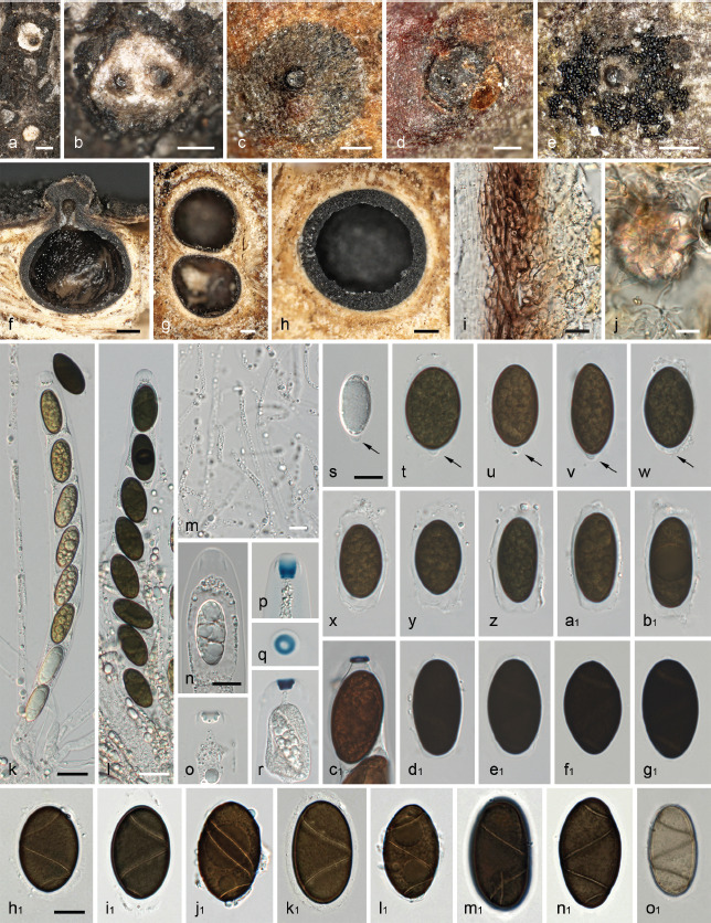
Fig. 14.

Leptomassaria simplex. a–b. Habit of young ostioles on host surface surrounded by white stromatic tissues; c–d. clypeus with ostiole; e. ostiole of mature ascoma surrounded by ejected ascospores; f–h. ascoma in vertical (f) and transverse (g–h) section, showing reduced, lighter brown entostroma surrounding the perithecia; i. peridium in transverse section (in 3 % KOH); j. loosely intertwined entostromatic hyphae with a crystal (in 3 % KOH); k–l. immature (k) and mature (l) ascus (aqueous NaCl solution); m. paraphyses in water; n–r, c1. apical apparatuses in water (n), 3 % KOH (o), Lugol’s solution (p–q) and Lugol’s solution after 3 % KOH pretreatment (r, c1); s–b1, d1–o1. immature (s, o1) and mature (t–b1, d1–n1) ascospores; arrows denoting basal cellular appendages (s–b1, h1–l1 in water; d1–g1, m1–o1 in 3 % KOH) (a–b, f. WU-MYC 0044028; c. WU-MYC 0044029; d, g, i–j, c1–g1, m1–o1. B 700017008 (lectotype); e, h. WU-MYC 0044027; k–b1, h1–l1. WU-MYC 0044025 (epitype)). — Scale bars: a–h = 200 μm; i–j, m–o1 = 10 μm; k–l = 20 μm.
