

Abstract
During extensive surveys of global Phytophthora diversity 14 new species detected in natural ecosystems in Chile, Indonesia, USA (Louisiana), Sweden, Ukraine and Vietnam were assigned to Phytophthora major Clade 10 based on a multigene phylogeny of nine nuclear and three mitochondrial gene regions. Clade 10 now comprises three subclades. Subclades 10a and 10b contain species with nonpapillate sporangia, a range of breeding systems and a mainly soil- and waterborne lifestyle. These include the previously described P. afrocarpa, P. gallica and P. intercalaris and eight of the new species: P. ludoviciana, P. procera, P. pseudogallica, P. scandinavica, P. subarctica, P. tenuimura, P. tonkinensis and P. ukrainensis. In contrast, all species in Subclade 10c have papillate sporangia and are self-fertile (or homothallic) with an aerial lifestyle including the known P. boehmeriae, P. gondwanensis, P. kernoviae and P. morindae and the new species P. celebensis, P. chilensis, P. javanensis, P. multiglobulosa, P. pseudochilensis and P. pseudokernoviae. All new Phytophthora species differed from each other and from related species by their unique combinations of morphological characters, breeding systems, cardinal temperatures and growth rates. The biogeography and evolutionary history of Clade 10 are discussed. We propose that the three subclades originated via the early divergence of pre-Gondwanan ancestors > 175 Mya into water- and soilborne and aerially dispersed lineages and subsequently underwent multiple allopatric and sympatric radiations during their global spread.
Citation: Jung T, Milenković I, Corcobado T, et al. 2022. Extensive morphological and behavioural diversity among fourteen new and seven described species in Phytophthora Clade 10 and its evolutionary implications. Persoonia 49: 1-57. https://doi.org/10.3767/persoonia.2022.49.01.
Keywords: allopatric, biogeography, evolution, Gondwana, Laurasia, Oomycete, phylogeny, radiation, sympatric
INTRODUCTION
The oomycete genus Phytophthora currently includes six obligate biotrophic unculturable species and 192 hemibiotrophic or necrotrophic culturable species (Chen et al. 2022). Most are soil-, water- or aerially dispersed plant pathogens, some causing severe diseases on host species in horticultural, forestry and natural ecosystems (Erwin & Ribeiro 1996, Yang et al. 2017, Jung et al. 2018b, Chen et al. 2022). Recent phylogenetic and phylogenomic studies have demonstrated that the c. 800 species of obligate biotrophic downy mildews reside as two distinct phylogenetic clades within the paraphyletic genus Phytophthora, suggesting their evolution from hemibiotrophic Phytophthora ancestors (Thines & Choi 2016, Jung et al. 2017d, McCarthy & Fitzpatrick 2017, Bourret et al. 2018, Fletcher et al. 2018, 2019, Scanu et al. 2021). Phytophthora currently resolves into 12 major phylogenetic clades with numerous subclades (Yang et al. 2017, Jung et al. 2017d, Chen et al. 2022). Recently Bourret et al. (2018) proposed additional Clades, 13 and 14, to accommodate the undescribed Phytophthora taxon mugwort and the obligate biotrophic P. cyperi which is more likely to be a downy mildew, and two further Clades, 15 and 16, comprising the 20 described genera of downy mildews. The evolutionary history of Phytophthora is characterised by an early divergence, most likely 175–210 million years ago (Mya) (Jung et al. 2017d), resulting in a main cluster comprising extant Phytophthora Clades 1–8, 11–13 and the downy mildew Clades 14–16, and a basal cluster containing extant Phytophthora Clades 9 and 10 (Yang et al. 2017, Jung et al. 2017d, Bourret et al. 2018, Scanu et al. 2021, Chen et al. 2022).
In the first ITS-based phylogeny of the genus Phytophthora Clade 10 included only a single species, P. boehmeriae, described in 1927 from Taiwan (Tucker 1931, Erwin & Ribeiro 1996, Cooke et al. 2000). However, another six Clade 10 species have been described since 2005 including, in chronological order, P. kernoviae, P. gallica, P. morindae, P. gondwanensis, P. intercalaris and P. afrocarpa (Brasier et al. 2005, Jung & Nechwatal 2008, Nelson & Abad 2010, Crous et al. 2015, Yang et al. 2016, Bose et al. 2021). Phytophthora boehmeriae, P. kernoviae and P. morindae are aerially dispersed pathogens producing caducous papillate sporangia in dense sympodia and infecting above-ground plant tissues. Phytophthora boehmeriae is noted for causing leaf blight of the herbaceous ramie (Boehmeria nivea) and boll rot of cotton (Gossypium spp.) in China and Taiwan and leaf blight of sweet pepper (Capsicum annuum) in India (Tucker 1931, Erwin & Ribeiro 1996, Chowdappa et al. 2014, Thorpe et al. 2021); P. kernoviae for causing aerial bleeding stem lesions on Fagus sylvatica trees and leaf and shoot blight of Rhododendron spp. and Drimys winteri in the UK and Ireland (Brasier et al. 2005, O’ Hanlon et al. 2016); and P. morindae for leaf blight and fruit rot of Morinda citrifolia var. citrifolia in Hawaii (Nelson & Abad 2010). Phytophthora gondwanensis has been recovered from soil samples around various host plants across Australia and exotic Eucalyptus smithii and Acacia mearnsii plantations in South Africa and Brazil, respectively, and is also a pathogen of Zanthoxylum piperitum in China and Ficus sp. in Papua New Guinea (Dos Santos et al. 2006, Crous et al. 2015, Burgess et al. 2021). Its caducous sporangia (Crous et al. 2015) indicate an aerial lifestyle. Another airborne taxon closely related to P. boehmeriae, Phytophthora taxon boehmeriae-like, was first isolated in 1939 causing brown rot of Citrus sinensis fruits in Argentina (Frezzi 1941, 1950, Erwin & Ribeiro 1996, Yang et al. 2017).
In contrast, the previously described Clade 10 species P. gallica, P. intercalaris and P. afrocarpa produce persistent nonpapillate sporangia with mainly internal proliferation (Jung & Nechwatal 2008, Yang et al. 2016, Bose et al. 2021). Phytophthora gallica and P. intercalaris were exclusively recovered from waterbodies or riparian forests in Europe and the USA (Jung & Nechwatal 2008, Jones et al. 2014, Sims et al. 2015, Yang et al. 2016, Redondo et al. 2018a, b) suggesting a lifestyle as litter decomposers and opportunistic root pathogens of riparian plants similar to primarily aquatic Phytophthora species from Clades 6 and 9 (Brasier et al. 2003, Jung et al. 2011, Yang et al. 2014). Phytophthora afrocarpa is currently known only from rhizosphere soil of the coniferous tree Afrocarpus falcatus in a temperate mountain forest in South Africa (Bose et al. 2021).
For Phytophthora taxon canthium, an undescribed Clade 10 taxon from temperate forest soil in South Africa related to P. boehmeriae, P. morindae and P. kernoviae, no information on hosts or morphology is available (Oh et al. 2013). While P. kernoviae is damaging as an invasive pathogen in Europe it is relatively benign in natural forests of Valdivia (Chile) and New Zealand suggesting long-term coevolution and, hence, a Southern Hemisphere origin (Gardner et al. 2015, Sanfuentes et al. 2016, Jung et al. 2018a). In 2014, two new Clade 10 taxa closely related to P. kernoviae, P. chilensis nom. prov. and P. pseudokernoviae nom. prov. (Jung et al. 2018a), and a third new Clade 10 taxon were isolated alongside P. kernoviae from forest streams and necrotic leaves of D. winteri in the Valdivian rainforests. In 2017, two new Phytophthora taxa related to P. gallica, informally designated as P. taxon gallica-like 1 and 2, were detected in a stream running through an evergreen cloud forest in the North of Vietnam (Jung et al. 2020). Recent surveys in Indonesia, USA (Louisiana), Sweden and Ukraine also produced Phytophthora isolates which, based on ITS sequence analysis, constituted another three unknown Clade 10 taxa, bringing the total number of new undescribed taxa in Clade 10 to fourteen.
In this study, morphological and physiological characteristics and DNA sequence data from nine nuclear and three mitochondrial gene regions were used to characterise the 14 new Phytophthora species from Clade 10; compare them morphologically and behaviourally to the known species in Clade 10 and describe them as P. celebensis, P. chilensis, P. javanensis, P. ludoviciana, P. multiglobulosa, P. procera, P. pseudochilensis, P. pseudogallica, P. pseudokernoviae, P. scadinavica, P. subarctica, P. tenuimura, P. tonkinensis and P. ukrainensis spp. nov.; and consider the implications of their morphological and behavioural properties and distribution for the evolution of the Clade.
TERMINOLOGY
Definitions of homothallism, heterothallism and sterility
Homothallism and heterothallism are somewhat archaic, quasimorphological terms used more to describe whether gametangia are formed in single or paired Phytophthora cultures rather than the process this represents. Because of their common historical usage in species descriptions we have also used the terms here, but we prefer a more Darwinian definition reflecting the underlying breeding system or breeding strategy. By homothallic we mean self-fertility in single culture; but this process does not preclude outbreeding in nature as a result of the fusion of antheridia and oogonia of different genotypes. By heterothallic we mean that two mating or compatibility types (A1 and A2) are required to initiate gametogenesis between otherwise largely self-incompatible individuals; but while this process promotes outcrossing, once initiated it can also lead to a significant frequency of self-fertilisation (selfing). By sterility we mean an isolate’s lack of the intrinsic ability to form gametangia whether in single culture or in pairings with A1 or A2 isolates; but this does not exclude the possibility that it may act as a ‘silent’ A1 or A2, inducing gametangial formation by selfing in an A2 or an A1 isolate of another species (cf. P. gonopodyides, Brasier et al. 2003).
Use of the terms Phytophthora taxon x and Phytophthora sp. x
The informal term ‘Phytophthora taxon x’ (cf. Brasier et al. 2003) was developed to cover situations where it was clear that a novel entity of some taxonomic level had been identified, but formal description was likely to be delayed pending further analysis to determine the level of taxonomic distinction (e.g., species, subspecies, variety etc; cf. Brasier & Rayner 1987) and because of the often considerable time required to produce the appropriate publication. This situation has arisen more frequently as more and more novel Phytophthora taxa are being discovered. In this regard, we do not concur with the use of the informal terminology ‘Phytophthora sp. x’ in the same context. A putative new taxon is not a species (or a ‘sp.’) until its correct hierarchical taxonomic status has been defined (as far as is reasonable), and its proposed name has been formally designated under the ICNafp (International Code of Nomenclature for algae, fungi, and plants; https://www.iapt-taxon.org) guidelines. On this basis we consider that a casual use of ‘Phytophthora sp. x’ in the case of a putative but only partially characterised new taxon is somewhat prejudicial to the final outcome. We have therefore confined ourselves to use the term ‘Phytophthora taxon x’ throughout this manuscript.
MATERIAL AND METHODS
Phytophthora isolates
Details of all isolates used in the phylogenetic, morphological and physiological studies are given in Table 1. Sampling and isolation methods from forest soil and river systems were described in Jung et al. (2017a, 2018a, 2020). The isolates of P. celebensis, P. chilensis, P. pseudochilensis, P. pseudogallica, P. javanensis, P. ludoviciana, P. multiglobulosa, P. procera, P. subarctica, P. tenuimura, P. tonkinensis and P. ukrainensis spp. nov. were recovered from forest streams in Valdivian rainforests in Chile, a montane cloud forest in Vietnam, tropical lowland rainforests in Indonesia, a swamp forest in Louisiana, boreal forests in Sweden and a riparian forest in Ukraine (Fig. 1; Table 1), by plating necrotic baiting leaves or naturally fallen floating leaves onto selective PARPNH-agar (Jung et al. 1996, 2020). All isolates of P. pseudokernoviae sp. nov. were isolated from necrotic Drimys winteri leaves in a Valdivian rainforest (Fig. 1d–e) whereas all isolates of P. scandinavica sp. nov. were baited in the lab from riverbank soil collected in the Kiruna area of northern Sweden using young Fagus sylvatica leaves as baits (Jung et al. 2019). In addition, for comparative studies isolates of four described Clade 10 species were used which had been obtained between 2014 and 2018: P. kernoviae from forest streams in Valdivian rainforests (Jung et al. 2018a) and from Rhododendron spp. in Ireland (O’Hanlon et al. 2016); P. gallica from forest streams and riparian ecosystems in Croatia, Czech Republic, Serbia, Sweden and Ukraine; P. intercalaris from a nursery seedling of Aronia in Croatia; and P. gondwanensis from a forest stream on Amami Island, Japan (Table 1). For all isolates, single hyphal tip cultures were produced under the stereomicroscope at ×20 from the margins of fresh cultures on V8-juice agar (V8A; 16 g agar, 3 g CaCO3, 100 mL Campbell’s V8 juice, 900 mL distilled water; Jung et al. 1999). Stock cultures were maintained on V8A and carrot juice agar (CA; 20 g agar, 3 g CaCO3, 100 mL carrot juice, 900 mL distilled water; Scanu et al. 2014) at 10 °C in the dark. All isolates of the 14 new Phytophthora spp. are preserved in the culture collection maintained at Mendel University in Brno. Ex-type cultures were deposited at the Agriculture Research Service (ARS) Culture Collection, Peoria, IL, USA, and/or the CBS Culture Collection (CBS) at the Westerdijk Institute, Utrecht, Netherlands (Table 1). Dried V8A cultures of the 14 ex-type isolates were deposited as holotypes in the herbarium of the Hungarian Natural History Museum, Budapest, Hungary.
Table 1.
Details of isolates from Phytophthora major Clades 7, 9 and 10 included in the phylogenetic, morphological and growth-temperature studies. GenBank accession numbers for sequences obtained in the present study are printed in italics.
| Phytophthora species | Isolate numbers a | Origin | GenBank accession numbers | |||||||
|---|---|---|---|---|---|---|---|---|---|---|
| International collections | Local collections | Host | Location; year; collector; reference | 28S LSU ITS | βtub hsp90 | tigA rpl10 | tef-1α enl | ras-ypt1 cox1 | nadh1 rps10 | |
| P. afrocarpa | CBS 147467ET | CMW54630b | Afrocarpus falcatus | South Africa; 2017; K. Sawada; J.Μ. Hulbert; Bose et al. 2021 | n.a. | MT762324 | n.a. | n.a. | n.a. | n.a. |
| MT762306 | MT762333 | n.a. | n.a. | MT762315 | n.a. | |||||
| CBS 147590 | CMW54631b | A. falcatus | South Africa; 2017; K. Sawada; J.Μ. Hulbert; Bose et al. 2021 | n.a. | MT762325 | n.a. | n.a. | n.a. | n.a. | |
| MT762307 | MT762334 | n.a. | n.a. | MT762316 | n.a. | |||||
| P. boehmeriae | CBS 291.29, IMI180614, ATCC 60238, WPC P6950ET | 45F9b | Boehmeria nivea | Taiwan; 1927; K. Sawada; Tucker 1931 | HQ665190 | EU080162 | EU080167 | EU080163 | MH974993 | DQ361200 |
| HQ643149 | EU080165 | EU080161 | EU080164 | HQ261251-KT183047 | JF770876 | |||||
| WPC P3963 | CPHST BL79b | Gossypium hirsutum | China; 1989; C.-Y. Shen; n.a. | JAAVTJ010 | JAAVTJ010 | JAAVTJ010 | JAAVTJ010 | JAAVTJ010 | JAAVTJ010 | |
| 000283 e | 000250 e | 000206 e | 000055 e | 000326 e | 000225 e | |||||
| JAAVTJ010 | JAAVTJ010 | JAAVTJ010 | JAAVTJ010 | JAAVTJ010 | JAAVTJ010 | |||||
| 000075 e | 000384 e | 000040 e | 000045 e | 000225 e | 000225 e | |||||
| SCRP23b | G. hirsutum | China; 1998; Y. Wang; n.a. | JAGDFL01 | JAGDFL01 | JAGDFL01 | JAGDFL01 | JAGDFL010 | JAGDFL010 | ||
| 0000638 e | 0000070 e | 0000109 e | 0000202 e | 000236 e | 000296 e | |||||
| JAGDFL01 | JAGDFL01 | JAGDFL01 | JAGDFL01 | JAGDFL010 | JAGDFL010 | |||||
| 0000638 e | 0000190 e | 0000393 e | 0000600 e | 000296 e | 000296 e | |||||
| WPC P7460b | Capsicum annuum | India, Madhya Pradesh; 1989; N.D. Sharma; n.a. | n.a. | n.a. | n.a. | n.a. | n.a. | n.a. | ||
| FJ801971 | n.a. | n.a. | n.a. | n.a. | n.a. | |||||
| OCPC4b | C. annuum; fruit rot | India, Bangalore Rural; 2011; S. Madhura; Chowdappa et al. 2014 | n.a. | n.a. | n.a. | n.a. | n.a. | n.a. | ||
| KC677806 | n.a. | n.a. | n.a. | n.a. | n.a. | |||||
| P. celebensis | CBS 148800ET | SL092bcd | Fallen bamboo leaf, forest stream R09 | Indonesia, Sulawesi; 2019; T. Jung, Μ. Junaid, N. Nasri; this study | ON000626 | OM975899 | OM974594 | OM984880 | ON024938 | OM976896 |
| ON000720 | OM976416 | OM974453 | OM976512 | ON013786 | OM976654 | |||||
| SL091bcd | Fallen bamboo leaf, forest stream R09 | Indonesia, Sulawesi; 2019; T. Jung, Μ. Junaid, N. Nasri; this study | ON000622 | OM975895 | OM974590 | OM984876 | ON024934 | OM976892 | ||
| ON000716 | OM976412 | OM974449 | OM976508 | ON013782 | OM976650 | |||||
| SL540bcd | Fallen bamboo leaf, forest stream R09 | Indonesia, Sulawesi; 2019; T. Jung, Μ. Junaid, N. Nasri; this study | ON000623 | OM975896 | OM974591 | OM984877 | ON024935 | OM976893 | ||
| ON000717 | OM976413 | OM974450 | OM976509 | ON013783 | OM976651 | |||||
| SL541bcd | Fallen bamboo leaf, forest stream R09 | Indonesia, Sulawesi; 2019; T. Jung, Μ. Junaid, N. Nasri; this study | ON000624 | OM975897 | OM974592 | OM984878 | ON024936 | OM976894 | ||
| ON000718 | OM976414 | OM974451 | OM976510 | ON013784 | OM976652 | |||||
| SL542bcd | Fallen bamboo leaf, forest stream R09 | Indonesia, Sulawesi; 2019; T. Jung, Μ. Junaid, N. Nasri; this study | ON000625 | OM975898 | OM974593 | OM984879 | ON024937 | OM976895 | ||
| ON000719 | OM976415 | OM974452 | OM976511 | ON013785 | OM976653 | |||||
| P. chilensis | CBS 148797, NRRL64353ET | CL165bcd | Baiting stream R04, Valdivian rainforest | Chile; 2014; T. Jung, A. Durán, E. Sanfuentes; Jung et al. 2018b | ON000632 | OM975905 | OM974600 | OM984886 | ON024944 | OM976902 |
| ON000726 | OM976422 | OM974459 | OM976518 | ON013792 | OM976660 | |||||
| CL166bcd | Baiting stream R04, Valdivian rainforest | Chile; 2014; T. Jung, A. Durán, E. Sanfuentes; Jung et al. 2018b | ON000627 | OM975900 | OM974595 | OM984881 | ON024939 | OM976897 | ||
| ON000721 | OM976417 | OM974454 | OM976513 | ON013787 | OM976655 | |||||
| CL169bcd | Baiting stream R04, Valdivian rainforest | Chile; 2014; T. Jung, A. Durán, E. Sanfuentes; Jung et al. 2018b | ON000628 | OM975901 | OM974596 | OM984882 | ON024940 | OM976898 | ||
| ON000722 | OM976418 | OM974455 | OM976514 | ON013788 | OM976656 | |||||
| CL170bcd | Baiting stream R04, Valdivian rainforest | Chile; 2014; T. Jung, A. Durán, E. Sanfuentes; Jung et al. 2018b | ON000629 | OM975902 | OM974597 | OM984883 | ON024941 | OM976899 | ||
| ON000723 | OM976419 | OM974456 | OM976515 | ON013789 | OM976657 | |||||
| CL171bcd | Baiting stream R05, Valdivian rainforest | Chile; 2014; T. Jung, A. Durán, E. Sanfuentes; Jung et al. 2018b | ON000630 | OM975903 | OM974598 | OM984884 | ON024942 | OM976900 | ||
| ON000724 | OM976420 | OM974457 | OM976516 | ON013790 | OM976658 | |||||
| CL172bcd | Baiting stream R05, Valdivian rainforest | Chile; 2014; T. Jung, A. Durán, E. Sanfuentes; Jung et al. 2018b | ON000631 | OM975904 | OM974599 | OM984885 | ON024943 | OM976901 | ||
| ON000725 | OM976421 | OM974458 | OM976517 | ON013791 | OM976659 | |||||
| P. cinnamomi | TW012c | Cinnamomum | Taiwan; 2013; T. Jung; Jung et al. 2017b | ON000634 | OM975907 | n.a. | OM984888 | n.a. | OM976904 | |
| micranthum | ON000728 | OM976424 | OM974461 | OM976519 | ON013794 | OM976662 | ||||
| TJ1123, MP74c | n.a. | Australia (WA); n.a.; CALMe; Hüberli 1995 | ON000633 | OM975906 | OM974601 | OM984887 | n.a. | OM976903 | ||
| ON000727 | OM976423 | OM974460 | n.a. | ON013793 | OM976661 | |||||
| P. constricta | CBS 125801 | VHS 16130, TJ306, 55C3b | Kwongan heathland | Australia, WA; 2006; VHS; Rea et al. 2011 | ON000635 | OM975908 | OM974602 | OM984889 | ON024945 | OM976905 |
| ON000729 | OM976425 | OM974462 | OM976520 | ON013795 | OM976663 | |||||
| P. fallax | CBS 119109, ICMP 15575ET | 46J2, WPC P10722, CPHST BL63, NZFS 310Lb | Eucalyptus salignae, leaf necrosis | New Zealand, Rotoehu Forst; 1992; C. Barr; Dick et al. 2006 | KX252573 | KX252569 | KX252574 | KX252570 | MH443229 | KX252570 |
| MG865489 | KX252572 | KX252568 | KX252571 | MH136885-KC733451 | JQ439192 | |||||
| ICMP 17563 | NZFS 310Kb | Eucalyptus delagatensis | New Zealand, Owaka Valley; 1997; R.E. Beever; Winkworth et al. 2020 | n.a. | n.a. | n.a. | n.a. | n.a. | MN 883608f | |
| MN883608f | MN 883608f | |||||||||
| P. gallica | CBS 111474ET | GAL1b | Quercus robur, riparian forest | France; 1998; T. Jung; Jung & Nechwatal 2008 | KX252594 | KX252590 | KX252595 | KX252591 | MH443236 | n.a. |
| KF317090 | KX252593 | KX252589 | KX252592 | MH136893-KF317112 | n.a. | |||||
| NRRL 66988 | SFB267bcd | Stream baiting, mountain forest | Serbia; 2017; I. Milenkovic; this study | ON000637 | OM975910 | OM974604 | OM984891 | ON024947 | OM976907 | |
| ON000731 | OM976427 | OM974464 | OM976522 | ON013797 | OM976665 | |||||
| MO130bcd | Stream baiting, broadleaved forest | Czech Republic; 2019; H. Czech Republic; 2019; Ďatková; Ďatková 2020 | ON000636 | OM975909 | OM974603 | OM984890 | ON024946 | OM976906 | ||
| ON000730 | OM976426 | OM974463 | OM976521 | ON013796 | OM976664 | |||||
| TJ582 = 20. VRBJOH 7/15bcd | Alnus glutinosa, swamp forest | Croatia; 2014; Z. Tomić; this study | ON000639 | OM975912 | OM974606 | OM984893 | ON024949 | OM976909 | ||
| ON000733 | OM976429 | OM974466 | OM976524 | ON013799 | OM976667 | |||||
| SW423b | Stream baiting, Pinus Silvestris forest | Sweden, SWSweden; 2017; I. Milenković, T. Corcobado; this study | ON000638 | OM975911 | OM974605 | OM984892 | ON024948 | OM976908 | ||
| ON000732 | OM976428 | OM974465 | OM976523 | ON013798 | OM976666 | |||||
| UA399b | A. glutinosa, soil | Ukraine, Transcarpathia region; 2019; I. Milenković, T. Corcobado; this study | ON000640 | OM975913 | OM974607 | n.a. | ON024950 | OM976910 | ||
| ON000734 | OM976430 | OM974467 | OM976525 | ON013800 | OM976668 | |||||
| 33-4-R.1b | Alnus rhombifolia rhizosphere | Oregon, USA; 2010; L.L. Sims; Sims et al. 2015 | n.a. | n.a. | n.a. | n.a. | n.a. | n.a. | ||
| KJ666760 | n.a. | n.a. | n.a. | n.a. | n.a. | |||||
| 76-P1b | Baiting of a waterbody | New York State, USA; 2010 or 2011 ; L.A. Jones; Jones et al. 2014 | n.a. | n.a. | n.a. | n.a. | n.a. | n.a. | ||
| KJ865228 | n.a. | n.a. | n.a. | n.a. | n.a. | |||||
| P. gondwanensis | CBS 139336ET | CMW42633, W1858b | Gondwana rainforest soil | Australia, NSW; 2014; K. Scarlett; Crous et al. 2015 | n.a. | KP070605 | n.a. | OK267378 | n.a. | n.a. |
| KP070695 | OL466916 | OK533441 | n.a. | OK185360 | n.a. | |||||
| NRRL 64120 | JP1308bcd | Fallen leaf, forest stream R37 | Japan, Amami Island; 2018; T. Jung, K. Kageyama, H. Masuya; this study | ON000641 | OM975914 | OM974608 | OM984894 | ON024951 | OM976911 | |
| ON000735 | OM976431 | OM974468 | OM976526 | ON013801 | OM976669 | |||||
| JP2350bcd | Fallen leaf, forest stream R37 | Japan, Amami Island; 2018; T. Jung, K. Kageyama, H. Masuya; this study | ON000642 | OM975915 | OM974609 | OM984895 | ON024952 | OM976912 | ||
| ON000736 | OM976432 | OM974469 | OM976527 | ON013802 | OM976670 | |||||
| JP2351bcd | Fallen leaf, forest stream R37 | Japan, Amami Island; 2018; T. Jung, K. Kageyama, H. Masuya; this study | ON000643 | OM975916 | OM974610 | OM984896 | ON024953 | OM976913 | ||
| ON000737 | OM976433 | OM974470 | OM976528 | ON013803 | OM976671 | |||||
| JP2352bcd | Fallen leaf, forest stream R37 | Japan, Amami Island; 2018; T. Jung, K. Kageyama, H. Masuya; this study | ON000644 | OM975917 | OM974611 | OM984897 | ON024954 | OM976914 | ||
| ON000738 | OM976434 | OM974471 | OM976529 | ON013804 | OM976672 | |||||
| JP2353bcd | Japan, Amami Island; 2018; T. Jung, K. Kageyama, H. Masuya; this study | ON000645 | OM975918 | OM974612 | OM984898 | ON024955 | OM976915 | |||
| ON000739 | OM976435 | OM974472 | OM976530 | ON013805 | OM976673 | |||||
| ATTC 46717 WPC P1257 bg | Ficus sp., soil | Papua New Guinea; before 1994; n.a. | n.a. | n.a. | n.a. | n.a. | n.a. | n.a. | ||
| FJ801722 | n.a. | n.a. | n.a. | HQ261253 | n.a. | |||||
| ALT-18bg | Acacia mearnsii, plantation | Brazil, Rio Grande do Sul; 2005; A.F. Dos Santos; Dos Santos et al. 2006 | n.a. | KX396337 | n.a. | KX396325 | n.a. | n.a. | ||
| KX396302 | n.a. | n.a. | n.a. | KX396277 | n.a. | |||||
| CBS 100410 | VHS 3488bg | Persoonia longifolia, soil | Australia, WA; n.a.; F. Tay; n.a. | n.a. | n.a. | n.a. | n.a. | n.a. | n.a. | |
| KJ755133 | n.a. | n.a. | n.a. | n.a. | n.a. | |||||
| CMW19441bg | Eucalyptus smithii, plantation | South Africa, KwaZulu-Natal; before 2007; B. Maseko; Maseko et al. 2007 | n.a. | DQ988246 | n.a. | n.a. | n.a. | n.a. | ||
| DQ988207 | n.a. | n.a. | n.a. | n.a. | n.a. | |||||
| ATCC MYA-3893, IMI 403470 | 22G7, S833, p43bg | n.a. | n.a.; n.a.; Schmitthenner? Yang et al. 2017 | KX252608 | KX252604 | KX252609 | KX252605 | n.a. | n.a. | |
| KT183035 | KX252607 | KX252603 | KX252606 | KT183046 | n.a. | |||||
| P. intercalaris | CBS 140632ET | 45B7b | Stream water | USA, Virginia; 2007; X. Yang; Yang et al. 2016 | KX252615 | KT163336 | KX252616 | KX252612 | n.a. | n.a. |
| KT163268 | KX252614 | KX252610 | KX252613 | OK185358-KT163315 | n.a. | |||||
| CBS 140631 | 49A7b | Yang et al. 2016 | KX252629 | KX252625 | KX252630 | KX252626 | n.a. | n.a. | ||
| KT163273 | KX252628 | KX252624 | KX252627 | KT163320 | n.a. | |||||
| NRRL 64027 | TJ567bcd | Ornamental nursery plant of Aronia sp. | Croatia; 2014; Z. Tomić; this study | ON000646 | OM975919 | OM974613 | OM984899 | ON024956 | OM976916 | |
| ON000740 | OM976436 | OM974473 | OM976531 | ON013806 | OM976674 | |||||
| P. Javanensis | CBS 149203, NRRL64129ET | JV025abcd | Fallen leaf, forest stream R01 | Indonesia, Java; 2019; T. Jung, Μ. Junaid, N. Nasri; this study | ON000656 | OM975929 | OM974623 | OM984909 | ON024966 | OM976926 |
| ON000750 | OM976446 | OM974483 | OM976541 | ON013816 | OM976684 | |||||
| JV025bbcd | Fallen leaf, forest stream R01 | Indonesia, Java; 2019; T. Jung, Μ. Junaid, N. Nasri; this study | ON000647 | OM975920 | OM974614 | OM984900 | ON024957 | OM976917 | ||
| ON000741 | OM976437 | OM974474 | OM976532 | ON013807 | OM976675 | |||||
| JV191bcd | Fallen leaf, forest stream R01 | Indonesia, Java; 2019; T. Jung, Μ. Junaid, N. Nasri; this study | ON000648 | OM975921 | OM974615 | OM984901 | ON024958 | OM976918 | ||
| ON000742 | OM976438 | OM974475 | OM976533 | ON013808 | OM976676 | |||||
| JV192bcd | Fallen leaf, forest stream R01 | Indonesia, Java; 2019; T. Jung, Μ. Junaid, N. Nasri; this study | ON000649 | OM975922 | OM974616 | OM984902 | ON024959 | OM976919 | ||
| ON000743 | OM976439 | OM974476 | OM976534 | ON013809 | OM976677 | |||||
| JV193bcd | Fallen leaf, forest stream R01 | Indonesia, Java; 2019; T. Jung, Μ. Junaid, N. Nasri; this study | ON000650 | OM975923 | OM974617 | OM984903 | ON024960 | OM976920 | ||
| ON000744 | OM976440 | OM974477 | OM976535 | ON013810 | OM976678 | |||||
| SL081bcd | Fallen leaf, forest stream R07 | Indonesia, Sulawesi; 2019; T. Jung, Μ. Junaid, N. Nasri; this study | ON000651 | OM975924 | OM974618 | OM984904 | ON024961 | OM976921 | ||
| ON000745 | OM976441 | OM974478 | OM976536 | ON013811 | OM976679 | |||||
| SL084bcd | Fallen leaf, forest stream R07 | Indonesia, Sulawesi; 2019; T. Jung, Μ. Junaid, N. Nasri; this study | ON000652 | OM975925 | OM974619 | OM984905 | ON024962 | OM976922 | ||
| ON000746 | OM976442 | OM974479 | OM976537 | ON013812 | OM976680 | |||||
| SL0537bcd | Fallen leaf, forest stream R07 | Indonesia, Sulawesi; 2019; T. Jung, Μ. Junaid, N. Nasri; this study | ON000653 | OM975926 | OM974620 | OM984906 | ON024963 | OM976923 | ||
| ON000747 | OM976443 | OM974480 | OM976538 | ON013813 | OM976681 | |||||
| SL0538bcd | Fallen leaf, forest stream R07 | Indonesia, Sulawesi; 2019; T. Jung, Μ. Junaid, N. Nasri; this study | ON000654 | OM975927 | OM974621 | OM984907 | ON024964 | OM976924 | ||
| ON000748 | OM976444 | OM974481 | OM976539 | ON013814 | OM976682 | |||||
| SL0539bcd | Fallen leaf, forest stream R07 | Indonesia, Sulawesi; 2019; T. Jung, Μ. Junaid, N. Nasri; this study | ON000655 | OM975928 | OM974622 | OM984908 | ON024965 | OM976925 | ||
| ON000749 | OM976445 | OM974482 | OM976540 | ON013815 | OM976683 | |||||
| P. kernoviae | IMI 393170, WPC P19827ET | CPHST BL 91, P1571b | Fagus sylvatica, bark canker | UK, Cornwall; 2004; C.M. Brasier; Brasier et al. 2005 | VKKV0100 | VKKV0100 | VKKV0100 | VKKV0100 | VKKV01000 | VKKV01000 |
| 0063 e | 0263 e | 0441 e | 0415-0416 e | 091 e | 418 e | |||||
| VKKV0100 | VKKV0100 | VKKV0100 | VKKV0100 | VKKV01000 | VKKV01000 | |||||
| 0063 e | 0311 e | 0402 e | 0008 e | 418 e | 418 e | |||||
| 00238/432b | Rhododendron ponticum | UK, Scotland; 2010; A. Schlenzig; Sambles et al. 2015 | AOFI02000 | AOFI02000 | AOFI03000 | AOFI02000 | AOFI030000 | AOFI020003 | ||
| 967 e | 572 e | 088 e | 453 e | 34 e | 41 e | |||||
| AOFI02000 | AOFI02000 | AOFI02000 | AOFI02000 | AOFI020003 | AOFI020003 | |||||
| 967 e | 890 e | 251 e | 244 e | 41 e | 41 e | |||||
| 00629/1b | R. ponticum | UK, Scotland; 2011; A. Schlenzig; Sambles et al. 2015 | AOFJ02000 | AOFJ02000 | AOFJ02000 | AOFJ02001 | AOFJ02000 | AOFJ02000 | ||
| 931 e | 547 e | 111 e | 008 e | 030 e | 333 e | |||||
| AOFJ02000 | AOFJ02000 | AOFJ02000 | AOFJ02000 | AOFJ02000 | AOFJ02000 | |||||
| 931 e | 791 e | 243 e | 237 e | 333 e | 333 e | |||||
| 00844/4b | R. ponticum | UK, Scotland; 2011; A. Schlenzig; Sambles et al. 2015 | AOFK0200 | AOFK0200 | AOFK0200 | AOFK0200 | AOFK02000 | AOFK02000 | ||
| 0947 e | 0552 e | 0104 e | 0492 e | 030 e | 336 e | |||||
| AOFK0200 | AOFK0200 | AOFK0200 | AOFK0200 | AOFK02000 | AOFK02000 | |||||
| 0947 e | 0796 e | 0229 e | 0248 e | 336 e | 336 e | |||||
| NRRL 64156 | CL155bcd | Baiting stream R01, Valdivian rainforest | Chile; 2014; T. Jung, A. Durán, E. Sanfuentes; Jung et al. 2018b | ON000657 | OM975930 | OM974624 | OM984910 | ON024967 | OM976927 | |
| ON000751 | OM976447 | OM974484 | OM976542 | ON013817 | OM976685 | |||||
| CL167bcd | Baiting stream R04, Valdivian rainforest | Chile; 2014; T. Jung, A. Durán, E. Sanfuentes; Jung et al. 2018b | ON000658 | OM975931 | OM974625 | OM984911 | ON024968 | OM976928 | ||
| ON000752 | OM976448 | OM974485 | OM976543 | ON013818 | OM976686 | |||||
| CL213bcd | Baiting stream R09, Valdivian rainforest | Chile; 2014; T. Jung, A. Durán, E. Sanfuentes; Jung et al. 2018b | ON000659 | OM975932 | OM974626 | OM984912 | ON024969 | OM976929 | ||
| ON000753 | OM976449 | OM974486 | OM976544 | ON013819 | OM976687 | |||||
| CL233bcd | Baiting stream R13, Valdivian rainforest | Chile; 2014; T. Jung, A. Durán, E. Sanfuentes; Jung et al. 2018b | ON000660 | OM975933 | OM974627 | OM984913 | ON024970 | OM976930 | ||
| ON000754 | OM976450 | OM974487 | OM976545 | ON013820 | OM976688 | |||||
| CL234bcd | Baiting stream R13, Valdivian rainforest | Chile; 2014; T. Jung, A. Durán, E. Sanfuentes; Jung et al. 2018b | ON000661 | OM975934 | OM974628 | OM984914 | ON024971 | OM976931 | ||
| ON000755 | OM976451 | OM974488 | OM976546 | ON013821 | OM976689 | |||||
| PR11-532, TJ1605bcd | Rhododendron sp. | Ireland, Kerry; 2011; R. O’Hanlon; O’Hanlon et al. 2016 | ON000663 | OM975936 | OM974630 | OM984916 | ON024973 | OM976933 | ||
| ON000757 | OM976453 | OM974490 | OM976548 | ON013823 | OM976691 | |||||
| PR12-106, TJ1604bcd | R. ponticum | Ireland, Cork; 2012; R. O’Hanlon; O’Hanlon et al. 2016 | ON000662 | OM975935 | OM974629 | OM984915 | ON024972 | OM976932 | ||
| ON000756 | OM976452 | OM974489 | OM976547 | ON013822 | OM976690 | |||||
| PR12-513, TJ1607bcd | Rhododendron sp. | Ireland, Kerry; 2011; R. O’Hanlon; O’Hanlon et al. 2016 | ON000664 | OM975937 | OM974631 | OM984917 | ON024974 | OM976934 | ||
| ON000758 | OM976454 | OM974491 | OM976549 | ON013824 | OM976692 | |||||
| Chile 1b | Drimys winteri leaf litter, Valdivian rainforest | Chile; 2014; E. Sanfuentes; Studholme et al. 2019 | MBAB0100 | MBAB0100 | MBAB0100 | MBAB0100 | MBAB01001 | MBAB01000 | ||
| 2152 e | 0005 e | 0353 e | 1709 e | 431 e | 528 e | |||||
| MBAB0100 | MBAB0100 | MBAB0100 | MBAB0100 | MBAB01000 | MBAB01000 | |||||
| 2152 e | 0009 e | 1629 e | 1403 e | 528 e | 528 e | |||||
| Chile 2b | D. winteri leaf litter, Valdivian rainforest | Chile; 2012; E. Sanfuentes; Studholme et al. 2019 | n.a. | MAYM0200 | MAYM0200 | MAYM0200 | MAYM0200 | MAYM0200 | ||
| n.a. | 2138 e | 1352 e | 2226 e | 0340 e | 2296 e | |||||
| MAYM0200 | MAYM0200 | MAYM0200 | MAYM0200 | MAYM0200 | ||||||
| 1247 e | 1485 e | 0769 e | 0148 e | 1662 e | ||||||
| Chile 4b | D. winteri leaf litter, Valdivian rainforest | Chile; 2012; E. Sanfuentes; Studholme et al. 2019 | MBDN0200 | MBDN0200 | MBDN0200 | MBDN0200 | MBDN02000 | MBDN02001 | ||
| 1104 e | 0444 e | 0007 e | 1933 e | 021 e | 413 e | |||||
| MBDN0200 | MBDN0200 | MBDN0200 | MBDN0200 | MBDN02000 | MBDN02000 | |||||
| 1104 e | 0637 e | 0110 e | 0197 e | 533 e | 817 e | |||||
| NZFS 2646b | Annona cherimola | New Zealand, Northland; 2005; n.a.; Studholme et al. 2016 | JPVW0200 | JPVW0200 | JPVW0200 | JPWV0200 | JPWV02000 | JPVW02000 | ||
| 0770 e | 0072 e | 0085 e | 1728 e | 032 e | 257 e | |||||
| JPVW0200 | JPVW0200 | JPVW0200 | JPWV0200 | JPWV02000 | JPVW02000 | |||||
| 0770 e | 0617 e | 0195 e | 0175 e | 257 e | 257 e | |||||
| NZFS 3630b | Pinus radiata | New Zealand, Tokoroa; 2011; n.a.; Studholme et al. 2016 | JPWU0200 | JPWU0200 | JPWU0200 | JPWU0200 | JPWU02000 | JPWU02000 | ||
| 0930 e | 0457 e | 0080 e | 0131 e | 011 e | 272 e | |||||
| JPWU0200 | JPWU0200 | JPWU0200 | JPWU0200 | JPWU02000 | JPWU02000 | |||||
| 0930 e | 0634 e | 0196 e | 0174 e | 272 e | 272 e | |||||
| P. ludoviciana | CBS 149205, NRRL 64143ET | LU057bcd | Fallen leaf, forest stream R01 | USA, Louisiana; 2020; T. Corcobado, T. Májek; this study | ON000666 | OM975939 | OM974633 | OM984919 | ON024976 | OM976936 |
| ON000760 | OM976456 | OM974493 | OM976551 | ON013826 | OM976694 | |||||
| LU038bcd | Fallen leaf, forest stream R01 | USA, Louisiana; 2020; T. Corcobado, T. Májek; this study | ON000665 | OM975938 | OM974632 | OM984918 | ON024975 | OM976935 | ||
| ON000759 | OM976455 | OM974492 | OM976550 | ON013825 | OM976693 | |||||
| P. morindae | CBS 121982ET | 62B5b | Morinda citrifolia; black flag disease | USA, Hawaii; 2005; S.C. Nelson; Nelson & Abad 2010 | KX252638 | KX252634 | KX252639 | KX252635 | MH988446 | n.a. |
| MH620178 | KX252637 | KX252633 | KX252636 | MH136936-KT183050 | n.a. | |||||
| P. multiglobulosa | CBS 148799ET | SL005bcd | Fallen leaf, forest stream R01 | Indonesia, Sulawesi; 2019; T. Jung, Μ. Junaid, N. Nasri; this study | ON000669 | OM975942 | OM974636 | OM984922 | ON024979 | OM976939 |
| ON000763 | OM976459 | OM974496 | OM976554 | ON013829 | OM976697 | |||||
| SL006bcd | Fallen leaf, forest stream R01 | Indonesia, Sulawesi; 2019; T. Jung, Μ. Junaid, N. Nasri; this study | ON000667 | OM975940 | OM974634 | OM984920 | ON024977 | OM976937 | ||
| ON000761 | OM976457 | OM974494 | OM976552 | ON013827 | OM976695 | |||||
| SL007bcd | Fallen leaf, forest stream R01 | Indonesia, Sulawesi; 2019; T. Jung, Μ. Junaid, N. Nasri; this study | ON000668 | OM975941 | OM974635 | OM984921 | ON024978 | OM976938 | ||
| ON000762 | OM976458 | OM974495 | OM976553 | ON013828 | OM976696 | |||||
| P. procera | CBS 149226 NRRL 64144ET | LU013bcd | Fallen leaf, forest stream R01 | USA, Louisiana; 2020; T. Corcobado, T. Májek; this study | ON000673 | OM975946 | OM974640 | OM984926 | ON024983 | OM976942 |
| ON000767 | OM976463 | OM974500 | n.a. | ON013833 | OM976701 | |||||
| LU007bcd | Fallen leaf, forest stream R01 | USA, Louisiana; 2020; T. Corcobado, T. Májek; this study | ON000670 | OM975943 | OM974637 | OM984923 | ON024980 | n.a. | ||
| ON000764 | OM976460 | OM974497 | OM976555 | ON013830 | OM976698 | |||||
| LU010bcd | Fallen leaf, forest stream R01 | USA, Louisiana; 2020; T. Corcobado, T. Májek; this study | ON000671 | OM975944 | OM974638 | OM984924 | ON024981 | OM976940 | ||
| ON000765 | OM976461 | OM974498 | OM976556 | ON013831 | OM976699 | |||||
| LU056bcd | Fallen leaf, forest stream R01 | USA, Louisiana; 2020; T. Corcobado, T. Májek; this study | ON000672 | OM975945 | OM974639 | OM984925 | ON024982 | OM976941 | ||
| ON000766 | OM976462 | OM974499 | OM976557 | ON013832 | OM976700 | |||||
| P. pseudochilensis | CBS 148798, NRRL 64352ET | CL168bcd | Baiting stream R04, Valdivian rainforest | Chile; 2014; T. Jung, A. Durán, E. Sanfuentes; Jung et al. 2018b | ON000679 | OM975952 | OM974646 | OM984932 | ON024989 | OM976948 |
| ON000773 | OM976469 | OM974506 | OM976563 | ON013839 | OM976707 | |||||
| CL335bcd | Baiting stream R04, Valdivian rainforest | Chile; 2014; T. Jung, A. Durán, E. Sanfuentes; Jung et al. 2018b | ON000674 | OM975947 | OM974641 | OM984927 | ON024984 | OM976943 | ||
| ON000768 | OM976464 | OM974501 | OM976558 | ON013834 | OM976702 | |||||
| CL336bcd | Baiting stream R04, Valdivian rainforest | Chile; 2014; T. Jung, A. Durán, E. Sanfuentes; Jung et al. 2018b | ON000675 | OM975948 | OM974642 | OM984928 | ON024985 | OM976944 | ||
| ON000769 | OM976465 | OM974502 | OM976559 | ON013835 | OM976703 | |||||
| CL337bcd | Baiting stream R04, Valdivian rainforest | Chile; 2014; T. Jung, A. Durán, E. Sanfuentes; Jung et al. 2018b | ON000676 | OM975949 | OM974643 | OM984929 | ON024986 | OM976945 | ||
| ON000770 | OM976466 | OM974503 | OM976560 | ON013836 | OM976704 | |||||
| CL338bcd | Baiting stream R04, Valdivian rainforest | Chile; 2014; T. Jung, A. Durán, E. Sanfuentes; Jung et al. 2018b | ON000677 | OM975950 | OM974644 | OM984930 | ON024987 | OM976946 | ||
| ON000771 | OM976467 | OM974504 | OM976561 | ON013837 | OM976705 | |||||
| CL339bcd | Baiting stream R04, Valdivian rainforest | Chile; 2014; T. Jung, A. Durán, E. Sanfuentes; Jung et al. 2018b | ON000678 | OM975951 | OM974645 | OM984931 | ON024988 | OM976947 | ||
| ON000772 | OM976468 | OM974505 | OM976562 | ON013838 | OM976706 | |||||
| P. pseudogallica | CBS 149206, NRRL 64136ET | VN861bcd | Fallen leaf, stream | Vietnam; 2017; T. Jung, N.M. Chi; Jung et al. 2020 | ON000680 | OM975953 | OM974647 | OM984933 | ON024990 | OM976949 |
| R02, cloud forest | ON000774 | OM976470 | OM974507 | OM976564 | ON013840 | OM976708 | ||||
| VN910bcd | Fallen leaf, stream | Vietnam; 2017; T. Jung, N.M. Chi; Jung et al. 2020 | ON000681 | OM975954 | OM974648 | OM984934 | ON024991 | OM976950 | ||
| R02, cloud forest | ON000775 | OM976471 | OM974508 | OM976565 | ON013841 | OM976709 | ||||
| VN920bcd | Fallen leaf, stream | Vietnam; 2017; T. Jung, N.M. Chi; Jung et al. 2020 | ON000682 | OM975955 | OM974649 | OM984935 | ON024992 | OM976951 | ||
| R02, cloud forest | ON000776 | OM976472 | OM974509 | OM976566 | ON013842 | OM976710 | ||||
| VN922bcd | Fallen leaf, stream | Vietnam; 2017; T. Jung, N.M. Chi; Jung et al. 2020 | ON000683 | OM975956 | OM974650 | OM984936 | ON024993 | OM976952 | ||
| R02, cloud forest | ON000777 | OM976473 | OM974510 | OM976567 | ON013843 | OM976711 | ||||
| P. pseudokernoviae | CBS 148796, NRRL 64351ET | CL012bcd | Fallen D. winteri leaf, Valdivian rainforest | Chile; 2014; T. Jung, A. Durán, E. Sanfuentes; Jung et al. 2018b | ON000686 | OM975959 | OM974653 | OM984939 | ON024996 | OM976955 |
| ON000780 | OM976476 | OM974513 | OM976570 | ON013846 | OM976714 | |||||
| CL013bcd | Fallen D. winteri leaf, Valdivian rainforest | Chile; 2014; T. Jung, A. Durán, E. Sanfuentes; Jung et al. 2018b | ON000684 | OM975957 | OM974651 | OM984937 | ON024994 | OM976953 | ||
| ON000778 | OM976474 | OM974511 | OM976568 | ON013844 | OM976712 | |||||
| CL156bcd | Leaf of D. winteri seedling, Valdivian rainforest | Chile; 2014; T. Jung, A. Durán, E. Sanfuentes; Jung et al. 2018b | ON000685 | OM975958 | OM974652 | OM984938 | ON024995 | OM976954 | ||
| ON000779 | OM976475 | OM974512 | OM976569 | ON013845 | OM976713 | |||||
| Chile 6bh | D. winteri leaf litter, Valdivian rainforest | Chile; 2014; E. Sanfuentes; Studholme et al. 2019 | MBD00200 | MBD00200 | MBD00200 | MBD00200 | MBD00200 | MBD00200 | ||
| 0514 e | 0455 e | 0053 e | 1754 e | 0016 e | 0923 e | |||||
| MBD00200 | MBD00200 | MBD00200 | MBD00200 | MBD00200 | MBD00200 | |||||
| 0514 e | 0600 e | 0109 e | 0427 e | 0562 e | 0811 e | |||||
| Chile 7bh | D. winteri leaf litter, Valdivian rainforest | Chile; 2014; E. Sanfuentes; Studholme et al. 2019 | MBAD0200 | MBAD0200 | MBAD0200 | MBAD0200 | MBAD02000 | n.a. | ||
| 1209 e | 2676 e | 0192 e | 0493 e | 695 e | ||||||
| MBAD0200 | MBAD0200 | MBAD0200 | MBAD0200 | MBAD02000 | n.a. | |||||
| 1209 e | 0923 e | 1760 e | 0835 e | 110e | ||||||
| P. scandinavica | CBS 149204, NRRL 66990ET | SW325bcd | Riverbank soil in boreal forest | Sweden, Kiruna; 2017; I. Milenković, T. Corcobado; this study | ON000692 | OM975965 | OM974659 | OM984945 | ON025002 | OM976961 |
| ON000786 | OM976482 | OM974519 | OM976576 | ON013852 | OM976720 | |||||
| SW314bcd | Riverbank soil in boreal forest | Sweden, Kiruna; 2017; I. Milenković, T. Corcobado; this study | ON000687 | OM975960 | OM974654 | OM984940 | ON024997 | OM976956 | ||
| ON000781 | OM976477 | OM974514 | OM976571 | ON013847 | OM976715 | |||||
| SW315bcd | Riverbank soil in boreal forest | Sweden, Kiruna; 2017; I. Milenković, T. Corcobado; this study | ON000688 | OM975961 | OM974655 | OM984941 | ON024998 | OM976957 | ||
| ON000782 | OM976478 | OM974515 | OM976572 | ON013848 | OM976716 | |||||
| SW316bcd | Riverbank soil in boreal forest | Sweden, Kiruna; 2017; I. Milenković, T. Corcobado; this study | ON000689 | OM975962 | OM974656 | OM984942 | ON024999 | OM976958 | ||
| ON000783 | OM976479 | OM974516 | OM976573 | ON013849 | OM976717 | |||||
| SW326bcd | Riverbank soil in boreal forest | Sweden, Kiruna; 2017; I. Milenković, T. Corcobado; this study | ON000690 | OM975963 | OM974657 | OM984943 | ON025000 | OM976959 | ||
| ON000784 | OM976480 | OM974517 | OM976574 | ON013850 | OM976718 | |||||
| SW327bcd | Riverbank soil in boreal forest | Sweden, Kiruna; 2017; I. Milenković, T. Corcobado; this study | ON000691 | OM975964 | OM974658 | OM984944 | ON025001 | OM976960 | ||
| ON000785 | OM976481 | OM974518 | OM976575 | ON013851 | OM976719 | |||||
| P. subarctica | CBS 148850, NRRL 64339ET | SW176bcd | Baiting stream R09, boreal forest | Sweden, Kiruna; 2017; I. Milenković, T. Corcobado; this study | ON000696 | OM975969 | OM974663 | OM984949 | ON025006 | OM976965 |
| ON000790 | OM976486 | OM974523 | OM976580 | ON013856 | OM976724 | |||||
| SW639bcd | Baiting stream R09, boreal forest | Sweden, Kiruna; 2017; I. Milenković, T. Corcobado; this study | ON000693 | OM975966 | OM974660 | OM984946 | ON025003 | OM976962 | ||
| ON000787 | OM976483 | OM974520 | OM976577 | ON013853 | OM976721 | |||||
| SW640bcd | Baiting stream R09, boreal forest | Sweden, Kiruna; 2017; I. Milenković, T. Corcobado; this study | ON000694 | OM975967 | OM974661 | OM984947 | ON025004 | OM976963 | ||
| ON000788 | OM976484 | OM974521 | OM976578 | ON013854 | OM976722 | |||||
| P. subarctica | SW641bcd | Baiting stream R09, boreal forest | Sweden, Kiruna; 2017; I. Milenković, T. Corcobado; this study | ON000695 | OM975968 | OM974662 | OM984948 | ON025005 | OM976964 | |
| ON000789 | OM976485 | OM974522 | OM976579 | ON013855 | OM976723 | |||||
| P. tenuimura | CBS 149227 | LU052bcd | Fallen leaf, forest stream R01 | USA, Louisiana; 2020; T. Corcobado, T. Májek; this study | ON000704 | OM975977 | OM974671 | OM984957 | ON025014 | OM976973 |
| NRRL 64142ET | ON000798 | OM976494 | OM974531 | OM976588 | ON013864 | OM976732 | ||||
| LU050bcd | Fallen leaf, forest stream R01 | USA, Louisiana; 2020; T. Corcobado, T. Májek; this study | ON000697 | OM975970 | OM974664 | OM984950 | ON025007 | OM976966 | ||
| ON000791 | OM976487 | OM974524 | OM976581 | ON013857 | OM976725 | |||||
| LU051bcd | Fallen leaf, forest stream R01 | USA, Louisiana; 2020; T. Corcobado, T. Májek; this study | ON000698 | OM975971 | OM974665 | OM984951 | ON025008 | OM976967 | ||
| ON000792 | OM976488 | OM974525 | OM976582 | ON013858 | OM976726 | |||||
| LU062bcd | Fallen leaf, forest stream R01 | USA, Louisiana; 2020; T. Corcobado, T. Májek; this study | ON000699 | OM975972 | OM974666 | OM984952 | ON025009 | OM976968 | ||
| ON000793 | OM976489 | OM974526 | OM976583 | ON013859 | OM976727 | |||||
| LU065bcd | Fallen leaf, forest stream R01 | USA, Louisiana; 2020; T. Corcobado, T. Májek; this study | ON000700 | OM975973 | OM974667 | OM984953 | ON025010 | OM976969 | ||
| ON000794 | OM976490 | OM974527 | OM976584 | ON013860 | OM976728 | |||||
| LU066bcd | Fallen leaf, forest stream R01 | USA, Louisiana; 2020; T. Corcobado, T. Májek; this study | ON000701 | OM975974 | OM974668 | OM984954 | ON025011 | OM976970 | ||
| ON000795 | OM976491 | OM974528 | OM976585 | ON013861 | OM976729 | |||||
| LU073bcd | Fallen leaf, forest stream R01 | USA, Louisiana; 2020; T. Corcobado, T. Májek; this study | ON000702 | OM975975 | OM974669 | OM984955 | ON025012 | OM976971 | ||
| ON000796 | OM976492 | OM974529 | OM976586 | ON013862 | OM976730 | |||||
| LU074bcd | Fallen leaf, forest stream R01 | USA, Louisiana; 2020; T. Corcobado, T. Májek; this study | ON000703 | OM975976 | OM974670 | OM984956 | ON025013 | OM976972 | ||
| ON000797 | OM976493 | OM974530 | OM976587 | ON013863 | OM976731 | |||||
| P. tonkinensis | CBS 148852, NRRL 64356ET | VN859bcd | Fallen leaf, stream R02, cloud forest | Vietnam; 2017; T. Jung, N.M. Chi; Jung et al. 2020 | ON000705 | OM975978 | OM974672 | OM984958 | ON025015 | OM976974 |
| ON000799 | OM976495 | OM974532 | OM976589 | ON013865 | OM976733 | |||||
| VN1106bcd | Fallen leaf, stream R02, cloud forest | Vietnam; 2017; T. Jung, N.M. Chi; Jung et al. 2020 | ON000706 | OM975979 | OM974673 | OM984959 | ON025016 | OM976975 | ||
| ON000800 | OM976496 | OM974533 | OM976590 | ON013866 | OM976734 | |||||
| VN1107bcd | Fallen leaf, stream R02, cloud forest | Vietnam; 2017; T. Jung, N.M. Chi; Jung et al. 2020 | ON000707 | OM975980 | OM974674 | OM984960 | ON025017 | OM976976 | ||
| ON000801 | OM976497 | OM974534 | OM976591 | ON013867 | OM976735 | |||||
| VN1108bcd | Fallen leaf, stream R02, cloud forest | Vietnam; 2017; T. Jung, N.M. Chi; Jung et al. 2020 | ON000708 | OM975981 | OM974675 | OM984961 | ON025018 | OM976977 | ||
| ON000802 | OM976498 | OM974535 | OM976592 | ON013868 | OM976736 | |||||
| VN1109bcd | Fallen leaf, stream R02, cloud forest | Vietnam; 2017; T. Jung, N.M. Chi; Jung et al. 2020 | ON000709 | OM975982 | OM974676 | OM984962 | ON025019 | OM976978 | ||
| ON000803 | OM976499 | OM974536 | OM976593 | ON013869 | OM976737 | |||||
| P. ukrainensis | CBS 148851, NRRL 64255ET | UA373bcd | Fallen Quercus leaf, Vereshchytsia River | Ukraine; 2019; I. Milenković, T. Corcobado, I. Matsiakh; this study | ON000711 | OM975984 | OM974678 | OM984964 | ON025021 | OM976980 |
| ON000805 | OM976501 | OM974538 | OM976595 | ON013871 | OM976739 | |||||
| UA376b | Fallen Quercus leaf, Vereshchytsia River | Ukraine; 2019; I. Milenković, T. Corcobado; this study | ON000712 | OM975985 | OM974679 | n.a. | ON025022 | OM976981 | ||
| ON000806 | OM976502 | OM974539 | OM976596 | ON013872 | OM976740 | |||||
| UA430bcd | Fallen Quercus leaf, Vereshchytsia River | Ukraine; 2019; I. Milenković, T. Corcobado, I. Matsiakh; this study | ON000713 | OM975986 | OM974680 | OM984965 | ON025023 | OM976982 | ||
| ON000807 | OM976503 | OM974540 | OM976597 | ON013873 | OM976741 | |||||
| UA431bcd | Fallen Quercus leaf, Vereshchytsia River | Ukraine; 2019; I. Milenković, T. Corcobado, I. Matsiakh; this study | ON000714 | OM975987 | OM974681 | OM984966 | ON025024 | OM976983 | ||
| ON000808 | OM976504 | OM974541 | OM976598 | ON013874 | OM976742 | |||||
| UA432bcd | Fallen Quercus leaf, Vereshchytsia River | Ukraine; 2019; I. Milenković, T. Corcobado, I. Matsiakh; this study | ON000715 | OM975988 | OM974682 | OM984967 | ON025025 | OM976984 | ||
| ON000809 | OM976505 | OM974542 | OM976599 | ON013875 | OM976743 | |||||
| SW154bcd | Baiting stream R08, boreal forest | Sweden, Kiruna; 2017; I. Milenković, T. Corcobado; this study | ON000710 | OM975983 | OM974677 | OM984963 | ON025020 | OM976979 | ||
| ON000804 | OM976500 | OM974537 | OM976594 | ON013870 | OM976738 | |||||
| P. taxon boehmeriae-like | CBS 357.52, IMI 32199, ATTC 60173, WPC P1378 | 45F8bg | Citrus sinensis, fruit brown rot | Argentina, Corrientes; 1939; M.J. Frazzi; Frazzi 1941 | KX252645 | KX252641 | KX252646 | KX252642 | n.a. | n.a. |
| KF317089 | KX252644 | KX252640 | KX252643 | KF317111-HQ261252 | n.a. | |||||
| P. taxon boehmeriae-like 2 | Psldbg | Zanthoxylum piperitum | China, Shannxi; ca 2014; W. Jiefei; n.a. | n.a. | n.a. | n.a. | n.a. | n.a. | n.a. | |
| KJ854387 | n.a. | n.a. | n.a. | n.a. | n.a. | |||||
| P. taxon boehmeriae-like 2 | Pme002bi | Medicago sativa | China; n.a.; C.Z. Lan; n.a. | n.a. | n.a. | n.a. | n.a. | n.a. | n.a. | |
| MG823393 | n.a. | n.a. | n.a. | n.a. | n.a. | |||||
| P. taxon canthium | CMW35236b | Forest soil | South Africa, Eastern Cape; ca 2013; E. Oh; Oh et al. 2013 | n.a. | n.a. | n.a. | n.a. | n.a. | n.a. | |
| KC855189 | n.a. | n.a. | n.a. | KC855134 | n.a. | |||||
| P. taxon gallica-like 3 | DGW18203.2bj | China, Hebei, Saihanba Forest; n.a. | n.a. | n.a. | n.a. | n.a. | n.a. | n.a. | ||
| OK083779 | n.a. | n.a. | n.a. | n.a. | n.a. | |||||
| P. taxon koreanensis | KACC 40173bg | Ailanthus altissima, leaf blight | Korea, Manchon Mountain; 1993; J.S. Kim; Kim & Kim 2004 | n.a. | n.a. | n.a. | n.a. | n.a. | n.a. | |
| AF228076 | n.a. | n.a. | n.a. | n.a. | n.a. | |||||
| P. taxon Maryland 6 | 808_GUN_1_cb | Stream baiting | USA, Maryland; 2009; Y. Baici; n.a. | n.a. | n.a. | n.a. | n.a. | n.a. | n.a. | |
| KC479202 | n.a. | n.a. | n.a. | n.a. | n.a. | |||||
n.a. = not available.
a Abbreviations of isolates and culture collections: ATCC = American Type Culture Collection, Manassas, USA; CBS = Centraalbureau voor Schimmelcultures, Utrecht, Netherlands; CMW= Forestry and Agricultural Biotechnology Institute (FABI), Pretoria, South Africa; CPHST BL = culture collection of IDPhy (https://idtools.org/id/phytophthora/); IMI = CABI Bioscience, UK; NRRL = Agriculture Research Service (ARS) Culture Collection, Peoria, IL, USA; SCRP = The James Hutton Institute, Dundee, UK; VHS = Vegetation Health Service Collection, Department of Environment and Conservation, Perth, Australia; WPC = World Phytophthora Collection, University of California Riverside, USA; other isolate names and numbers are as given by the collectors and on GenBank, respectively.ET indicates ex-type strains.
b Isolates used in the phylogenetic studies.
c Isolates used in the morphological studies.
d Isolates used in the temperature-growth studies.
e Genome sequence sourced from the GenBank Whole-Genome Shotgun contigs.
f Mitochondrial genome sequence.
g Originally identified as P boehmeriae.
h Originally identified as P. kernoviae.
i Originally identified as P. medicaginis.
j Originally identified as P. gallica.
Fig. 1.

Natural habitats of known and new Phytophthora species from phylogenetic Clade 10. a. Flooded swamp forest near Archie in Louisiana, USA (P. ludoviciana, P. procera, P. tenuimura); b. river running through a boreal forest near Kiruna in northern Sweden (P. scandinavica, P. ukrainensis); c–e. Valdivian rainforest in Parque Oncol near Valdivia, Chile (P. chilensis, P. kernoviae, P. pseudochilensis, P. pseudokernoviae); d–e. fallen leaves and seedling of Drimys winteri with necrotic lesions (arrows) from which P. pseudokernoviae was isolated; f. stream running through a montane cloud forest at the Fansipan in northern Vietnam (P. pseudogallica, P. tonkinensis); g. waterfall in a submontane tropical rainforest in Java, Indonesia (P. javanensis); h. tropical hill rainforest in Sulawesi, Indonesia (P. multiglobulosa); i. submontane tropical rainforest in Sulawesi, Indonesia (P. celebensis).
DNA isolation, amplification and sequencing
For all Phytophthora isolates from Clade 10 obtained in this study and for the ex-type isolate CBS 125801 of P. constricta from Clade 9 DNA was extracted from c. 15–100 mg of mycelium scraped from 1–3-wk-old V8A cultures, placed into 2 mL homogenisation tubes (Lysis Matrix A; MP Biomedicals, Irvine, USA) and disrupted using a Precellys Evolution instrument (Bertin Technologies, Montigny-le-Bretonneux, France) until the mixture was homogenous. DNA was purified using the Monarch Genomic DNA Purification Kit (New England Biolabs, Ipswich, USA) and treated with RNase A following manufacturer’s protocol for tissue samples. DNA was eluted with 100 μL of pre-warmed elution buffer and preserved at -80 °C for long-term storage.
Nine nuclear gene regions, i.e., the internal transcribed spacer region (ITS1–5.8S–ITS2) of the ribosomal RNA gene (ITS), the 5’ terminal domain of the large subunit (28S-LSU) of the nuclear ribosomal RNA, heat shock protein 90 (hsp90), β-tubulin (βtub), 60S ribosomal protein L10 (rpl10), TIGA gene fusion protein (genes encoding triose-phosphate isomerase (TPI) and glyceraldehyde-3-phosphate dehydrogenase (GAPDH) fused and forming a single transcriptional unit: tigA), translation elongation factor 1 alpha (tef-1α), enolase (enl), Ras-like GTP-binding protein YPT1 (ras-ypt1), and the three mitochondrial genes cytochrome-c oxidase 1 (cox1), subunit 1 of NADH dehydrogenase (nadh1) and 40S ribosomal protein S10 (rps10) were amplified and sequenced (Table 2). PCR amplifications were performed using a LightCycler 480 II instrument (Roche, Basel, Switzerland) or Eppendorf Mastercycler nexus GSX1 (Eppendorf, Hamburg, Germany). As demonstrated by Yang & Hong (2018), PCR success rate is highly variable for each locus. Therefore, the FM83 primer (cox1; Martin & Tooley 2003) and the tigA primers (Blair et al. 2008) were re-designed and the new reverse Ypt_820 primer (ras-ypt1) was designed in order to obtain a longer fragment of the coding sequence (Table 2).
Table 2.
PCR conditions and primers used for amplification and sequencing of Phytophthora isolates from Clade 10.
| Locus | Primer names | Primer sequences (5’-3’) | Orientation | Annealing temperature (°C) | Extension time (s) | Reference for primer sequences |
|---|---|---|---|---|---|---|
| βtub a,b | TUBUF2 | CGGTAACAACTGGGCCAAGG | Forward | 68 | 12 | Kroon et al. (2004) |
| TUBUR1 | CCTGGTACTGCTGGTACTCAG | Reverse | ||||
| Btub_F1A | GCCAAGTTCTGGGARGTSAT | Forward | 66 | 15 | Blair et al. (2008) | |
| Btub_R1A | CCTGGTACTGCTGGTAYTCMGA | Reverse | ||||
| cox1 d | OomCoxI-Levup a | TCAWCWMGATGGCTTTTTTCAAC | Forward | 60 | 10 | Robideau et al. (2011) |
| OomCoxI-Levlo a | CYTCHGGRTGWCCRAAAAACCAAA | Reverse | ||||
| COXF4N c | GTATTTCTTCTTTATTAGGTGC | Forward | 50 | 65 | Kroon et al. (2004) | |
| COXR4N c | CGTGAACTAATGTTACATATAC | Reverse | ||||
| OomCoxI-Levup c | TCAWCWMGATGGCTTTTTTCAAC | Forward | 50 | 80 | Robideau et al. (2011), this study. | |
| FM83 Oom c | CHCCNATAAARAATAACCARAARTG | Reverse | ||||
| FM84 c | TTTAATTTTTAGTGCTTTTGC | Forward | 50 | 95 | Martin & Tooley (2003), this study. | |
| FM83 Oom c | CHCCNATAAARAATAACCARAARTG | Reverse | ||||
| tef-1α a | EF1A_FL | GGTCACCTGATCTACAAGTGC | Forward | 60 | 15 | Blair et al. (2008) |
| EF1A_RL | CCTTCTTGTTCACCGACTTG | Reverse | ||||
| enl e | Enl_for d | CTTTGACTCGCGTGGCAAC | Forward | 55–58 | 90 | Blair et al. (2008) |
| Enl_FY d | CAACCCSACCGTYGAGGT | Forward | ||||
| Enl_rev | CCTCCTCAATACGMAGAAGC | Reverse | ||||
| hsp90 a | HSP90_F1int | CAAGGTGATCCCGGACAAGGC | Forward | 63–66 | 15 | Blair et al. (2008) |
| HSP90R1 | ACACCCTTGACRAACGACAG | Reverse | ||||
| ITS a | ITS1 | TCCGTAGGTGAACCTGCGG | Forward | 63–65 | 12 | White et al. (1990), Cooke et al. (2000) |
| ITS4 f | TCCTCCGCTTATTGATATGC | Reverse | ||||
| ITS6 f | GAAGGTGAAGTCGTAACAAGG | Forward | ||||
| LSU a,g | CTB6 | GCATATCAATAAGCGGAGG | Forward | 53 | 20 | Garbelotto et al. (1997), Hopple & Vilgalys (1994) |
| LR3 h | CCGTGTTTCAAGACGGG | Reverse | ||||
| LR3R h | GTCTTGAAACACGGACC | Forward | ||||
| LR7 | TACTACCACCAAGATCT | Reverse | ||||
| nadh1 c | NADHF1 | CTGTGGCTTATTTTACTTTAG | Forward | 50 | 65 | Kroon et al. (2004) |
| NADHR1 | CAGCAGTATACAAAAACCAAC | Reverse | ||||
| ras-ypt1 a | Ypt1F | CGACCATYGGYGTKGACTTT | Forward | 60–62 | 7 | Chen & Roxby (1996), this study |
| Ypt_820 | CCATCATCATGAADGCYTTYTCR | Reverse | ||||
| rpl10 a | 60SL10_for | GCTAAGTGTTACCGTTTCCAG | Forward | 62–64 | 7 | Martin & Tooley (2003) |
| 60SL10_rev | ACTTCTTGGAGCCCAGCAC | Reverse | ||||
| rps10 i | rps10_DB_FOR | GTTGGTTAGAGYARAAGACT | Forward | 48 | 30 | Foster et al. (2022) |
| rps10_DB_REV | RTAYACTCTAACCAACTGAGT | Reverse | ||||
| tigA c | Tig_FY_Oom | TYGTGGGCGGHAAYTGGAA | Forward | 60–63 | 120 | This study |
| G3P_for_Oom h | TBGCBATYAAYGGHTTYGG | Forward | ||||
| Tig_rev_Oom h | CCRAADCCRTTRATVGCVA | Reverse | ||||
| G3PDH_rev_Oom | DCCCCACTCRTTGTCRTACCAM | Reverse |
a PCR protool 1: 20 µl volume containing 10.4 pl H2O, 4 µl Q5 Reaction Buffer (5X), 1 µl of each primer (10 µM), 0.4 µl deoxynucleotide (dNTP) mixture (Meridian Bioscience, Memphis, USA) (2.5 mM each), 0.2 µl of Q5 High-Fidelity DNA Polymerase (2 U/µl) (New England Biolabs, Ipswich, USA), and 3 µl of gDNA. Initial denaturation for 30 s at 98 °C ; 35 cycles consisting of 5 s at 98 °C, 20 s at optimised annealing temperafore for each primer set optimised length of extension at 72 °C ; 2 min at 72 °C for final extension.
b Two primer pairs Were used separately: TUBUF2/TUBUR1 or Btub_F1A/Btub_R1A.
c PCR protocol 2: 20 µl volume containing 10 µl H2O, 4 µl PrimeSTAR GXL Buffer (5X), 0.8 µl of each primer, 1.6 µl dNTP mixture, 0.4 µl PrimeSTAR GXL DNA Polymerase (1.25 U/µl) (TaKaRa Bio, Kusatsu, Shiga, Japan), and 3 µl of gDNA. Initial denaturation for 5s at 98 °C; 35 cycles consisting of 10 s at 98 °C, 15 s at optimised annealing temperature, optimised length of extension at 68 °C; 5 min at 68 °C for final extension.
d COX4FN/COX4RN primers were used to obtain the amplicons for sequencing. For samples that did not amplify with COX4N primers, two sets of alternative primers (OomCoxI-Levup/FM83_Oom; FM84/FM83_Oom) were used.
e Two primer combinations were used separately: Enl_for/Enl_rev or Enl_FY/Enl_rev.
f Two primer combinations were used separately: ITS1/ITS4 or ITS6/ITS4.
g Double concentration of Q5 polymerase.
h Primers used exclusively for sequencing.
i PCR protocol 3: 20 µl volume containing 6.2 µl H2O, 10 µl OneTaq Hot Start Quick-Load 2X Master Mix with Standard Buffer (New England Biolabs, Ipswich, USA) 0.4 µl of each primer, and 3 µl of gDNA. Initial denaturation for 5 s at 98 °C; 35 cycles consistin g of 30 s at 98 °C, 30 s at optimised ann ealing temperature, optimised length of extension at 72 °C; 7 min at 72 °C for final extension.
Global alignments of cox1, tigA and ras-ypt1 complete sequences were produced, including both sequences obtained from GenBank and own unpublished sequences, representing all described species and undescribed taxa of Phytophthora and Halophytophthora (if available) and Nothophytophthora, and selected species from other oomycete genera. Each nucleotide of each primer was carefully checked whether it is conserved and, if necessary, replaced by a degenerate nucleotide. All primers were synthesized by Elizabeth Pharmacon spol. s.r.o. (Brno, Czech Republic). Their annealing temperatures were estimated using Tm calculator (http://tmcalculator.neb.com/#!/main) and adjusted empirically, according to observed PCR amplification rates. Table 2 provides a comprehensive overview of the PCR conditions and the primers used.
PCR products were visualised by gel electrophoresis (300 V; 5 min) using 2 % agarose gel stained by DNA Stain G (SERVA, Heidelberg, Germany). All amplicons were purified and sequenced in both directions by Eurofins Genomics GmbH (Cologne and Ebersberg, Germany) using the amplification primers, except for the LSU and tigA amplicons which required each two additional primers (Table 2). Electropherograms were quality checked and forward and reverse reads were compiled using Geneious Prime® v. 2022.0.2 (Biomatters Ltd., Auckland, New Zealand). Pronounced double peaks were considered as heterozygous positions and labelled according to the IUPAC (International Union of Pure and Applied Chemistry; https://iupac.org) coding system. All sequences generated in this study were deposited in GenBank and accession numbers are given in Table 1.
Phylogenetic analysis
For phylogenetic analyses, the sequences obtained in this study were complemented with publicly available sequences of isolates from Phytophthora Clade10 sourced from the GenBank Nucleotide Collection and GenBank Whole-Genome Shotgun contigs (Table 1). The sequences of all loci used in the analyses were aligned using the MAFFT v. 7 (Katoh & Standley 2013) plugin within the Geneious Prime® software by the E-INS-I strategy (ITS) or the G-INS-I strategy (all other loci). The ITS alignments in this study were manually edited and adjusted.
Since for the informally designated Clade 10 taxa P. taxon canthium and P. taxon Maryland 6 and several isolates of P. boehmeriae and P. gallica only sequences from the ITS and partly a few other gene regions were available at GenBank, they could not be included in the multigene phylogenetic analyses of this study. To assess the phylogenetic positions of P. taxon canthium and P. taxon Maryland 6 within Clade 10 and to confirm the identity of a geographically representative range of isolates designated at GenBank as P. boehmeriae and P. gallica, respectively, an 892-characters ITS-dataset was analysed. It included 74 isolates of the 14 new Phytophthora species, 53 isolates belonging to the seven described species and three informally designated taxa of Clade 10, and two isolates of the Clade 9 species P. constricta (CBS 125801) and P. fallax (CBS 119109) as outgroup taxa.
The relative phylogenetic positions of the 14 new and eight described or informally designated Phytophthora species from Clade 10 were assessed with a 12-partition (LSU, ITS, βtub, hsp90, tigA, rpl10, tef-1α, enl, ras-ypt1, cox1, nadh1, rps10) dataset (11 259 characters) which included 88 Clade 10 isolates representative of genetic variation and geographic distribution and P. constricta (CBS 125801) and P. fallax as outgroup taxa. For P. fallax, sequences from two isolates (CBS 119109 and ICMP 17563) were combined since for neither of them all 12 loci were available at GenBank (the two isolates shared an identical cox1 sequence). For P. taxon boehmeriae-like and P. morindae ras-ypt1, nadh1 and rps10 genes were not available. Their 9-gene alignments included in the 12-partition dataset contained 9 650 characters. For P. afrocarpa only ITS, βtub, hsp90 and cox1 sequences (3 141 characters) could be included in the 12-partition dataset. The 90-isolates datasets of the 12 loci were also analysed separately.
For Maximum Likelihood (ML) analyses best-fit substitution models were selected using ModelFinder (Kalyaanamoorthy et al. 2017) based on the corrected Akaike Information Criterion (AICc). The phylogeny was reconstructed with IQ-TREE v. 1.6.12 (Nguyen et al. 2015) using 2 000 standard nonparametric bootstrap replicates. As a summarizing tree the 50 % majority rule consensus tree was built with SumTrees v. 4.4.0 within the Python library DendroPy v. 4.4.0 (Sukumaran & Holder 2010). Edge lengths of the summarizing tree were calculated as mean lengths for the corresponding edges in the input set of trees.
Bayesian Inference (BI) analyses were performed using BEAST 2 (Bouckaert et al. 2014). For all BI analyses Metropolis coupled MCMC (MC3) implemented in the CoupledMCMC package (Müller & Bouckaert 2019) was used with four chains – three heated and one cold. The chain length was always set to 20 000 000, except for the concatenated 12-loci dataset where it was 40 000 000, and every 5 000th state was sampled. Target switch probability was set to the recommended value of 0.234 (Kone & Kofke 2005, Atchadé et al. 2011). Site models for individual partitions were automatically selected by model averaging implemented in the bModelTest package (Bouckaert & Drummond 2017). As a clock model the uncorrelated lognormal relaxed molecular clock model (Drummond et al. 2006) was used in all cases. The unit of branch lengths of the sampled trees was set to be substitutions per site. Parameter estimates were summarized with TreeAnnotator v. 2.6.0 (part of BEAST 2) and mapped onto the 50 % majority-rule consensus tree created by SumTrees v. 4.4.0 (Sukumaran & Holder 2010). The option ‘force-rooted’ was set for SumTrees telling the program to treat all the trees as rooted. The posterior estimates of the parameters were summarised with Tracer (Rambaut et al. 2018). The quality of the parameter estimates was assessed based on visual analysis of the trace plots and ESS values. The minimum ESS value for the parameter estimates to be considered properly sampled was 200 (standard setting). The likelihood and most of the other parameters of all the final trees were higher than 200. In all BI analyses a 25 % burn-in was used.
Phylogenetic trees were visualised in TreeGraph2 v. 2.15.0-887 beta (Stöver & Müller 2010) and/or MEGA 11 v. 11.0.11 (Tamura et al. 2021) and edited in figure editor programs. All datasets and trees deriving from BI and ML analyses were deposited in the Dryad Digital Repository (https://datadryad.org; https://doi.org/10.5061/dryad.41ns1rngw).
Morphology of asexual and sexual structures
Morphological features of sporangia, oogonia, oospores, antheridia, chlamydospores, hyphal swellings and aggregations of all isolates of the 14 new species and selected isolates of related species from Clade 10 were compared with each other.
To induce the formation of sporangia, two 12–15 mm square discs were cut from the growing edge of a 3–7-d-old V8A colony and flooded in a 90 mm diam Petri dish with non-sterile soil extract (50 g of filtered oak forest soil in 1000 mL of distilled water, filtered after 24 h) just above the surface of the aerial mycelium (Jung et al. 1996). The Petri dishes were incubated at 20 °C and natural daylight near a window and the soil extract changed after c. 6 h. Shape, type of apex, caducity and special features of sporangia and the formation of hyphal swellings and aggregations were recorded after 24–48 h. For each isolate 50 sporangia were measured at ×400 using a compound microscope (Zeiss Imager.Z2), a digital camera (Zeiss Axiocam ICc3) and a biometric software (Zeiss ZEN).
The formation of chlamydospores, gametangia (oogonia and antheridia) and their characteristic features were examined on V8A after 21–30 d growth at 20 °C in the dark. Self-sterile isolates of P. ludoviciana, P. procera, P. pseudogallica, P. subarctica and P. ukrainensis were paired on V8A with known A1 and A2 mating type tester strains of P. cinnamomi (A1: TW12; A2: MP74) and examined after 4 wk incubation at 20 °C in the dark in order to determine their mating type (Jung et al. 2017c). For each isolate of homothallic species each 50 oogonia, oospores and antheridia chosen at random were measured under a compound microscope at ×400 as described before. The oospore wall index was calculated according to Dick (1990). In addition, if present, the diameters of 50 chlamydospores per isolate were measured.
Colony morphology, growth rates and cardinal temperatures
Colony growth patterns of all 14 new Clade 10 species and P. gallica, P. gondwanensis, P. intercalaris and P. kernoviae were described from 10–14-d-old cultures grown at 20 °C in the dark on V8A, CA and potato-dextrose agar (PDA; HiMedia, Mumbai, India). Colony morphologies were described according to patterns observed previously (Erwin & Ribeiro 1996, Brasier et al. 2005, Jung & Nechwatal 2008, Jung et al. 2011, 2017b, c, d).
For temperature-growth relationships, representative isolates of all 14 new Clade 10 species, P. gallica, P. gondwanensis, P. intercalaris and P. kernoviae (Table 1) were sub-cultured onto V8A in 90 mm Petri dishes and incubated for 24 h at 20 °C to stimulate onset of growth (Jung et al. 2002). Then three replicate plates per isolate were transferred to 10, 15, 20, 25, 27.5, 30, 32.5 and 35 °C. Radial growth was recorded after 7–14 d, before colonies reached the margin of the Petri dishes, along two lines intersecting the centre of the inoculum at right angles and the mean growth rates (mm/d) were calculated. Plates showing no growth at 25, 27.5, 30, 32.5 or 35 °C were returned to 20 °C to determine the lethal temperatures.
RESULTS
Phylogenetic analysis
The relative phylogenetic positions of the designated Clade 10 taxa P. taxon canthium and P. taxon Maryland 6 and the identity of a geographically representative range of isolates designated at GenBank as P. boehmeriae and P. gallica was studied using an 892-characters ITS-dataset (129 isolates). The ML bootstrap best tree and the 50 % majority consensus rule tree derived from the BI analysis showed nearly identical topology, and the latter is presented here with both BI Posterior Probability and ML bootstrap values included (Fig. 2; Dryad dataset: https://doi.org/10.5061/dryad.41ns1rngw). In both phylogenetic analyses of the ITS dataset the deeper phylogeny within Clade 10 could not be resolved and was characterised by a strongly supported polytomy of three clusters comprising all soil- and waterborne taxa with nonpapillate persistent sporangia (Subclades 10a and 10b) and one large cluster of airborne species with papillate caducous sporangia (Subclade 10c) (Fig. 2). Isolates 33-4-R.1 from Oregon and 76-P1 from New York State were confirmed to belong to P. gallica. However, isolate DGW182032 from north-eastern China, originally assigned to P. gallica, resided on a separate branch differing from its nearest relatives P. gallica and P. subarctica at 7–8 positions, and is, hence, re-designated here as Phytophthora taxon gallica-like 3. Differing from the extype isolate of P. intercalaris from Virginia at 10 positions isolate 808_GUN_1_c of the informally designated P. taxon Maryland 6 from a stream in Maryland clustered in the BI analysis with P. intercalaris while in the ML analysis it resided with strong support in sister position to the latter (Fig. 2; Dryad dataset: https://doi.org/10.5061/dryad.41ns1rngw). Phytophthora taxon canthium from the Cape Province in South Africa clustered together with the airborne P. morindae from Hawaii in sister position to a polytomy containing the four airborne species which constitute the P. kernoviae complex, i.e., P. kernoviae, P. chilensis, P. pseudochilensis and P. pseudokernoviae (Fig. 2), differing from the P. kernoviae complex and P. morindae at 39–42 and 44 positions, respectively. Phytophthora boehmeriae isolates WPC P3963 and SCRP23 from cotton in China and isolates WPC P7460 and OCPC4 from sweet pepper in India were confirmed to belong to P. boehmeriae. Conversely, isolates ATTC 46717 from Ficus soil in Papua New Guinea, ALT-18 from an Acacia mearnsii plantation in Brazil, CBS 100410 from Persoonia longifolia in Western Australia, and CMW 19441 from a Eucalyptus smithii plantation in South Africa, all originally identified as P. boehmeriae, resided within and, hence, were re-designated as P. gondwanensis. Isolate KACC 40173 from Ailanthus altissima in Korea, originally also identified as P. boehmeriae, clustered with P. boehmeriae but resided on a considerably longer branch. Differing from P. boehmeriae isolates at 11–12 positions isolate KACC 40173 most likely belongs to an undescribed sister species of P. boehmeriae which is designated here as Phytophthora taxon koreanensis. The unnamed isolate Psld from Zanthoxylum piperitum in China and isolate Pme002 from Medicago sativa in China, erroneously identified as P. medicaginis, clustered in sister position to P. taxon boehmeriae-like from orange plantations in Argentina. Differing from the latter at 9 and 14 positions, respectively, isolates Psld and Pme002 most likely constitute an unknown sister species of P. taxon boehmeriae-like which is designated here as Phytophthora taxon boehmeriae-like 2.
Fig. 2.

Fifty percent majority rule consensus phylogram derived from Bayesian inference analysis of an ITS dataset of Phytophthora major Clade10. Bayesian posterior probabilities and Maximum Likelihood bootstrap values (in %) are indicated but not shown below 0.80 and 70 %, respectively. Phytophthora constricta and P. fallax from Clade 9 were used as outgroup taxa (not shown). Subclades are indicated as in the multigene phylogeny in Fig. 3. (T) designates ex-type isolates. — Scale bar indicates 0.05 expected changes per site per branch.
Phytophthora taxon canthium was also included in the BI and ML analyses of a 1 346 characters cox1 dataset which included 114 isolates of the 14 new and the seven described species from Clade 10, and P. constricta and P. fallax as outgroup taxa. The overall topology of the cox1 trees (not shown; Dryad dataset: https://doi.org/10.5061/dryad.41ns1rngw) was similar to the ITS trees. Phytophthora taxon canthium formed a polytomy together with two clusters containing all 11 papillate airborne Clade 10 taxa. Across a sequence length of 855 characters P. taxon canthium differed from the latter at 29–38 positions (genetic distance = 3.4–4.4 %).
The relative phylogenetic positions of the 14 new Clade 10 species and the phylogenetic structure of Clade 10 were studied using a 12-partition (LSU, ITS, βtub, hsp90, tigA, rpl10, tef-1a, enl, ras-ypt1, cox1, nadhl, rps10) dataset of 90 isolates from the 14 new and eight described or informally designated Phytophthora taxa from Clade 10 with P. constricta and P. fallax from Clade 9 as outgroup taxa. In both the BI and ML analyses support for most nodes was strong and equivalent. The ML bootstrap best tree and the fifty percent majority rule consensus tree derived from the BI analysis showed nearly identical topology amongst species with the exception that in the BI analysis the P. intercalaris - P. scandinavica cluster resided within Subclade 10b whereas in the ML analysis it resided in a sister position to Subclade 10c but with low bootstrap support. The BI tree is presented here with both Bayesian Posterior Probability values and Maximum Likelihood bootstrap values included (Fig. 3; Dryad dataset: https://doi.org/10.5061/dryad.41ns1rngw). Both BI and ML analyses revealed 22 discrete and fully supported lineages within Clade 10 unambiguously corresponding to the seven described species P. afrocarpa, P. boehmeriae, P. gallica, P. gondwanensis, P. intercalaris, P. kernoviae and P. morindae; the 14 new species P. celebensis, P. chilensis, P. javanensis, P. ludoviciana, P. multiglobulosa, P. procera, P. pseudochilensis, P. pseudogallica, P. pseudokernoviae, P. scandinavica, P. subarctica, P. tenuimura, P. tonkinensis and P. ukrainensis; and the informally designated P. taxon boehmeriae-like (Fig. 3). The overall structure of Clade 10 was characterised by two Subclades (10a and 10b) which contained the 11 water- or soilborne species with nonpapillate persistent sporangia, and the large Subclade 10c comprising all 11 airborne taxa with papillate caducous sporangia. Subclades 10a and 10c had strong support values whereas in Subclade 10b the relative phylogenetic positions of the P. intercalaris-P. scandinavica cluster and the cluster containing the other six species could not be resolved (Fig. 3). The early diverged Subclade 10a which included P. procera and the sister species P. ludoviciana and P. tenuimura, all isolated from an inundated swamp forest in Louisiana, resided in a strongly supported basal position to a large cluster containing the other two subclades. Within Subclade 10b the distantly related P. scandinavica from riverbank soil in Northern Sweden and P. intercalaris from waterbodies in Eastern North America grouped together in sister position to a large cluster containing the other six species. However, due to low support values and inconsistent grouping in the BI and ML analyses the relative phylogenetic positions of the two clusters remained ambiguous (Fig. 3). Although for P. afrocarpa only ITS, βtub, hsp90 and cox1 sequences were available its phylogenetic position was unambiguously resolved in both BI and ML analyses. Phytophthora afrocarpa resided in a distinct well supported basal position of a cluster containing three predominantly aquatic species from Europe, i.e., P. ukrainensis and the sister species P. gallica and P. subarctica, and the two sister species P. pseudogallica and P. tonkinensis from a mountain stream in Northern Vietnam (Fig. 3). The fully supported Subclade 10c comprised three clusters of airborne species with papillate caducous sporangia. The two sister species P. javanensis and P. multiglobulosa grouped with P. celebensis forming the P. celebensis complex. Despite their close relationship the separation of these three species was well supported in both BI and ML analyses (Fig. 3). The P. celebensis complex resided in sister position to a cluster containing P. gondwanensis from Australia and Japan, P. boehmeriae from China and Taiwan and P. taxon boehmeriae-like from Argentina. The third cluster of Subclade 10c comprised the four species from the P. kernoviae complex, i.e., P. kernoviae, P. pseudokernoviae and the two sister species P. chilensis and P. pseudochilensis. The separation of the closely related P. chilensis, P. pseudochilensis and P. pseudokernoviae was strongly supported (Fig. 3). Phytophthora morindae from Hawaii resided in sister position to the P. kernoviae complex but was quite distinct from all other lineages. Due to low support values (BI 0.58, ML 0.87) the relative phylogenetic positions of Subclades 10b and 10c could not be unambiguously resolved.
Fig. 3.

Fifty percent majority rule consensus phylogram derived from Bayesian inference analysis of a concatenated twelve-locus (LSU, ITS, βtub, hsp90, tigA, rpl10, tef-1α, enl, ras-ypt1, cox1, nadh1, rps10) dataset of Phytophthora major Clade10. Bayesian posterior probabilities and Maximum Likelihood bootstrap values (in %) are indicated but not shown below 0.80 and 70 %, respectively. Phytophthora constricta and P. fallaxfrom Clade 9 were used as outgroup taxa (not shown). (T) designates ex-type isolates. — Scale bar indicates 0.01 expected changes per site per branch.
Across the 11 259-character multigene alignment the 21 Phytophthora species from Clade 10 differed from each other at 36–1179 positions (Table 3) corresponding to sequence differences of 0.32–10.49 % (Table 4). The three species from the early diverged P. ludoviciana - P. procera - P. tenuimura complex (= Subclade 10a) were very distinct differing from all other Clade 10 species at 972–1179 positions (= 8.48–10.57 %). The sister species P. ludoviciana with P. tenuimura, and P. gallica with P. subarctica showed differences at 155–162 and 144–148 positions (Table 3), respectively, corresponding to sequence differences of 1.39–1.45 and 1.28–1.31 % (Table 4), respectively. Within the P. kernoviae-P. chilensis-P. pseudochilensis-P. pseudokernoviae complex the individual species were discriminated by 45–217 bp (= 0.4–1.94 %) (Table 3, 4). Phytophthora celebensis, P. javanensis and P. multiglobulosa differed from each other at 36–102 positions (0.32–0.92 %) (Table 3, 4). There were in total 2 667 polymorphic sites (23.7 %) within Clade 10 of which 770 (28.8 %) were species-specific. With 59.2 and 40.5 %, respectively, the ras-ypt1 and ITS alignments had the highest proportions of polymorphic sites while the LSU and rpl10 regions (9.0 and 15.5 % polymorphic sites, respectively) were most conserved. The six known Clade 10 species P. boehmeriae, P. gallica, P. gondwanensis, P. intercalaris, P. kernoviae, P. morindae and P. taxon boehmeriae-like had 65, 33, 64, 78, 44, 77 and 39 unique polymorphisms shared by most or all isolates of the respective species (Table 5). In addition, individual isolates of P. boehmeriae, P. gallica, P. gondwanensis, P. intercalaris and P. kernoviae had another 4, 27, 10, 30 and 7 polymorphisms, respectively (Table 5). The 14 new Clade 10 species P. celebensis, P. chilensis, P. javanensis, P. ludoviciana, P. multiglobulosa, P. procera, P. pseudochilensis, P. pseudogallica, P. pseudokernoviae, P. scandinavica, P. subarctica, P. tenuimura, P. tonkinensis and P. ukrainensis had 28, 11, 15, 56, 8, 73, 23, 82, 20, 100, 53, 46, 125 and 64 unique polymorphisms, respectively, shared by most or all isolates of the respective species (Table 5). In individual isolates of P. ludoviciana, P. procera, P. pseudogallica, P. pseudokernoviae, P. tenuimura and P. ukrainensis additional 1, 18, 1, 1, 19 and 1 polymorphisms were present (Table 5). In addition, the sister species P. ludoviciana with P. tenuimura, P. gallica with P. subarctica, P. chilensis with P. pseudochilensis, and P. javanensis with P. multiglobulosa shared 48, 50,10 and 8 unique polymorphisms. Moreover, 251, 73 and 116 unique polymorphisms were shared by the individual species from Subclade 10a, the P. kernoviae complex and the P. celebensis complex, respectively (Table 5). The LSU, ITS, hsp90, ras-ypt1 and rps10 alignments contained one or multiple indels of in total 3, 106, 24, 146 and 6 characters, respectively, which were partly shared between related Clade 10 species. Noteworthy, the ras-ypt1 sequences of P. ludoviciana, P. procera and P. tenuimura shared 89 unique indel positions. The hsp90 sequence of P. pseudochilensis was characterised by a unique 18-character insertion at positions 433–451 (Table 5) while the hsp90 sequences of the four species from the P. kernoviae complex shared a unique 3-character insertion at positions 467–469 (Table 5). Heterozygous positions were present in all nine nuclear gene regions except of LSU, and in all Clade 10 taxa except of P. boehmeriae. The frequencies of heterozygous sites varied considerably between and within species and between different lifestyles (Table 5). Across the 11259 characters alignment the soilborne P. scandinavica was heterozygous at only 2 positions. Likewise, including additional heterozygous positions present only in individual isolates, the 11 airborne species had only 0–12 (on average 5.7) heterozygous positions (Table 5). In contrast, the nine primarily aquatic species were heterozygous at 9–70 (on average 43.7) positions (Table 5). With 55, 60, 63 and 59 heterozygous positions, respectively, P. gallica, P. intercalaris, P. ludoviciana and P. subarctica might be of hybrid origin. The mitochondrial cox1, nadh1 and rps10 genes contained no heterozygous sites.
Table 3.
Pairwise numbers of different positions along a 11259 character multigene (LSU, ITS, βtub, hsp90, tigA, rpl10, tef-1α, enl, ras-ypt1, cox1, nadhl, rps10) sequence alignment among 21 described Phytophthora species and P. taxon boehmeriae-like from Clade 10.
| Phytophthora species | P. ludoviciana | P. tenuimura | P. procera | P. afrocarpa a | P. gallica | P. subarctica | P. intercalaris | P. pseudogallica | P. scandinavica | P. tonkinensis | P. ukrainensis | P. boehmeriae | P. t. boehmeriae-likec | P. morindae c | P. gondwanensis | P. kernoviae | P. chilensis | P. pseudochilensis | P. pseudokernoviae | P. celebensis | P. Javanensis | P. multiglobulosa |
|---|---|---|---|---|---|---|---|---|---|---|---|---|---|---|---|---|---|---|---|---|---|---|
| P. ludoviciana | 0-3 | 155-162 | 318-320 | 322 | 990-993 | 996 | 1087 | 1039-1042 | 1077 | 1065-1066 | 987-996 | 1121-1128 | 827-828 | 857-858 | 1129-1130 | 1144-1156 | 1153-1156 | 1163-1164 | 1148-1155 | 1153-1154 | 1135-1143 | 1126-1127 |
| P. tenuimura | 2-27 | 324-332 | 320-325 | 990-1000 | 999-1004 | 1091-1097 | 1046-1052 | 1077-1081 | 1063-1072 | 988-1002 | 1119-1131 | 817-822 | 860-864 | 1130-1135 | 1160-1177 | 1161-1168 | 1175-1179 | 1158-1168 | 1147-1154 | 1131-1146 | 1125-1132 | |
| P. procera | 14-44 | 305-309 | 975-982 | 988-990 | 1070-1073 | 1030-1031 | 1064-1067 | 1064-1065 | 972-981 | 1106-1112 | 815 | 855 | 1098-1099 | 1141-1150 | 1142-1146 | 1156-1157 | 1141-1146 | 1119 | 1104-1111 | 1095 | ||
| P. afrocarpa a | 0 | 211-216 | 216 | 264-268 | 255 | 257 | 279 | 218 | 305 | 297 | 279 | 305 | 278-280 | 275-276 | 294 | 279-280 | 281 | 284-285 | 283b | |||
| P. gallica | 18/36 | 144-148 | 816-820 | 569-577 | 802-808 | 655-660 | 374-391 | 939-947 | 697-704 | 731-737 | 932-941 | 976-987 | 984-991 | 995-1000 | 978-988 | 954-962 | 932-942 | 925-933 | ||||
| P. subarctica | 0 | 832 | 580-581 | 824 | 659 | 387-394 | 951-955 | 707 | 740 | 947 | 987-993 | 994-997 | 1002 | 983-988 | 957 | 940-941 | 933 | |||||
| P. intercalaris | 0 | 927-928 | 488 | 987 | 817-825 | 998-1001 | 758 | 757 | 999 | 1033-1041 | 1021-1023 | 1034 | 1011-1017 | 1009 | 991-998 | 987 | ||||||
| P. pseudogallica | 0-1 | 907-908 | 438-439 | 593-598 | 1028-1035 | 806-807 | 806-807 | 1031-1032 | 1039-1050 | 1033-1038 | 1046-1047 | 1031-1035 | 1015-1016 | 1002-1006 | 992-993 | |||||||
| P. scandinavica | 0 | 943 | 795-800 | 944-948 | 718 | 722 | 953 | 979-987 | 977-982 | 992 | 973-975 | 957 | 936-946 | 937 | ||||||||
| P. tonkinensis | 0 | 656-660 | 1067-1074 | 823 | 837 | 1076 | 1083-1089 | 1078-1079 | 1089 | 1072-1077 | 1084 | 1070-1077 | 1067 | |||||||||
| P. ukrainensis | 0-20 | 952-963 | 735-742 | 744-752 | 952-959 | 980-995 | 972-984 | 984-990 | 966-981 | 952-961 | 933-949 | 928-937 | ||||||||||
| P. boehmeriae | 12-13 | 268-273 | 536-542 | 311-314 | 753-769 | 749-764 | 757-767 | 735-750 | 622-629 | 599-619 | 596-607 | |||||||||||
| P. t. boehmeriae-like c | 0 | 507 | 223 | 545-551 | 534-535 | 548 | 525-531 | 426 | 416-420 | 413 | ||||||||||||
| P. morindae c | 0 | 529 | 370-374 | 356-358 | 367 | 344-348 | 508 | 496-501 | 492 | |||||||||||||
| P. gondwanensis | 0-41 | 744-751 | 725-728 | 737 | 719-725 | 606 | 580-589 | 580 | ||||||||||||||
| P. kernoviae | 0-24 | 198-208 | 210-217 | 180-192 | 686-693 | 677-690 | 675-680 | |||||||||||||||
| P. chilensis | 0-19 | 45-56 | 76-86 | 680-684 | 676-690 | 672-676 | ||||||||||||||||
| P. pseudochilensis | 0 | 93-97 | 691 | 688-696 | 682 | |||||||||||||||||
| P. pseudokernoviae | 4-13 | 672-675 | 664-679 | 661-666 | ||||||||||||||||||
| P. celebensis | 0 | 91-102 | 98 | |||||||||||||||||||
| P. Javanensis | 0-32 | 36-50 | ||||||||||||||||||||
| P. multiglobulosa | 0 |
a For P. afrocarpa only four gene regions (ITS, βtub, hsp90, cox1) with a combined alignment length of 3286 bp were available.
b The overlap between the cox1 sequences of P. afrocarpa and P. multiglobulosa was only 434 bp; the pairwise alignment length was 3125 bp.
c For P. taxon boehmeriae-like and P. morindae ras-ypt1, nadhl and rps10 genes were not available; the 9-gene alignment length was 9650 bp.
Due to the occurrence of intraspecfic variation pairwise differences between species also showed slight variations which is indicated by two numbers separated by a hyphen. Pairwise numbers of different positions between sister species and among species complexes are shown in bold type.
Table 4.
Pairwise sequence differences (%) along a 11259 character multigene (LSU, ITS, βtub, hsp90, tigA, rpl10, tef-1α, enl, ras-ypt1, cox1, nadhl, rps10) sequence alignment among 21 described Phytophthora species and P taxon boehmeriae-like from Clade 10.
| Phytophthora species | P. ludoviciana | P. tenuimura | P. procera | P. afrocarpa a | P. gallica | P. subarctica | P. intercalaris | P. pseudogallica | P. scandinavica | P. tonkinensis | P. ukrainensis | P. boehmeriae | P. t. boehmeriae-likec | P. morindae c | P. gondwanensis | P. kernoviae | P. chilensis | P. pseudochilensis | P. pseudokernoviae | P. celebensis | P. Javanensis | P. multiglobulosa |
|---|---|---|---|---|---|---|---|---|---|---|---|---|---|---|---|---|---|---|---|---|---|---|
| P. ludoviciana | 0.03 | 1.39-1.45 | 2.85-2.86 | 9.8 | 8.86-8.89 | 8.92 | 9.73 | 9.3-9.33 | 9.68 | 9.67-9.68 | 8.85-8.94 | 10.03-10.1 | 8.65-8.66 | 8.96-8.97 | 10.11-10.57 | 10.24-10.37 | 10.32-10.35 | 10.41-10.42 | 10.28-10.34 | 10.48 | 10.31-10.38 | 10.23-10.24 |
| P. tenuimura | 0.02-0.24 | 2.81-2.97 | 9.74-9.89 | 8.81-8.9 | 8.89-8.93 | 9.71-9.76 | 9.31-9.36 | 9.63-9.66 | 9.6-9.68 | 8.81-8.94 | 9.96-10.06 | 8.48-8.54 | 8.93-8.97 | 10.06-10.54 | 10.34-10.49 | 10.33-10.39 | 10.46-10.49 | 10.3-10.39 | 10.36-10.42 | 10.21-10.35 | 10.16-10.22 | |
| P. procera | 0.13-0.44 | 9.28-9.41 | 8.73-9.94 | 8.84-8.97 | 9.58-9.78 | 9.22-9.43 | 9.57-9.84 | 9.66-9.81 | 8.72- 9.0 | 9.9-9.99 | 8.48-8.52 | 8.9-8.94 | 9.83-10.49 | 10.23-10.4 | 10.22-10.32 | 10.35-10.44 | 10.21-10.28 | 10.17-10.24 | 10.03-10.21 | 9.95-10.06 | ||
| P. afrocarpa a | 0 | 6.42-6.58 | 6.58 | 8.04-8.16 | 7.76 | 7.82 | 8.49 | 6.64 | 9.28 | 9.04 | 8.49 | 9.28 | 8.46-8.52 | 8.37-8.4 | 8.95 | 8.49-8.52 | 8.55 | 8.65-8.68 | 9.06b | |||
| P. gallica | 0.16- 0.31 | 1.28-1.31 | 7.25-7.28 | 5.05-5.12 | 7.15-7.21 | 5.9-5.95 | 3.33-3.47 | 8.31-8.42 | 7.22-7.3 | 7.58-7.64 | 8.28-8.66 | 8.68-8.83 | 8.74-8.8 | 8.84-8.88 | 8.69-8.78 | 8.6-8.67 | 8.4-8.49 | 8.33-8.41 | ||||
| P. subarctica | 0 | 7.39 | 5.15-5.16 | 7.35 | 5.94 | 3.44-3.51 | 8.45-8.48 | 7.33 | 7.67 | 8.41-8.66 | 8.78-8.89 | 8.83-8.84 | 8.9 | 8.73-8.78 | 8.63 | 8.47-8.48 | 8.41 | |||||
| P. intercalaris | 0 | 8.23-8.24 | 4.35 | 8.89 | 7.27-7.34 | 8.86-8.89 | 7.85 | 7.84 | 8.87-9.2 | 9.2-9.29 | 9.07-9.09 | 9.18 | 8.98- 9.03 | 9.09 | 8.93- 9.0 | 8.89 | ||||||
| P. pseudogallica | 0-0.01 | 8.09-8.1 | 3.95-3.96 | 5.27-5.32 | 9.13-9.19 | 8.35-8.36 | 8.35-8.36 | 9.16-9.51 | 9.25-9.36 | 9.17-9.22 | 9.29-9.3 | 9.16-9.19 | 9.15-9.16 | 9.03-9.07 | 8.94-8.95 | |||||||
| P. scandinavica | 0 | 8.53 | 7.09-7.14 | 8.42-8.46 | 6.4 | 6.44 | 8.5-8.61 | 8.76-8.85 | 8.72-8.76 | 8.85 | 8.68-8.7 | 8.66 | 8.47-8.56 | 8.48 | ||||||||
| P. tonkinensis | 0 | 5.92-5.96 | 9.61-9.68 | 8.67 | 8.82 | 9.69-9.97 | 9.76-9.89 | 9.71-9.72 | 9.81 | 9.66-9.7 | 9.77 | 9.64-9.71 | 9.61 | |||||||||
| P. ukrainensis | 0-0.18 | 8.47-8.56 | 6.54-6.6 | 7.73-7.81 | 8.47-8.76 | 8.73-8.91 | 8.65-8.75 | 8.76- 8-81 | 8.6-8.73 | 8.6-8.67 | 8.43-8.56 | 8.38-8.46 | ||||||||||
| P. boehmeriae | 0.11- 0.12 | 2.78-2.83 | 5.55-5.62 | 2.72-2.79 | 6.69-6.88 | 6.65-6.79 | 6.72-6.81 | 6.53-6.66 | 5.61-5.67 | 5.4-5.58 | 5.37-5.47 | |||||||||||
| P. t. boehmeriae-like c | 0 | 5.25 | 2.01-2.31 | 5.65-5.73 | 5.53-5.54 | 5.68 | 5.44-5.5 | 4.49 | 4.4-4.44 | 4.35 | ||||||||||||
| P. morindae c | 0 | 5.13-5.48 | 3.83-3.9 | 3.69-3.71 | 3.8 | 3.56-3.61 | 5.36 | 5.25-5.3 | 5.18 | |||||||||||||
| P. gondwanensis | 0-0.42 | 6.61-6.79 | 6.44-6.48 | 6.55-6.59 | 6.37-6.45 | 5.42-5.46 | 5.23-5.4 | 5.23-5.31 | ||||||||||||||
| P. kemoviae | 0.01-0.19 | 1.76-1.86 | 1.87-1.94 | 1.6-1.72 | 6.18-6.29 | 6.13-6.29 | 6.08-6.17 | |||||||||||||||
| P. chilensis | 0-0.17 | 0.4- 0.5 | 0.68- 0.76 | 6.13-6.15 | 6.09-6.20 | 6.06-6.09 | ||||||||||||||||
| P. pseudochilensis | 0 | 0.83- 0.86 | 6.23 | 6.2-6.27 | 6.15 | |||||||||||||||||
| P. pseudokernoviae | 0.04- 0.12 | 6.06- 6.08 | 5.98-6.12 | 5.96- 6.0 | ||||||||||||||||||
| P. celebensis | 0 | 0.82-0.92 | 0.88 | |||||||||||||||||||
| P. Javanensis | 0-0.28 | 0.32-0.45 | ||||||||||||||||||||
| P. multiglobulosa | 0 |
a For P. afrocarpa only four gene regions (ITS, βtub, hsp90, cox1) with a combined alignment length of 3286 bp were available.
b The overlap between the cox1 sequences of P. afrocarpa and P. multiglobulosa was only 434 bp; the pairwise alignment length was 3125 bp.
c For P. taxon boehmeriae-like and P. morindae ras-ypt1, nadhl and rps10 genes were not available; the 9-gene alignment length was 9650 bp.
Due to the occurrence of intraspecfic variation pairwise differences between species also showed slight variations which is indicated by two numbers separated by a hyphen. Pairwise sequence differences between sister species and among species complexes are shown in bold type.
Table 5.
Numbers of unique polymorphic positions, heterozygous positions and indels along a 11259 character multigene (LSU, ITS, βtub, hsp90, tigA, rpl10, tef-1α, enl, ras-ypt1, cox1, nadhl, rps10) sequence alignment for 21 described Phytophthora species and P. taxon boehmeriae-like from Clade 10.
| Nonpapillate species | Airborne papillate species | Sister species and species complexes | |||||||||||||||||||||||||||||||
|---|---|---|---|---|---|---|---|---|---|---|---|---|---|---|---|---|---|---|---|---|---|---|---|---|---|---|---|---|---|---|---|---|---|
| Primarily aquatic | Soil-borne | ||||||||||||||||||||||||||||||||
| P. gallica | P. intercalaris | P. ludoviciana | P. procera | P. pseudogallica | P. subarctica | P. tenuimura | P. tonkinensis | P. ukrainensis | P. scandinavica | P. boehmeriae | P. taxon boehmeriae-likec | P. morindaec | P. gondwanensis | P. kernoviae | P. chilensis | P. pseudochilensis | P. pseudokernoviae | P. celebensis | P. javanensis | P. multiglobulosad | P. gallica + P. subarctica | P. ludoviciana + P. tenuimura | P. ludoviciana, P. procera + P. tenuimura | P. chilensis + P. pseudochilensis | P. chilensis + P. pseudokernoviae | P. pseudochilensis + P. pseudokernoviae | P. chilensis, P. pseudochilensis + P. pseudokernoviae | P. chilensis, P. pseudochilensis, P. pseudokernoviae + P. kernoviae | P. celebensis + P. javanensis | P. celebensis + P. multiglobulosa | P. javanensis + P. multiglobulosa | P. celebensis + P. javanensis + P. multiglobulosa | |
| Total no. of unique polymorphic positions present in > 50 % of isolates | 33 | 78 | 56 | 73 | 82 | 53 | 46 | 125 | 64 | 100 | 65 | 39 | 77 | 64 | 44 | 11 | 23 | 20 | 28 | 15 | 8 | 50 | 48 | 251 | 10 | 3 | 2 | 16 | 73 | 4 | 0 | 8 | 116 |
| No. of unique heterozygous positions present in > 50 % of isolates a | 11 | 17 | 39 | 8 | 23 | 45 | 11 | 6 | 21 | 1 | 0 | 12 | 0 | 4 | 2 | 3 | 3 | 5 | 4 | 7 | 2 | 10 | 9 | 2 | 0 | 1 | 1 | 0 | 0 | 0 | 0 | 1 | 0 |
| No. of unique indel positions present in > 50 % of isolates a | 0 | 0 | 1 | 1 | 1 | 0 | 1 | 3 | 4 | 8 | 0 | 0 | 1 | 0 | 0 | 0 | 18e | 0 | 0 | 0 | 0 | 16 | 3 | 89 | 0 | 0 | 0 | 1 | 3 | 0 | 0 | 0 | 11 |
| Total no. of additional unique polymorphic positions present in individual isolates | 27 | 30 | 1 | 18 | 1 | 0 | 19 | 0 | 1 | 0 | 4 | 0 | 0 | 10 | 7 | 0 | 0 | 1 | 0 | 0 | 0 | n.a. | n.a | n.a. | n.a | n.a | n.a | n.a | n.a. | n.a | n.a | n.a | n.a |
| No. of additional unique heterozygous positions present in individual isolates b | 24 | 29 | 1 | 14 | 1 | 0 | 11 | 0 | 1 | 0 | 0 | 0 | 0 | 4 | 4 | 0 | 0 | 1 | 0 | 0 | 0 | n.a. | n.a | n.a. | n.a | n.a | n.a | n.a | n.a. | n.a | n.a | n.a | n.a |
| Total no. of heterozygous positions present in > 50 % of isolates | 25 | 18 | 60 | 13 | 32 | 59 | 34 | 9 | 26 | 2 | 0 | 12 | 1 | 5 | 3 | 4 | 5 | 6 | 5 | 8 | 1 | n.a. | n.a | n.a. | n.a | n.a | n.a | n.a | n.a. | n.a | n.a | n.a | n.a |
| No. of additional heterozygous positions present in individual isolates | 30 | 42 | 3 | 19 | 1 | 0 | 18 | 0 | 5 | 0 | 0 | 0 | 0 | 7 | 7 | 0 | 0 | 1 | 0 | 0 | 0 | n.a | n.a | n.a. | n.a | n.a | n.a | n.a | n.a. | n.a | n.a | n.a | n.a |
a The no. of unique heterozygous positions and the number of unique indels are included in the total no. of unique polymorphic positions present in the majority of isolates of a species.
b The no. of unique heterozygous positions is included in the total no. of unique polymorphic positions present in individual isolates of a species.
c For P. taxon boehmeriae-like and P. morindae ras-ypt1, nadhl and rps10 genes were not available; 9-gene alignment length 9 650 bp.
d Due to a shorter length of the cox1 sequence the length of the 12-gene alignment of P. multiglobulosa; was 11098 bp.
e all indels in hsp90.
f all indels in ras-ypt1.
Due to the occurrence of intraspecfic variation some unique polymorphisms were only present in individual isolates of a species.
Nucleotides missing from the terminal part(s) of partial sequences and undetermined bases (N) were not considered as polymorphisms.
TAXONOMY
Morphological and physiological characters and morphometric data of the 14 new Phytophthora species and related species from Clade 10 are given in the comprehensive Tables 6–8.
Table 6.
Morphological characters and dimensions (mean ± SD; μm), cardinal temperatures (°C) and temperature-growth relations (mm/d) on V8-juice agar of Phytophthora species from Subclades 10a and 10b. Most discriminating characters are highlighted in bold. Percentages in brackets are ranges of isolate means. – means character not observed.
| Subclade 10a | Subclade 10b | |||||||
|---|---|---|---|---|---|---|---|---|
| P. ludoviciana | P. procera | P. tenuimura | P. afrocarpa | P. gallica | P. intercalaris | P. pseudogallica | P. scandinavica | |
| No. of isolates/source | 2a | 4a | 7a | Bose et al. (2021) | Jung & Nechwatal (2008) | Yang et al. (2016) | 4a | 5a |
| Sporangia | 67 % ovoid-elongated ovoid, 20.5 % tubular, 11 % ellipsoid-elong. ellipsoid (obpyriform) | 50% ovoid-elong. ovoid, 30.7% ellipsoid-elong. ellipsoid, 8% tubular, 8% limoniform (obpyrif.) | 55.2% ovoid-elong. ovoid, 40.8 ellipsoid-elong. ellipsoid (tubular, pyriform, obovoid) | ovoid, elongated ovoid, rarely obpyriform | 35 % obpyriform, 24 % ovoid, 17 % peanut-shaped, 12 % limoniform | ovoid, ellipsoid, pyriform, obpyriform, limoniform | 94.5 % ovoid to broad-ovoid, 4.5 % globose to subglobose (obpyriform) | 70.8 % ovoid, 29.2 % obpyriform |
| apex | nonpapillate | nonpapillate | nonpapillate | nonpapillate | nonpapillate | nonpapillate | nonpapillate | nonpapillate |
| Ixb mean | 41.6 ± 4.7x26.3 ±2.6 | 60.2±11.3x 25.2±3.9 | 45.7±8.4 X 25.0±2.3 | 28±1.2x 18.6±0.7 | 52.5±11 x27±5 | 38.7±5.0 X 27.0±2.9 | 25.2±2.7 x 21.3±2.5 | 52.9±6.6 X 37.1±4.2 |
| range of isolate means | 41.2–42.1 X 25.4–27.1 | 49.3-70.2 X 21.3-27.7 | 40.1-54.5 x 23.6-25.8 | n.a. | 51-54 X 25-29 | n.a. | 24.3-26.7 X 20.1-22.8 | 51.0-55.9 x 36.1-38.3 |
| total range | 30.5–68.9 X 18.3–30.8 | 29.1-92.6 X 12.7-33.5 | 30.6-84.9 X 15.6-30.4 | 13.3-49.5x7.4-31.2 | 30-100 X 19-47.5 | 25.2-52.5 X 19.2-37.0 | 19.4-33.7 X 13.0-29.5 | 35.5-72.4 x 28.3-50.8 |
| l/b ratio | 1.59 ± 0.17 | 2.45 ±0.61 | 1.83 ± 0.34 | 1.5 ±0.01 | 2 ± 0.5 | 1.43 | 1.2 ±0.1 | 1.43 ± 0.14 |
| caducity | – | – | – | – | – | – | – | – |
| pedicels | – | – | – | – | – | – | – | – |
| internal proliferation | extended (rarely nested) | nested and extended | nested and extended | only extended | nested and extended | nested and extended | nested and extended | nested and extended |
| exitpores | 10.4 ± 1.9 | 10.3 ±1.8 | 9.7 ± 2.0 | 7.3 ± 0.7 | 11.5 ± 3 | n.a. | 8.6 ± 1.5 | 13.2 ± 2.6 |
| sympodia | very rare, lax | rare, dense or lax | very rare, lax | frequent | - | infrequent, lax | rare, lax | rare, lax |
| zoospore cysts | 9.2 ± 1.0 | 10.2 ±1.2 | 8.9 ± 1.2 | 5.2-12.6 | n.a. | n.a. | 11.5 ± 1.7 | 13.7 ± 1.4 |
| Breeding system Oogonia | sterile | sterile | homothallic golden-brown with age | sterile | sterile | heterothallic (only A1) all ornamented | sterile | homothallic |
| mean diam | – | – | 42.2 ± 6.3 | – | – | 40.8 ±6.5 | – | 40.5 ± 6.4 |
| range of isolate means | – | – | 39.9-44.7 | – | – | n.a. | – | 38.8-42.5 |
| total range | – | – | 26.1-60.5 | – | – | n.a. | – | 19.1-64.4 |
| tapering base | – | – | 2.0% (0-4%) | – | – | n.a. | – | 5.6% (2-10%) |
| elongated/excentric/ comma-shaped | – | – | 21.5% (12-38%) | – | – | n.a. | – | 33.6% (28-40%) |
| Oospores | – | – | – | – | – | |||
| plerotic oospores | – | – | 98% (96-100%) | – | – | n.a. | – | 13.2% (6-18%) |
| mean diam | – | – | 39.7 ± 5.8 | – | – | 35.8 ±5.2 | – | 39.2 ± 6.8 |
| Total range | – | – | 24.4-54.2 | – | – | n.a. | – | 16.8-51.7 |
| wall diam | – | – | 1.3 ±0.2 | – | – | n.a. | – | 3.1 ±0.9 |
| oospore wall index | – | – | 0.20 ± 0.03 | – | – | n.a. | – | 0.45 ± 0.06 |
| Abortion rate | – | – | 11.0% (6-16%) | – | – | 100% | – | 20% (12-24%) |
| Antheridia | – | – | 100% paragynous | – | – | 100% amphigynous | – | 100% paragynous |
| size | – | – | 15.1±3.5 x 9.7±6.0 | – | – | 20.3 x 15.5 | – | 15.1±2.9 x 11.7±2.1 |
| Chlamydospores | – | – | – | globose, in clusters; dark-brown when mature | globose, pyriform, club-shaped, irregular | globose | 91% globose, 9% sub-globose; golden-brown; multiple lipid globules | - |
| size | – | – | – | 26.2 ± 0.46 | 47.5 ± 7 | 49.8 ±7 | 47.3 ±11.7 | – |
| Hyphal swellings | limoniform, rare; undulating hyphae | limoniform, rare; undulating hyphae | -; undulating hyphae | globose or subglobose; some catenulate | in water; globose or irregular | limoniform or irregular | – | elongated, irregular, limoniform, subglobose |
| Hyphal aggregations | + | – | – | n.a. | n.a. | n.a. | + | - |
| Maximum temperature | >32.5 – <35 | >32.5-<35 | >30-<32.5 | 30 | >30-<32.5 | >32.5-<35 | >25-<27.5 | 27.5-30 (30-32.5) |
| Optimum temperature | 27.5 | 25 | 27.5 | 25 | 20 | 25 | 20 | 20 |
| Growth rate at 20 °C | 0.95 + 0.09 | 0.92 + 0.11 | 0.92 + 0.14 | n.a. | 1.3 + 0.22 | 2.5 | 1.63 + 0.18 | 4.7 ± 0.06 |
| Growth rate at 25 °C | 1.37 ± 0.07 | 1.07 ±0.08 | 1.07 ± 0.19 | n.a. | 1.0 ±0.19 | 3.1 | 1.53 ± 0.23 | 4.6 ± 0.04 |
a Numbers of isolates included in the growth tests: P. ludoviciana = 2; P. procera = 4; P. tenuimura = 7; P. gallica = 3; P. intercalaría = 1 ; P. pseudogallica = 4; P. scandinavica = 5.
Table 8.
Morphological characters and dimensions (mean ± SD; μm), cardinal temperatures (°C) and temperature-growth relations (mm/d) on V8-juice agar of Phytophthora species from Subclade 10c. Most discriminating characters are highlighted in bold. Percentages in brackets are ranges of isolate means. – indicates character not observed.
| Subclade 10c | ||||||||
|---|---|---|---|---|---|---|---|---|
| P. kernoviae (Chile) | P. kernoviae (Ireland) | P. chilensis | P. pseudochilensis | P. pseudokernoviae | P. celebensis | P. javanensis | P. multiglobulosa | |
| No. of isolates/source | 5a | 3a | 6a | 6a | 4a | 5a | 10a | 3a |
| Sporangia | 91.2 % broad-ovoid to elongated-ovoid, 5.2 % limoniform (subglobose, ellipsoid, mouse-shaped) | 82.7% broad-ovoid to elongated-ovoid, 13.3% limoniform (subglobose, mouse-shaped) | 91.6% broad-ovoid to elongated-ovoid, 3.3% limoniform (obovoid, obpyriform, ellipsoid) | 74% ovoid to elongated ovoid, 9.5% obpyriform, 8% limoniform-elongated limoniform (ellipsoid, obovoid) | 88% ovoid, 12% limoniform | 52.8% ovoid to broadovoid, 41.6 limoniform, (ellipsoid, subglobose, pyriform) | 53.6% ovoid, 43% limoniform (obovoid, subglobose, ellipsoid) | 57.3% ovoid, 42% limoniform (obpyriform) |
| apex | papillate | papillate, semipapillate | papillate | papillate | papillate | papillate | papillate, few bi papillate | papillate |
| Ixb mean | 44.0 ± 6.1 x 30.8 ± 3.9 | 38.5±4.5 x 25.8±2.7 | 43.3±6.1 x 29.5±3.5 | 48.8±8.2 x 2 7.0 ± 3.1 | 42.7±4.0x29.7±2.8 | 38.7 ± 5.0x26.2 ± 3.3 | 38.3 ± 4.5x23.2 ± 2.9 | 43.1 ±6.2 x 27.1 ± 3.0 |
| range of isolate means | 39.2–49.6 x 27.3–33.7 | 37.1-39.3 x 24.4-26.6 | 41.3-45.5 x 27.3-31.8 | 43.3-54.9 x 25.7-29.3 | 41.9-43.4 x 28.6-31.5 | 37.3–40.7 x 25.0–27.9 | 33.6-41.3 x 20.5-25.6 | 41.1-46.5 x 26.1-28.2 |
| total range | 27.4-59.4 x 19.0-40.0 | 26.7-52.1 x 17.4-32.1 | 20.7-60.3 x 13.4-42.1 | 25.6-69.1 x 17.4-39.4 | 30.5-55.0 x 21.3-35.7 | 26.3–64.3 x 17.6–40.7 | 19.3-62.6 x 9.2-36.7 | 28.3-64.9 x 21.1-36.5 |
| l/b ratio | 1.43 ± 0.13 | 1.50 ± 0.12 | 1.47 ± 0.15 | 1.81 ± 0.24 | 1.44 ± 0.14 | 1.48 ± 0.12 | 1.66 ± 0.19 | 1.60 ± 0.18 |
| caducity | + | + | + | + | + | + | + | + |
| pedicels | 7.8 ± 4.1 | 6.9 ± 3.8 | 4.0 ± 1.4 | 4.9 ± 1.6 | 3.6 ± 1.0 | 9.9 ± 2.7 | 7.4 ± 2.1 | 7.7 ± 3.3 |
| internal proliferation | – | – | – | – | – | – | – | – |
| exitpores | 4.7 ± 0.9 | 3.7 ± 0.5 | 5.3 ± 0.6 | 4.4 ± 0.9 | 5.6 ± 0.7 | 3.7 ± 0.7 | 3.3 ± 0.6 | 3.6 ± 0.5 |
| sympodia | dense, 2-4 (5) sporangia | dense, 2-4 (5) sporangia | dense, 2-4 (5) sporangia | dense, 2-10 sporangia | dense, 2-4 (5) sporangia | dense, 2-5 sporangia | dense, 2-6 sporangia | dense, 2-5 sporangia |
| zoospore cysts | 9.4 ± 0.9; diplanetism | 9.6 ± 1.4; diplanetism | 9.6 ± 1.4 | 9.9 ± 1.2; diplanetism | 9.9 ± 1.6; diplanetism | 8.0 ± 0.8; diplanetism | 8.4 ± 0.7; diplanetism | 9.2 ± 0.8; diplanetism |
| Breeding system Oogonia | homothallic | homothallic | homothallic | homothallic | homothallic golden-brown with age | homothallic | homothallic | homothallic |
| mean diam | 26.0 ± 2.6 | 23.6 ± 2.5 | 24.9 ± 2.6 | 27.3 ± 3.4 | 30.1 ± 4.3 | 24.9 ± 3.1 | 28.1 ± 4.0 | 25.4 ± 3.5 |
| range of isolate means | 25.4-26.9 | 23.0-24.3 | 24.1-25.5 | 25.9-28.5 | 28.1-34.2 | 23.6-25.8 | 27.1-29.2 | 23.2-25.5 |
| total range | 18.2-34.7 | 16.1-31.1 | 14.6-35.4 | 16.3-35.3 | 18.6-39.3 | 13.1-36.1 | 15.6-42.7 | 17.0-38.4 |
| tapering base | 12.8% (0-28%) | 9.3% (6-14%) | 47.7% (22-76%) | 87.5% (72-96%) | 86% (81-88%) | 8.4% (4-14%) | 12.4% (4-22%) | - |
| comma-shaped | 39.6% (20-56%) | 30.0% (24-38%) | 26.7% (12-48%) | 20.5% (14-28%) | 35.3% (24-42%) | 22.4% (16-32%) | 25.7% (10-40%) | 18.7% (12-26%) |
| elongated/excentric | 17.6% (10-30%) | 15.3% (12-22%) | 2.3% (0-4%) | 11.5% (4-18%) | 16.0% (6-28%) | 7.6% (4-14%) | 16.4% (10-24%) | 6.0% (4-10%) |
| Oospores | ||||||||
| plerotic oospores | 99.6% (98-100%) | 99.7% (99-100%) | 88.0% (80-92%) | 91% (86-96%) | 99.6% (98-100%) | 100% | 80.3% (58-92%) | 100% |
| mean diam | 22.7 ± 2.1 | 20.7 ± 2.3 | 22.8 ± 2.4 | 24.4 ± 3.0 | 27.0 ± 3.7 | 21.9 ± 2.9 | 25.0 ± 3.6 | 22.5 ± 3.2 |
| Total range | 18.1-27.2 | 14.2-26.5 | 13.6-32.6 | 15.4-32.1 | 16.3-35.8 | 12.3-31.5 | 13.5-39.4 | 15.0-35.2 |
| wall diam | 1.96 ± 0.34 | 1.92 ±0.45 | 1.8 ± 0.26 | 1.61 ± 0.33 | 2.07 ± 0.33 | 1.6 ± 0.27 | 1.75 ± 0.34 | 1.5 ± 0.23 |
| oospore wall index | 0.43 ± 0.05 | 0.45 ± 0.06 | 0.40 ± 0.04 | 0.34 ± 0.07 | 0.40 ± 0.06 | 0.38 ± 0.04 | 0.36 ±0.04 | 0.35 ± 0.04 |
| Abortion rate | 20.6% (4-46%) | 40.0% (19-82%) | 8.0% (6-10%) | 65.5% (56-80%) | 7.7% (6-10%) | 24.4% (6-48%) | 16.8% (8-32%) | 7.3% (2-12%) |
| Antheridia | 99.6% amphigynous | 93.3% amphigynous | 100% amphigynous | 100% amphigynous | 95.3% amphigynous | 100% amphigynous | 100% amphigynous | 100% amphigynous |
| size | 11.6±2.0 x 9.4±1.3 | 10.9±2.0 x 8.5±1.2 | 12.7±2.2x9.5±1.1 | 13.7±3.6x9.8±1.7 | 14.6±2.9 x 9.6±1.3 | 11.4±2.1 x 9.8±1.3 | 12.2±2.2 x 10.1 ±1.6 | 11.4 ± 2.1 x 9.9 ± 1.4 |
| Chlamydospores | – | – | – | – | – | – | – | – |
| size | – | – | – | – | – | – | – | – |
| Hyphal swellings | rare; ovoid, subglobose, limoniform; 12.1 ± 0.4 | – | rare; subglob., irregular, limoniform, 10.4 ± 2.4 | rare; subglob., irregular, limoniform, 10.3 ± 2.7 | rare; (sub)globose, limoniform; 9.9 ± 2.1 | – | – | – |
| Hyphal aggregations | + | + | + | + | + | + | + | + |
| Maximum temperature | 20-<25 | 20-<25 | 20-<25 | 20-<25 | 20-<25 | >27.5-<30 | >27.5-<30 | >27.5-<30 |
| Optimum temperature | 20 | 20 | 20 | 15 | 15 | 25 | 20 | 20 |
| Growth rate at 20 °C | 3.08 ± 0.42 | 3.69 ± 0.34 | 2.77 ± 0.07 | 1.57 ± 0.43 | 2.45 ± 0.41 | 2.77 ± 0.19 | 3.36 ± 0.55 | 1.9 ± 0.09 |
| Growth rate at 25 °C | 0 | 0 | 0 | 0 | 0.39 ± 0.13 | 2.87 ± 0.23 | 2.37 ±0.29 | 1.5 ± 0.31 |
aNumbers of isolates included in the growth tests: P. kernoviae (Chile) = 5; P. kernoviae (Ireland) = 3; P. chilensis = 6; P. pseudochilensis = 6; P. pseudokernoviae =3; P. celebensis = 5; P. javanensis = 10; P multiglobulosa = 3.
Phytophthora ludoviciana T. Jung, T. Májek, M. Ferreira & I. Milenković, sp. nov. — MycoBank MB 842943; Fig. 4
Fig. 4.

Morphological structures of Phytophthora ludoviciana. a–m. Sporangia formed on V8-agar (V8A) flooded with soil extract; a–j. nonpapillate sporangia with a flat apex; a–c. ovoid; d. same ovoid sporangium as in c, releasing zoospores; e. elongated-ovoid with a conspicuous basal plug and widening of the sporangiophore towards the sporangial base; f. ellipsoid; g. elongated-ellipsoid; h. tubular with tapering base; i. tubular with curved apex; j. tubular with curved apex, a conspicuous basal plug and widening of the sporangiophore towards the sporangial base; k–m. tubular sporangia with conspicuous basal plugs and widening of the sporangiophores towards the sporangial base, empty after zoospore release; k. with curved asymmetric apex and sombrero-like basal plug; l. with sombrero-like basal plug and beginning internal extended proliferation (arrow); m. with curved base and internal extended proliferation; n. limoniform hyphal swelling with branching (arrow) of the sporangiophore; o. dense colony of undulating hyphae in V8A; p-q. compact hyphal aggregations in V8A. — Scale bar = 20 μm, applies to a–q.
Etymology. Name refers to the origin of all known isolates (ludoviciana Lat. = from Louisiana).
Typus. USA, Louisiana, Archie, isolated from a naturally fallen leaf in a flooded swamp forest. Mar. 2020, T. Corcobado & T. Májek (holotype HNHM-MYC-009703, dried culture on V8A, Herbarium of the Hungarian Natural History Museum, Budapest, Hungary; ex-type culture CBS 149205 = NRRL 64143 = LU057). ITS and cox1 sequences GenBank ON000760 and ON013826, respectively.
Sporangia, hyphae & chlamydospores (Fig. 4a–q) — Sporangia of P. ludoviciana were not observed on solid agar but were produced readily in non-sterile soil extract after 2–3 d. Sporangia were non-caducous with a nonpapillate flat apex (Fig. 4a–c, e–j) which was sometimes curved or asymmetric (Fig. 4i–k). Sporangia were borne terminally on unbranched sporangiophores which often widened towards to the sporangial base (Fig. 4e, j–m). Sporangial shapes were ovoid to elongated-ovoid (67 %; Fig. 4a–e), ellipsoid to elongated ellipsoid (11 %; Fig. 4f–g), tubular (20.5 %; Fig. 4h–m) or less frequently obpyriform (1.5 %). Sporangia often had a conspicuous, sometimes sombrero-like basal plug (Fig. 4j–m) and proliferated almost exclusively internal in an extended way (Fig. 4l–m). Internal nested or external proliferation were only rarely observed. Sporangial dimensions averaged 41.6 ± 4.7 × 26.3 ± 2.6 μm with an overall range of 30.5–68.9 × 18.3–30.8 μm and isolate means of 41.2–42.1 × 25.4–27.1 μm. The length/breadth ratio of the sporangia averaged 1.59 ± 0.17 with a range of isolate means of 1.56–1.63. Zoospores were discharged through an exit pore 7.1–16.2 μm wide (av. 10.4 ± 1.9 μm) (Fig. 4d, k–m). They were limoniform to reniform whilst motile (Fig. 4d), becoming spherical (av. diam = 9.2 ± 1.0 μm) on encystment. Sporangiophores sometimes branched at limoniform swellings (Fig. 4n). Hyphae were usually undulating and formed dense cultures (Fig. 4o) and small compact aggregations (Fig. 4p–q). Chlamydospores were not observed.
Oogonia, oospores & antheridia — All isolates of P. ludoviciana were sterile and did neither produce nor stimulate oogonia formation in mating tests with A1 and A2 tester strains of P. cinnamomi.
Colony morphology, growth rates & cardinal temperatures (Fig. 20, 23) — Colonies of P. ludoviciana were submerged and stellate on V8A, radiate with limited aerial mycelium on CA, and stoloniferous and dense felty on PDA (Fig. 20). On V8A maximum growth temperature was between 32.5 and < 35 °C. The isolates did not grow at 35 °C and did not resume growth when plates incubated for 5 d at 35 °C were transferred to 20 °C. Phytophthora ludoviciana had an optimum of growth at 27.5 °C with an average radial growth of 1.37 ± 0.07 mm/d but grew only slightly slower at 25 °C (Fig. 23). At 20 °C radial growth rates on V8A, CA and PDA were 0.95 ± 0.09 mm/d, 0.86 ± 0.06 mm/d and 0.44 ± 0.02 mm/d, respectively.
Fig. 20.

Colony morphology of Phytophthora ludoviciana, P. procera, P. tenuimura, P. gallica, P. intercalaris and P. pseudogallica after 14 d growth (from left to right) at 20 °C on V8 agar, carrot juice agar and potato-dextrose agar (from top to bottom).
Fig. 23.

Mean radial growth rates of two known and eight new Phytophthora species from Subclades 10a, i.e., P. ludoviciana (2 isolates), P. procera (4 isolates) and P. tenuimura (7 isolates), and 10b, i.e., P. gallica (3 isolates), P. intercalaris (1 isolate), P. pseudogallica (4 isolates), P. scandinavica (5 isolates), P. subarctica (4 isolates), P. tonkinensis (5 isolates) and P. ukrainensis (4 isolates), on V8 agar at different temperatures.
Additional specimen. USA, Louisiana, Archie, isolated from a naturally fallen leaf in a flooded swamp forest. Mar. 2020, T. Corcobado & T. Májek, LU038.
Phytophthora procera T. Jung, T. Corcobado, S. Raghuwinder & I. Milenković, sp. nov. — MycoBank MB 842944; Fig. 5
Fig. 5.

Morphological structures of Phytophthora procera. a–u. Sporangia formed on V8-agar (V8A) flooded with soil extract; a–o, r. nonpapillate sporangia with a flat apex; a–c. ovoid; a–b. borne terminally; c. borne laterally; d. ellipsoid, borne terminally; e. ellipsoid, borne laterally; f. limoniform; g. ovoid empty sporangium after zoospore release, with a conspicuous basal plug, externally proferating with an elongated-ovoid sporangium; h. obpyriform; i. elongated-obpyriform, asymmetric, with a conspicuous basal plug; j. elongated club-shaped to pyriform; k. tubular with conspicuous basal plug and widening of the sporangiophore towards the sporangial base; l–m. tubular with curved apices and conspicuous basal plugs; n. elongated-ellipsoid with a conspicuous basal plug and widening of the sporangiophore towards the sporangial base; o. elongated-ovoid, differentiating zoospores; p–q. same sporangium as in o, releasing zoospores; r–u. empty sporangia after zoospore release; r. internal nested proliferation with new ellipsoid sporangium and relics of the evanescent vesicle still present (arrow); s–t. internal nested and extended proliferation with internal sporangiophore originating aside of the conspicuous basal plug (arrows); t. internal sporangium protruding out of the old sporangium; u. internal extended proliferation with internal sporangiophore originating aside of the conspicuous, sombrero-like basal plug (arrow) and branching inside the sporangium; v–w. dense undulating hyphal growth in V8A; w. sympodial hyphal branching (arrow). — Scale bar = 20 μm, applies to a–w.
Etymology. Name refers to the slender shape of many sporangia (procera Lat. = slender).
Typus. USA, Louisiana, Archie, isolated from a naturally fallen leaf in a flooded swamp forest. Mar. 2020, T. Corcobado & T. Májek (holotype HNHM-MYC-009704, dried culture on V8A, Herbarium of the Hungarian Natural History Museum, Budapest, Hungary; ex-type culture CBS 149226 = NRRL 64144 = LU013). ITS and cox1 sequences GenBank ON000767 and ON013833, respectively.
Sporangia, hyphae & chlamydospores (Fig. 5a–w) — Sporangia of P. procera were not observed on solid agar but were produced readily in non-sterile soil extract after 2–3 d. Sporangia were persistent with a nonpapillate flat apex (Fig. 5a–o, r) which was sometimes slightly curved (9.3 %; Fig. 5l–m). Sporangia were usually borne terminally on unbranched sporangiophores or infrequently laterally (Fig. 5c, e, g). Sporangiophores sometimes widened towards the sporangial base (Fig. 5k, n, s). Sporangial shapes were variable ranging from ovoid to elongated-ovoid (50 %; Fig. 5a–c, g, o–q), ellipsoid to elongated ellipsoid (30.7 %; Fig. 5d–e, n, r), tubular (8 %; Fig. 5k–m) and limoniform to elongated limoniform (8%; Fig. 5f) or less frequently obpyriform to elongated-obpyriform (2.0 %; Fig. 5h–i), clubshaped, ampulliform and pyriform (1.3 %; Fig. 5j). Sporangia often had a conspicuous (0.7–7.8 μm thickness), sometimes sombrero-like basal plug (Fig. 5g, i, k, m–n, s–u). Sporangial proliferation occurred usually internally in a nested (Fig. 5r–t), sometimes with the new sporangium protruding out of the previous sporangium (Fig. 5t), and/or extended way (Fig. 5s–u). Peculiarly, the sporangiophores for internal extended proliferation usually originated from beside the basal plug (Fig. 5s–u) and sometimes already branched inside the empty sporangium (Fig. 5u). Sometimes external proliferation of sporangia was also observed (Fig. 5g). Sporangial dimensions averaged 60.2 ± 11.3 × 25.2 ± 3.9 μm with an overall range of 29.1–92.6 × 12.7–33.5 μm and isolate means of 49.3–70.2 × 21.3–27.7 μm. The length/breadth ratio of the sporangia averaged 2.45 ± 0.61 with a range of isolate means of 1.88–2.89. Zoospores were discharged through an exit pore 6.7–15.8 μm wide (av. 10.3 ± 1.8 μm) (Fig. 5p–q, s–u). They were globose, limoniform or reniform whilst motile (Fig. 5p–q), becoming globose (av. diam = 10.2 ± 1.2 μm) on encystment. Hyphal swellings were very rarely formed and limoniform. Hyphae often branched sympodially and were usually undulating forming dense cultures (Fig. 5v–w). Chlamydospores were not observed.
Oogonia, oospores & antheridia — All isolates of P. procera were sterile and did neither produce nor stimulate oogonia formation in mating tests with A1 and A2 tester strains of P. cinnamomi.
Colony morphology, growth rates & cardinal temperatures (Fig. 20, 23) — Colonies of P. procera were submerged and radiate on V8A, petaloid with limited aerial mycelium on CA, and dense-felty, rosaceous with submerged margins on PDA (Fig. 20). On V8A the four tested isolates of P. procera had similar cardinal temperatures and growth rates and a broad optimum of growth at 20, 25 and 27.5 °C with average radial growth rates of 0.92 ± 0.11 mm/d, 1.07 ± 0.08 mm/d and 0.97 ± 0.19 mm/d, respectively. Minimum and maximum growth temperatures were below 10 °C and slightly above 32.5 °C, respectively (Fig. 23). None of the seven isolates was growing at 35 °C and they did not resume growth when plates incubated for 5 d at 35 °C were transferred to 20 °C. At 20 °C radial growth rates on CA and PDA were 0.89 ± 0.12 mm/d and 0.35 ± 0.11 mm/d, respectively.
Additional specimens. USA, Louisiana, Archie, isolated from naturally fallen leaves in a flooded swamp forest, Mar. 2020, T. Corcobado & T. Májek, LU007, LU010, LU056.
Phytophthora tenuimura T. Jung, T. Corcobado, T. Májek & M. Ferreira, sp. nov. — MycoBank MB 842945; Fig. 6
Fig. 6.

Morphological structures of Phytophthora tenuimura. a–o. Sporangia formed on V8-agar (V8A) flooded with soil extract; a–k. nonpapillate sporangia with a flat apex; a–b. ovoid; c–e. elongated-ovoid; f. elongated-ovoid, slightly asymmetric with a curved apex; g. tubular with a curved apex; h. ellipsoid, laterally attached sporangiophore; i. ellipsoid with widening of the sporangiophore towards the sporangial base, shortly before release of the already differentiated zoospores; j. ellipsoid; k. elongated-ellipsoid; l–o. internal sporangial proliferation; l. nested proliferation with the new pyriform sporangium protruding out of the empty sporangium; m. nested and extended proliferation with undulating new sporangiophore (arrow); n. extended proliferation and conspicuous basal plug; o. extended proliferation with undulating new sporangiophore (arrow) and conspicuous basal plug; p–v. mature oogonia containing thick-walled plerotic oospores with large ooplasts, and paragynous antheridia, formed in single culture in V8A; p. globose oogonium with undulating stalk and terminal antheridium; q. globose oogonium with terminal antheridium (arrow); r. globose oogonium with wavy wall, intricated stalk and inconspicuous antheridium (arrow); s. globose oogonium with short tapering base, golden-brown colour of the oospore and oogonial base, and a tangentially aligned intercalary antheridium (arrow); t. excentric elongated oogonium with curved stalk, excentric elongated oospore and intercalary antheridium (arrow); u. globose oogonium with short tapering base, golden-brown colour of the oospore and oogonial base and inconspicuous antheridium (arrow); globose oogonium and ellipsoid oogonium with ellipsoid oospore; w. dense undulating hyphal growth in V8A. — Scale bar = 20 μm, applies to a–w.
Etymology. Name refers to the thin oospore walls (tenuis Lat. = thin; mura Lat. = wall).
Typus. USA, Louisiana, Archie, isolated from a naturally fallen leaf in a flooded swamp forest. Mar. 2020, T. Corcobado & T. Májek (holotype HNHM-MYC-009709, dried culture on V8A, Herbarium of the Hungarian Natural History Museum, Budapest, Hungary; ex-type culture CBS 149227 = NRRL 64142 = LU052). ITS and cox1 sequences GenBank ON000798 and ON013864, respectively.
Sporangia, hyphae & chlamydospores (Fig. 6a–o, w) — Sporangia of P. tenuimura were not observed on solid agar but were produced after 2–3 d in non-sterile soil extract. Sporangia were mostly borne terminally on unbranched sporangiophores (Fig. 6a–o) or very rarely laterally on short hyphae. Sporangia were non-caducous and nonpapillate with a flat apex (Fig. 6a–l). Sporangial shapes were ovoid to elongated-ovoid (55.2 %; Fig. 6a–f), ellipsoid to elongated-ellipsoid (40.8 %; Fig. 6h–k) or infrequently tubular (2.0 %; Fig. 6g), pyriform (1.2 %; Fig. 6l) and obovoid (0.8 %). Special features like a curved or asymmetric apex (15.6 %; Fig. 6f–g), lateral attachment of the sporangiophore to the sporangium (8.8 %; Fig. 6h) and a widening of the sporangiophore towards the sporangial base (0.4 %; Fig. 6i) were infrequently observed. Sporangia usually had an inconspicuous basal plug (0.7–2.9 μm thickness) but conspicuous basal plugs were infrequently observed (Fig. 6n–o). Sporangia proliferated internally in a nested way (Fig. 6l–m), sometimes with the new sporangium protruding out of the empty sporangium (Fig. 6l), and in an extended way (Fig. 6m, o), often with the new sporangiophore undulating (Fig. 6m, o). Zoospores were discharged through an exit pore 7.0–16.2 μm wide (av. 9.7 ± 2.0 μm) (Fig. 6m–o). They were usually limoniform to reniform whilst motile, becoming spherical (av. diam = 8.9 ± 1.2 μm) on encystment. Sporangial dimensions of five isolates averaged 45.7 ± 8.4 × 25.0 ± 2.3 μm (overall range 30.6–84.9 × 15.6–30.4 μm) with a range of isolate means of 40.1–54.5 × 23.6–25.8 μm. The length/breadth ratio averaged 1.83 ± 0.34 with a range of isolate means of 1.69–2.12. Hyphae showed undulating growth and formed dense colonies (Fig. 6w). Hyphal swellings and chlamydospores were not observed.
Oogonia, oospores & antheridia (Fig. 6p–v) — In single culture on V8A all seven isolates of P. tenuimura produced oogonia readily. Oogonia were borne terminally or laterally and had smooth (Fig. 6p–q, s–v) or sometimes slightly wavy walls (Fig. 6r). Oogonial shapes were globose to subglobose (78.5 %; Fig. 6p–s, u–v) or slightly excentric to elongated-ellipsoid (21.5 %; Fig. 6t, v), infrequently with a short tapering (2.0 %; Fig. 6s, u) or curved base (7.5 %; Fig. 6t). Oogonial stalks were sometimes intricate (Fig. 6r). Oogonia diameters of five isolates averaged 42.2 ± 6.3 μm with a wide overall range of 26.1–60.5 μm and a range of isolate means of 39.9–44.7 μm. Oospores were almost exclusively plerotic (98.0 %; Fig. 6p–v), globose (Fig. 6p–s, u–v) or ellipsoid (Fig. 6t, v), contained a large ooplast and often turned golden-brown with age, sometimes even staining the oogonial base (Fig. 6s, u). They had a mean diameter of 39.7 ± 5.8 μm (total range 24.4–54.2 μm), relatively thin walls (Fig. 6p–v) with an average thickness of 1.3 ± 0.2 μm (total range 0.9–1.6 μm) and a mean oospore wall index of 0.20 ± 0.03. With 11 % (6–16 %), mean oogonial abortion rate was low. Antheridia were formed terminally (Fig. 6p–q) or intercalary (Fig. 6s–t), in the latter case sometimes with the antheridial stalk tangentially aligning to the oogonium (Fig. 6s), and were exclusively paragynous (Fig. 6p–u), averaging 15.1 ± 3.5 × 9.7 ± 6.0 μm, with shapes ranging from clavate, subglobose to cylindrical.
Colony morphology, growth rates & cardinal temperatures (Fig. 20, 23) — Colonies of P. tenuimura had very limited aerial mycelium on V8A and CA with a radiate pattern on V8A and a chrysanthemum pattern on CA whereas colonies on PDA were dense-felty and stoloniferous (Fig. 20). Temperature-growth relations are shown in Fig. 23. All seven tested isolates had similar growth rates and cardinal temperatures. The maximum growth temperature was 30 – < 32.5 °C. Lethal temperature was 32.5 °C. The average radial growth rate at the optimum temperature of 27.5 °C was 1.17 ± 0.2 mm/d, but all isolates grew only slightly slower at 25 and 20 °C. At 20 °C radial growth rates on V8A, CA and PDA were 0.92 ± 0.14 mm/d, 0.94 ± 0.12 mm/d and 0.4 ± 0.05 mm/d, respectively.
Additional specimens. USA, Louisiana, Archie, isolated from naturally fallen leaves in a flooded swamp forest, Mar. 2020, T. Corcobado & T. Májek, LU050, LU051, LU065, LU066, LU073, LU074.
Phytophthora pseudogallica T. Jung, N.M. Chi, Brasier & I. Milenković, sp. nov. — MycoBank MB 842950; Fig. 7
Fig. 7.

Morphological structures of Phytophthora pseudogallica. a–m. Sporangia formed on V8 agar (V8A) flooded with soil extract; a–f. nonpapillate with flat apex; a–b. broad-ovoid with swollen apex before release of the already differentiated zoospores; c–d. ovoid. e. ovoid with slightly tapering base and swollen apex before release of the already differentiated zoospores; f. globose sporangium externally proliferating with an obpyriform sporangium; g. internal nested proliferation and release of zoospores, arrow points at flagellae; h–i. internal nested proliferation; j. internal nested proliferation of a laterally borne ovoid sporangium; k. internal nested proliferation and sporangiophore with conspicuous constriction (arrow); l. internal nested proliferation and undulating sporangiophore; m. ovoid sporangium internally proliferating in an extended way forming a new ovoid sporangium; n–s. golden-brown, thick-walled chlamydospores with multiple oil globules formed in V8A; n–p. globose intercalary; q. globose terminal (top) and subglobose intercalary (bottom); r. subglobose intercalary with a small hyphal swelling; s. globose, intercalary and catenulate; t. small dense hyphal aggregation. — Scale bar = 20 μm, applies to a–t.
Etymology. Name refers to the morphological similarity to P. gallica.
Typus. Vietnam, Sapa, isolated from a fallen leaf in a stream running through an evergreen cloud forest, 2017, T. Jung & N.M. Chi (holotype HNHM-MYC-009706, dried culture on V8A, Herbarium Hungarian Natural History Museum; ex-type culture CBS 149206 = NRRL 64136 = VN861). ITS and cox1 sequences GenBank ON000774 and ON013840, respectively.
Sporangia, hyphae & chlamydospores (Fig. 7a–t) — Sporangia of P. pseudogallica were not formed on solid agar but were produced after 2–3 d in non-sterile soil extract. Sporangia were non-caducous, nonpapillate with a flat apex (Fig. 7a–f, j–k, m) and an inconspicuous basal plug (0.8–1.8 μm thickness). They were borne terminally on unbranched sporangiophores (Fig. 7a–e, g–i, k–m) or occasionally on short lateral branches (Fig. 7j) or in small sympodia (Fig. 7f). Sporangiophores sometimes had a conspicuous constriction (Fig. 7k) and were occasionally undulating (Fig. 7l). Sporangial shapes were mostly ovoid to broad-ovoid (94.5 %; Fig. 7a–e, j–k, m) and less frequently globose to subglobose (4.5 %; Fig. 7f) or obpyriform (1.0 %; Fig. 7f). Sporangia commonly proliferated internally in both a nested (Fig. 7g–l) and extended way (Fig. 7m). External proliferation was occasionally observed (Fig. 7f). Sporangia were small with mean dimensions of 25.2 ± 2.7 × 21.3 ± 2.5 μm (overall range 19.4–33.7 × 13.0–29.5 μm) and a range of isolate means of 24.3–26.7 × 20.1–22.8 μm. The length/breadth ratio averaged 1.2 ± 0.1 with a range of isolate means of 1.18–1.21. Zoospores of P. pseudogallica were discharged through an exit pore 5.0–13.0 μm wide (8.6 ± 1.5 μm). They were subglobose, limoniform or reniform whilst motile, becoming spherical (av. diam = 11.5 ± 1.7 μm) on encystment. Small hyphal swellings were only rarely observed (Fig. 7r). All isolates formed in V8A occasionally small dense hyphal aggregations (Fig. 7t) and abundantly chlamydospores. Chlamydospores were globose (91 %; Fig. 7n-q, s) to subglobose (9 %; Fig. 7q–s), mostly borne intercalary, often in chains (catenulate), (Fig. 7n–s) or less frequently terminal (Fig. 7q). They usually contained multiple lipid globules and turned golden-brown with age. Chlamydospores were large averaging 47.3 ± 11.7 μm diam (total range 20.2–78.2 μm) and had relatively thick walls of 1.8 ± 0.4 μm.
Oogonia, oospores & antheridia — All isolates of P. pseudogallica were sterile and did neither produce nor stimulate oogonia formation in mating tests with A1 and A2 tester strains of P. cinnamomi.
Colony morphology, growth rates & cardinal temperatures (Fig. 20, 23) — Colonies were striate on V8A with white aerial mycelium in the centre and appressed to submerged margins, uniform and dense-felty on CA, and stoloniferous dense-felty on PDA (Fig. 20). Temperature-growth relations are shown in Fig. 23. Phytophthora pseudogallica had a maximum growth temperature between 25 and < 27.5 °C and an optimum for growth at 20 °C with a radial growth rate of 1.63 ± 0.18 mm/d but isolates were growing only slightly slower at 15 and 25 °C with radial growth rates of 1.51 ± 0.16 and 1.53 ± 0.23 mm/d, respectively. All isolates resumed growth when plates incubated for 5 d at 27.5 were transferred to 20 °C. Lethal temperature was between 27.5 and 30 °C. On CA and PDA radial growth rates at 20 °C were 0.43 ± 0.01 mm/d and 0.4 ± 0.07 mm/d, respectively.
Additional specimens. Vietnam, Sapa, isolated from fallen leaves in a stream running through an evergreen cloud forest, 2017, T. Jung & N.M. Chi, VN910, VN920, VN922.
Phytophthora scandinavica T. Jung, I. Milenković, M.A. Redondo, T. Corcobado, sp. nov. — MycoBank MB 842951; Fig. 8
Fig. 8.

Morphological structures of Phytophthora scandinavica. a–j. Sporangia formed on V8 agar (V8A) flooded with soil extract; a–e. nonpapillate; a–c. ovoid with flat apex; d. ovoid with pointed apex; e. obpyriform with flat apex; f. obpyriform with swollen apex before zoospore release; g. ovoid with pointed and swollen apex before zoospore release; h. ovoid, releasing zoospores; i. ovoid, releasing zoospores, with internal nested and external (arrow) proliferation; j. obpyriform, internal nested proliferation; k–l. irregular-tubular hyphal swellings in V8A; k. intercalary; l. lateral; m–t. mature smooth-walled oogonia formed in single culture in V8A, with thick-walled oospores containing large ooplasts and paragynous antheridia; m. globose oogonium with near-plerotic oospore; n–p. globose to subglobose oogonia with aplerotic oospores; q–r. excentric oogonia with aplerotic oospores; s–t. excentric comma-shaped oogonia with aplerotic oospores. — Scale bar = 25 μm, applies to a–t.
Etymology. Name refers to the origin of all known isolates in the Scandinavian country Sweden (scandinavica Lat. = from Scandinavia).
Typus. Sweden, Kiruna area, isolated from riverbank soil, Sept. 2017, I. Milenković & T. Corcobado (holotype HNHM-MYC-009708, dried culture on V8A, Herbarium of the Hungarian Natural History Museum, Budapest, Hungary; ex-type culture CBS 149204 = NRRL 66990 = SW325). ITS and cox1 sequences GenBank ON000786 and ON013852, respectively.
Sporangia, hyphal swellings & chlamydospores (Fig. 8a–l) — Sporangia of P. scandinavica were not observed on solid agar but were produced abundantly after 1–2 d in non-sterile soil extract. Sporangia were non-caducous with a nonpapillate (Fig. 8a–e, j), sometimes pointed apex (Fig. 8d, g), developing a swollen apex before zoospore release (Fig. 8f–g). Sporangia were ovoid to broad-ovoid (70.8 %; Fig. 8a–d, g–h) or obpyriform (29.2 %; Fig. 8e–f, j) and usually borne terminally on unbranched sporangiophores or infrequently on short lateral hyphae. In all isolates internal nested proliferation (Fig. 8i–j) was common and external proliferation occurred occasionally (Fig. 8i) whereas internal extended proliferation was not observed. Sporangial dimensions of P. scandinavica averaged 52.9 ± 6.6 × 37.1 ± 4.2 μm (overall range 35.5–72.4 × 28.3–50.8 μm) with isolate means of 51.0–55.9 × 36.1–38.3 μm. The length/breadth ratio of the sporangia averaged 1.43 ± 0.14 with a range of isolate means of 1.38–1.48. Zoospores were discharged through exit pores of 8.6–20.0 μm width (av. 13.2 ± 2.6 μm; Fig. 8h–j). They were limoniform to reniform whilst motile, becoming spherical (av. diam = 13.7 ± 1.4 μm) on encystment. In both liquid culture and solid agar inflated, tubular, irregular to coralloid hyphal swellings were intercalary and laterally formed (Fig. 8k–l). Chlamydospores were not observed.
Oogonia, oospores & antheridia (Fig. 8m–t) — All isolates of P. scandinavica produced oogonia in single culture on V8A which matured within 2–3 wk. Oogonia were smooth-walled, borne terminally or laterally and had globose to subglobose (66.4 %; Fig. 8m–p) or excentric, elongated or comma-like (33.6 %; Fig. 8q–t) shapes, sometimes with a short tapering base (5.6 %; Fig. 8r–s). Oogonial diameters averaged 40.5 ± 6.4 μm (overall range 19.1–64.4 μm and range of isolate means 38.8–42.5 μm). Oospores were predominantly aplerotic (86.8 %; Fig. 8n–t) or less frequently near-plerotic (13.2 %; Fig. 8m), globose to slightly subglobose, contained a large ooplast and turned slightly golden-brown with age (Fig. 8m–t). They had a mean diameter of 39.2 ± 6.8 μm (total range 16.8–51.7 μm), thick walls (av. 3.1 ± 0.9 μm, total range 1.3–5.3 μm) and a mean oospore wall index of 0.45 ± 0.06. Mean oogonial abortion rate was 20 % (12–24 %). Antheridia were formed terminally or laterally (Fig. 8m) and were exclusively paragynous, usually inserted very close to the oogonial base (Fig. 8m–t), averaging 15.1 ± 2.9 × 11.7 ± 2.1 μm, with shapes ranging from cubic, subglobose to cylindrical, sometimes with finger-like hyphal projections (10 %).
Colony morphology, growth rates & cardinal temperatures (Fig. 21, 23) — All five P. scandinavica isolates examined formed on V8A and CA faintly petaloid colonies with submerged margins and limited aerial mycelium in the centre, and stoloniferous, dense-felty colonies on PDA (Fig. 21). On V8A, the maximum temperature for growth was in three of the five tested isolates between 27.5 and 30 °C while the other two isolates showed very slow growth at 30 °C. None of the isolates was growing at 32.5 °C (Fig. 23) but they resumed growth when plates incubated for 5 d at 32.5 °C were transferred to 20 °C. Lethal temperature was between 32.5 and 35 °C. The average radial growth rate was 4.7 ± 0.06 mm/d at the optimum temperature of 20 °C and only marginally lower at 25 °C (4.6 ± 0.04 mm/d) (Fig. 23). On CA and PDA radial growth rates at 20 °C were 2.96 ± 0.17 mm/d and 1.49 ± 0.12 mm/d, respectively.
Fig. 21.

Colony morphology of Phytophthora tonkinensis, P. subarctica and P. ukrainensis after 14 d growth, and of P. scandinavica, P. gondwanensis and P. kernoviae after 10 d growth (from left to right) at 20 °C on V8 agar, carrot juice agar and potato-dextrose agar (from top to bottom).
Additional specimens. Sweden, Kiruna area, isolated from riverbank soil, Sept. 2017, I. Milenković & T. Corcobado, SW314, SW315, SW316, SW326, SW327.
Phytophthora subarctica T. Jung, T. Corcobado, J. Oliva & I. Milenković, sp. nov. — MycoBank MB 843134; Fig. 9
Fig. 9.

Morphological structures of Phytophthora subarctica. a–t. Sporangia formed on V8-agar (V8A) flooded with soil extract; a–l, n–o. nonpapillate sporangia with a flat apex; a. ovoid; b. ovoid, before release of zoospores; c. broad-ovoid, on a short lateral hypha; d. ovoid; e. obpyriform; f. obpyriform, before release of zoospores; g. limoniform; h. elongated-limoniform; i. elongated-ovoid; j. ellipsoid; k. elongated-obpyriform; l. ampulliform with a thick basal plug containing a conspicuous constriction (arrow); m. with conspicuous basal plug, after release of all but three zoospores, caducous having been shed at a sporangiophore constriction (arrow); n. elongated-ampulliform to tubular; o. obpyriform sporangium with long pedicel, a constriction in the basal plug of the pedicel (arrow), and external proliferation just below the basal plug; p. sporangium with conspicuous basal plug releasing zoospores through evanescent vesicle (arrow); q. sporangium with conspicuous basal plug releasing incompletely differentiated cytoplasm through evanescent vesicle (arrow); r. empty sporangium with conspicuous basal plug after zoospore release, with evanescent vesicle (arrow); s. internal nested proliferation; t. internal nested and extended proliferation and conspicuous basal plug (arrow); u. released, incompletely differentiated cytoplasm with multiple flagella with coiled ends (arrows); v. limoniform hyphal swelling. — Scale bar = 20 μm, applies to a–v.
Etymology: Name refers to the origin of all known isolates from the subarctic zone in northern Sweden.
Typus. Sweden, Kiruna area, isolated from a forest stream using a Fagus sylvatica leaf as bait, Sept. 2017, I. Milenković & T. Corcobado (holotype HNHM-MYC-020632, dried culture on V8A, Herbarium of the Hungarian Natural History Museum, Budapest, Hungary; ex-type culture CBS 148850 = NRRL 64339 = SW176). ITS and cox1 sequences GenBank ON000790 and ON013856, respectively.
Sporangia, hyphae & chlamydospores (Fig. 9a–v) — Sporangia of P. subarctica were not observed on solid agar but were produced readily in non-sterile soil extract after 1–2 d. Sporangia were non-caducous with a nonpapillate and mostly flat apex (Fig. 9a–l, n–o). Sporangia were usually borne terminally on unbranched sporangiophores (Fig. 9a–b, d–l) or infrequently on short lateral sporangiophores (Fig. 9c). Sporangiophores sometimes showed a conspicuous constriction near the sporangial base (Fig. 9l, o) where they were easily shed (Fig. 9m). Sporangial shapes were ovoid (37.2 %; Fig. 9a–b, d), broadovoid (20.0 %; Fig. 9c), elongated-ovoid (15.6 %; Fig. 9i), limoniform to elongated limoniform (7.2 %; Fig. 9g–h), ampulliform to elongated ampulliform (7.2 %; Fig. 9l, n), obpyriform to elongated obpyriform (6.1 %; Fig. 9e–f, k, o), pyriform (3.3 %) or ellipsoid to elongated ellipsoid (2.8 %; Fig. 9j). After zoospore release a conspicuous basal plug was observed in 62.2 % of sporangia (Fig. 9m, p–t). Sporangia proliferated internally, in both a nested (Fig. 9s–t) or an extended way (Fig. 9t) often with the new sporangiophore emerging beside the conspicuous basal plug, and infrequently externally (Fig. 9o). Sporangial dimensions averaged 53.8 ± 11.5 × 31.6 ± 4.1 μm with an overall range of 32.7–127.6 – ×16.3–39.5 μm and isolate means of 43.5–58.2 × 29.2–34.1 μm. The length/breadth ratio of the sporangia averaged 1.73 ± 0.46 with a range of isolate means of 1.49–1.90. Zoospores were discharged through an exit pore 7.4–15.0 μm wide (av. 10.4 ± 1.7 μm) (Fig. 9m, p–t) and an evanescent vesicle (Fig. 9p–t). They were limoniform to reniform whilst motile (Fig. 9m, p), becoming spherical (av. diam = 10.6 ± 0.9 μm) on encystment. Sometimes sporangia released incompletely differentiated cytoplasm (Fig. 9q) which immediately became spherical and had multiple flagella, often with coiled ends (Fig. 9u). Infrequently limoniform swellings were found on sporangiophores (Fig. 9v). Chlamydospores were not observed.
Oogonia, oospores & antheridia — All isolates of P. subarctica were sterile and did neither produce nor stimulate oogonia formation in mating tests with A1 and A2 tester strains of P. cinnamomi.
Colony morphology, growth rates & cardinal temperatures (Fig. 21, 23) — Colonies of P. subarctica were appressed to submerged with limited aerial mycelium in the centre on V8A and CA, radiate on V8A and faintly radiate on CA, and stoloniferous and dense felty with very slow growth on PDA (Fig. 21). On V8A P. subarctica had an optimum of growth at 25 °C with an average radial growth of 1.31 ± 0.16 mm/d (Fig. 23). Maximum growth temperature was between 30 and 32.5 °C. None of the isolates was growing at 32.5 °C and they did not resume growth when plates incubated for 5 d at 32.5 °C were transferred to 20 °C. At 20 °C radial growth rates on V8A, CA and PDA were 1.21 ± 0.14 mm/d, 1.15 ± 0.17 mm/d and 0.2 ± 0.11 mm/d, respectively.
Additional specimens. Sweden, Kiruna area, isolated from a forest stream using F. sylvatica leaves as baits, Sept. 2017, I. Milenković & T. Corcobado, SW639, SW640, SW641.
Phytophthora tonkinensis T. Jung, N.M. Chi, Scanu & I. Milenković, sp. nov. — MycoBank MB 843135; Fig. 10
Fig. 10.

Morphological structures of Phytophthora tonkinensis. a–n. Sporangia and zoospores formed on V8 agar (V8A) flooded with soil extract; a–d. nonpapillate sporangia with flat apex; a–b. ovoid, terminal; c. subglobose, lateral; d. ovoid, on lateral hypha; e–i. nonpapillate terminal sporangia with swollen apex before release of the already differentiated zoospores; e–g. ovoid; h. subglobose; i. ellipsoid; j. sporangium releasing zoospores; k. binucleate zoospores with long flagella (arrow); l. terminal sporangia with internal nested proliferation; m. lateral sporangium with beginning nested proliferation; n. sporangium with internal extended proliferation; o–u. mature oogonia with oospores containing large ooplasts, formed in single culture in V8A; o–p. with amphigynous unicellular antheridia with finger-like projections; o. globose, smooth-walled with plerotic oospore; p. slightly excentric, smooth-walled with near-plerotic oospore; q–u. with paragynous antheridia; q. globose, smooth-walled with tapering base and plerotic oospore; r. elongated, smooth-walled with curved base and near-plerotic oospore; s. excentric, with wavy wall, tapering base, aplerotic oospore and intercalary antheridium; t. globose, with wavy wall and plerotic oospore; u. globose, with wavy golden-brown wall and aplerotic oospore; v. coralloid hyphae; w. dense hyphal aggregation. — Scale bar = 20 μm, applies to a–w.
Etymology. Name refers to the origin of all known isolates in northern Vietnam (Tonkin is a previous name for the three northernmost regions of Vietnam).
Typus. Vietnam, Sapa, isolated from a fallen leaf in a stream running through an evergreen cloud forest, 2017, T. Jung & N.M. Chi (holotype HNHM-MYC-009701, dried culture on V8A, Herbarium Hungarian Natural History Museum, Budapest, Hungary; ex-type culture CBS 148852 = NRRL 64356 = VN859). ITS and cox1 sequences GenBank ON000799 and ON013865, respectively.
Sporangia, hyphae & chlamydospores (Fig. 10a–n, v–w) — Sporangia of P. tonkinensis were not observed on solid agar but were produced after 2–4 d in non-sterile soil extract. Sporangia were borne on unbranched sporangiophores, mostly terminally (Fig. 10a–b, d–h, j, l, n) or less frequently laterally on short hyphae (Fig. 10c, i, m). Sporangia were non-caducous, nonpapillate with a flat apex (Fig. 10a–i). Sporangial shapes were ovoid to broad-ovoid (61 %; Fig. 10a–c, e–g), subglobose (36.4 %; Fig. 10d, h) or infrequently obpyriform (1.3 %) and ellipsoid (1.3 %; Fig. 10i). Sporangia usually proliferated internally in both a nested (Fig. 10l–m) and extended way (Fig. 10n). Zoospores were discharged through an exit pore 5.8–14.7 μm wide (av. 9.8 ± 1.7 μm) (Fig. 10j, l–n). They were usually binucleate, limoniform to reniform whilst motile, becoming spherical (av. diam = 10.4 ± 1.0 μm) on encystment (Fig. 10j–k). Sporangial dimensions of four isolates averaged 29.0 ± 3.2 × 23.7 ± 3.1 μm (overall range 19.5–37.1 × 14.5–31.6 μm) with a range of isolate means of 27.8–29.8 × 23.0–24.0 μm. The length/breadth ratio averaged 1.23 ± 0.15 with a range of isolate means of 1.16–1.30. Hyphae were often irregular and coralloid (Fig. 10v) sometimes forming dense aggregations (Fig. 10w). Hyphal swellings and chlamydospores were not observed.
Oogonia, oospores & antheridia (Fig. 10o–u) — In single culture on V8A all four isolates of P. tonkinensis produced oogonia mainly in the centre of the colony and in fertile patches. Oogonia were borne terminally or laterally and had globose to subglobose (77.0 %; Fig. 10o, q–r, t–u) or slightly excentric or elongated (23.0 %; Fig. 10p, s) shapes, sometimes with a tapering (11.0 %; Fig. 10q–s) or curved base (13.5 %; Fig. 10r). Oogonial diameters averaged 46.2 ± 6.2 μm (overall range 30.3–59.1 μm and range of isolate means 45.1–48.4 μm). Oospores were near-plerotic to plerotic (69.0 %; Fig. 10o, q–r, t) or partly to fully aplerotic (31.0 %; Fig. 10p, s, u), globose, contained a large ooplast and often turned golden-brown with age (Fig. 10o–u). They had a mean diameter of 42.9 ± 5.5 μm (total range 28.4–56.9 μm), smooth (Fig. 10o–r) to slightly wavy walls (Fig. 10s–u) with an average thickness of 1.6 ± 0.3 μm (total range 1.1–3.0 μm) and a mean oospore wall index of 0.24 ± 0.03. With 6.3 % (4–8 %), mean oogonial abortion rate was low. Antheridia were formed terminally or laterally (Fig. 10o–r, t–u), often with a finger-like projection (Fig. 10o–p), or sometimes intercalary (Fig. 10s) and were predominantly paragynous (98.0 %; Fig. 10i–m) or infrequently amphigynous and unicellular (2.0 %; Fig. 10o–p), averaging 14.1 ± 2.2 × 10.8 ± 2.0 μm, with shapes ranging from clavate, subglobose to cylindrical.
Colony morphology, growth rates & cardinal temperatures (Fig. 21, 23) — Colonies on V8A and CA were appressed with very limited aerial mycelium and irregular margins, uniform on V8A and with faintly striate and dendroid sectors on CA; on PDA almost no growth (Fig. 21). Temperature-growth relations are shown in Fig. 23. All four isolates had similar growth rates and cardinal temperatures. The maximum growth temperature was between 20 and 25 °C. All isolates resumed growth when plates incubated for 5 d at 27.5 °C were transferred to 20 °C. Lethal temperature was between 27.5 and 30 °C. The average radial growth rate at the optimum temperature of 20 °C was 1.13 ± 0.1 mm/d but all isolates grew only slightly slower at 10 and 15 °C. On CA and PDA radial growth rates at 20 °C were 1.02 ± 0.2 mm/d and 0.17 ± 0.03 mm/d, respectively.
Additional specimens. Vietnam, Sapa, isolated from fallen leaves in a stream running through an evergreen cloud forest, 2017, T. Jung & N.M. Chi, VN1106, VN1107, VN1108, VN1109.
Phytophthora ukrainensis I. Milenković, T. Jung, T. Corcobado, I. Matsiakh, sp. nov. — MycoBank MB 843136; Fig. 11
Fig. 11.

Morphological structures of Phytophthora ukrainensis. a–l. Sporangia formed on V8-agar (V8A) flooded with soil extract; a–c, e–j. nonpapillate sporangia; a–b. ovoid; c. elongated-ovoid; d. obpyriform, with swollen apex before release of zoospores; e. obpyriform with a thick basal plug containing a conspicuous constriction (arrow); f. ellipsoid; g. peanut-shaped; h. ampulliform; i. ovoid sporangium and subglobose hyphal swelling; j. internal nested proliferation and release of zoospores; k. beginning internal nested proliferation with the new sporangiophore originating beside a conspicuous basal plug (arrow); l. internal extended proliferation with the new sporangiophore originating beside a conspicuous basal plug; m. hyphal aggregation; n–o. inflated coralloid hyphae. — Scale bar = 20 μm, applies to a–o.
Etymology. Name refers to the origin of most known isolates in Ukraine.
Typus. Ukraine, Lviv region, Ivano-Frankove, isolated from a naturally fallen Quercus leaf in the Vereshchytsia River, Aug. 2019, I. Milenković, T. Corcobado & I. Matsiakh (holotype HNHM-MYC-009710, dried culture on V8A, Herbarium of the Hungarian Natural History Museum, Budapest, Hungary; ex-type culture CBS 148851 = NRRL 64255 = UA373). ITS and cox1 sequences GenBank ON000805 and ON013871, respectively.
Sporangia, hyphae & chlamydospores (Fig. 11a–o) — Sporangia were not observed on solid agar but were produced abundantly within 1–2 d in non-sterile soil extract. Sporangia were usually borne terminally on unbranched sporangiophores (Fig. 11a–j) and proliferated internally in both a nested (Fig. 11j–k) and extended way (Fig. 11l) with the new sporangiophore originating aside of a conspicuous basal plug (Fig. 11 k–l). External proliferation was only rarely observed. Sporangia were non-caducous and nonpapillate (Fig. 11a–j), often becoming semipapillate-like before releasing zoospores (Fig. 11d). Sporangia were ovoid, elongated-ovoid or broad-ovoid (73.6 %; Fig. 11a–c, i–l), obpyriform (23.6 %; Fig. 11d–e) and infrequently ellipsoid (0.8 %; Fig. 11f), subglobose (0.8 %), peanut-like (0.6 %; Fig. 11g) or ampulliform (0.6 %; Fig. 11h). Some sporangia had a thick basal plug containing a conspicuous constriction (Fig. 11e). Sporangia were small averaging 35.9 ± 4.7 × 26.2 ± 3.4 μm with an overall range of 26.3–58.2 × 18.7–36.3 μm and isolate means of 34.0–38.1 × 23.5—29.9 μm. Mean length/breadth ratio of the sporangia was 1.38 ± 0.19 with a range of isolate means of 1.28–1.60. Zoospores were discharged through exit pores 5.3–15.0 μm wide (av. 8.4 ± 1.5 μm) (Fig. 11j–l). They were limoniform to reniform whilst motile, becoming spherical (av. diam = 10.4 ± 1.1 μm) on encystment. In liquid culture all isolates formed occasionally subglobose to limoniform swellings on sporangiophores (av. diam = 9.1 ± 1.5 μm; Fig. 11i). In solid V8A hyphae were often inflated and coralloid (Fig. 11n–o) and sometimes formed small hyphal aggregations (Fig. 11m).
Colony morphology, growth rates & cardinal temperatures (Fig. 21, 23) — All five P. ukrainensis isolates examined formed on V8A and CA colonies with limited aerial mycelium in the centre, striate on V8A and radiate on CA, and stoloniferous, dense-felty colonies on PDA (Fig. 21). Temperature-growth relations on V8A are shown in Fig. 23. The isolate from northern Sweden had slightly lower maximum and optimum temperatures for growth than the Ukrainian isolates. The Ukrainian isolates had their maximum between 32.5 and 35 °C and the isolates did not resume growth when plates incubated for 5 d at 35 °C were transferred to 20 °C. Their optimum temperature was 32.5 °C with a radial growth rate of 3.5 ± 0.12 mm/d. The Swedish isolate had a maximum temperature between 30 and 32.5 °C with 32.5 °C being lethal and an optimum temperature of 30 °C with a radial growth rate of 3.1 ± 0.28 mm/d (Fig. 23). At 20 °C radial growth rates of the Ukrainian and Swedish isolates on V8A, CA and PDA were 1.48 ± 0.21 mm/d, 2.62 ± 0.08 mm/d and 0.84 ± 0.37 mm/d, respectively.
Additional specimens. Sweden, Kiruna area, isolated from a forest stream using a Quercus roburleaf as bait, Sept. 2017, I. Milenković & T. Corcobado, SW154. – Ukraine, Lviv region, Ivano-Frankove, isolated from naturally fallen Quercus leaves in the Vereshchytsia River, Aug. 2019, I. Milenković, T. Corcobado & I. Matsiakh, UA376, UA430, UA431, UA432, UA433.
Phytophthora chilensis T. Jung, M. Horta Jung, A. Durán & I. Milenković, sp. nov. — MycoBank MB 842946; Fig. 12
Fig. 12.

Morphological structures of Phytophthora chilensis. a–k. Sporangia with short pedicels formed on V8 agar (V8A) flooded with soil extract; a–j. papillate sporangia; a. ovoid terminal; b–d. ovoid terminal with external proliferation; e. ovoid, lateral-sessile with conspicuous basal plug protruding into the sporangiophore; f. dense sympodium of three sporangia; g. slightly obpyriform, lateral-sessile sporangium with small ovoid hyphal swelling beside it and ovoid caducous sporangium with conspicuous basal plug (arrow), after release of most zoospores; h. elongated-ovoid caducous sporangium; i. ovoid caducous sporangia. j. ovoid caducous sporangium, differentiating zoospores; k. same sporangium as in j, releasing zoospores; l–u. mature smooth-walled oogonia formed in single culture in solid V8A, with oospores, containing large ooplasts, and amphigynous antheridia; j–t. globose oogonia with plerotic oospores; l. non-tapering base, bicellular antheridium; m–n. non-tapering bases, unicellular antheridia; o–s. tapering funnel-like bases, unicellular antheridia; t. slightly comma-shaped, tapering base and bicellular antheridium; u. excentric slightly comma-shaped oogonium with tapering base, aplerotic oospore and bicellular antheridium (left), and globose oogonium with tapering base, plerotic oospore and unicellular antheridium (right); v. dense compact hyphal aggregation in solid V8A. — Scale bar = 20 μm, applies to a–v.
Etymology. Name refers to the origin of all known isolates in Chile.
Typus. Chile, Parque Oncol, isolated from a stream running through a Valdivian rainforest using a Rhododendron leaf as bait, Nov. 2014, T. Jung, A. Durán & E. Sanfuentes (holotype HNHM-MYC-009700, dried culture on V8A, Herbarium of the Hungarian Natural History Museum, Budapest, Hungary; ex-type culture CBS 148797 = NRRL 64353 = CL165). ITS and cox1 sequences GenBank ON000726 and ON013792, respectively.
Sporangia, hyphae & chlamydospores (Fig. 12a–k, v) — Sporangia of P. chilensis were observed on solid agar and were produced abundantly in non-sterile soil extract after 1–2 d. Sporangia were borne terminally (Fig. 12a–d, f) or laterally in dense sympodia of 2–5 sporangia (Fig. 12f) although a few lateral-sessile sporangia were present in all isolates (Fig. 12e, g). Sporangia were papillate (Fig. 12a–j) and caducous (Fig. 12g–k) with short pedicels averaging 4.0 ± 1.4 μm (Fig. 12a–d, f–k). Sporangial shapes ranged from ovoid (64.0 %; Fig. 12a–b, e–g, i–j), broad-ovoid (23.3 %; Fig. 12c–d) and elongated ovoid (4.3 %; Fig. 12h) to limoniform (3.3 %), obpyriform (2.1 %; Fig. 12g) or subglobose, ellipsoid and obovoid (each 1.0 %). Most sporangia had a conspicuous basal plug which protruded into the empty sporangium (Fig. 12g). Sporangia proliferated exclusively externally (Fig. 12a–d, f). Sporangial dimensions of six isolates of P. chilensis averaged 43.3 ± 6.1 × 29.5 ± 3.5 μm (overall range 20.7–60.3 × 13.4–42.1 μm) with a range of isolate means of 41.3–45.5 × 27.3–31.8 μm. The length/breadth ratio averaged 1.47 ± 0.15 with a range of isolate means of 1.39– 1.71. Zoospores of P. chilensis were discharged directly through a narrow exit pore 3.2–6.9 μm wide (av. 5.3 ± 0.6 μm) (Fig. 12g, k). They were limoniform to reniform whilst motile, becoming spherical (av. diam = 9.6 ± 1.4 μm) on encystment. Small subglobose, limoniform or irregular swellings of 10.4 ± 2.4 μm were infrequently observed on sporangiophores (Fig. 12g). All isolates formed dense hyphal aggregations (Fig. 12v). Chlamydospores were not produced by any isolate.
Oogonia, oospores & antheridia (Fig. 12l–u) — Gametangia were readily produced in single culture by all isolates of P. chilensis on V8A within 1 wk and matured within 2–3 wk. Oogonia were borne terminally or laterally, smooth-walled, with round base and thin or thick stalk (Fig. 12l–n) or with tapering sometimes funnel-like base (on av. 44.7 %; Fig. 12o–u). They were predominantly globose to subglobose (av. 96.7 %; Fig. 12l–u) or sometimes excentric (av. 2.3 %; Fig. 12u). On average 26.7 % of oogonia were slightly comma-shaped (Fig. 12t–u). Oogonial diameters averaged 24.9 ± 2.6 μm (overall range 14.6–35.4 μm and range of isolate means 24.1–25.5 μm). Oospores had a mean diameter of 22.8 ± 2.4 μm (total range 13.6–32.6 μm), were globose and mostly plerotic (88 %; Fig. 12l–u) or less frequently aplerotic (12 %; Fig. 12u) and contained a large ooplast (Fig. 12l–u). The oospores had walls with a thickness of 1.8 ± 0.26 μm and an oospore wall index of 0.40 ± 0.04. Oogonial abortion rate was low (on. av. 8 %; 6–10 %). The antheridia averaged 12.7 ± 2.2 × 9.5 ± 1.1 μm were exclusively amphigynous with cylindrical or irregular shapes (Fig. 12l–u), unicellular (Fig. 12m–s) or less frequently bicellular with the basal cell being much smaller (Fig. 12l, t–u).
Colony morphology, growth rates & cardinal temperatures (Fig. 22, 24) — All six P. chilensis isolates formed after 10 d similar colonies on the three different agar media (Fig. 22). Colonies on V8A and CA were appressed with limited aerial mycelium, faintly striate to radiate with irregular margins on V8A and faintly radiate on CA, while colonies on PDA were dense-felty with radiating raised lobes separated by irregular trenches. The temperature-growth relations on V8A are shown in Fig. 24. Five of the six isolates had similar growth rates at all temperatures and an optimum temperature of 20 °C while isolate CL171 had an optimum temperature of 15 °C. The maximum growth temperature for all isolates was between 20 and 25 °C. None of the isolates was able to grow at 25 °C. Lethal temperature was 25 °C for isolates CL165, CL169, CL170 and CL171, and 30 °C for isolates CL166 and CL172. At 20 °C radial growth rates on V8A, CA and PDA were 2.77 ± 0.07 mm/d, 2.43 ± 0.1 mm/d and 0.62 ± 0.14 mm/d, respectively.
Fig. 22.
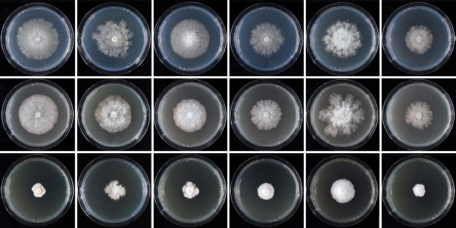
Colony morphology of Phytophthora chilensis, P. pseudochilensis, P. pseudokernoviae, P. celebensis, P. javanensis and P. multiglobulosa after 10 d growth (from left to right) at 20 °C on V8 agar, carrot juice agar and potato-dextrose agar (from top to bottom).
Fig. 24.

Mean radial growth rates of two known and six new Phytophthora species from Subclade 10c, i.e., P. celebensis (5 isolates), P. chilensis (6 isolates), P. gondwanensis (5 isolates), P. javanensis (5 isolates from Java, 5 isolates from Sulawesi), P. kernoviae (8 isolates), P. multiglobulosa (3 isolates), P. pseudochilensis (6 isolates) and P. pseudokernoviae (3 isolates), on V8 agar at different temperatures.
Additional specimens. Chile, Parque Oncol, isolated from streams running through Valdivian rainforests using Rhododendron leaves as baits, Nov. 2014, T. Jung, A. Durán & E. Sanfuentes, CL166, CL169, CL170, CL171, CL172.
Phytophthora pseudochilensis T. Jung, M. Horta Jung, E. Sanfuentes & I. Milenković, sp. nov. – MycoBank MB 842947; Fig. 13
Fig. 13.

Morphological structures of Phytophthora pseudochilensis. a–j. Sporangia with short pedicels formed on V8 agar (V8A) flooded with soil extract; a. ovoid, papillate, terminal, with external proliferation; b. obpyriform, semipapillate, terminal; c. dense sympodium of two liminoform (arrows) and three ovoid papillate sporangia; d. dense sympodium of ten mainly ovoid or limoniform sporangia except of one ellipsoid sporangium which has already been shed (arrow); e–i. caducous sporangia; e–f. elongated-ovoid, semipapillate; g. elongated-ovoid, papillate with slightly asymmetric apex and inclined papilla; h. elongated-ovoid, semipapillate with a laterally displaced apex; i. elongated-ellipsoid sporangium with slightly asymmetric apex which failed to release all zoospores; j. sympodium of two elongated-ovoid sporangia, one mature and semipapillate and one releasing zoospores; k–r. mature smooth-walled oogonia formed in single culture in solid V8A, with oospores, containing large ooplasts, and amphigynous unicellular antheridia; k. non-tapering base; l–r. tapering funnel-like oogonial bases; l. near plerotic oospore and antheridium with finger-like projection (arrow); m–n. plerotic oospore; o. aplerotic oospore and antheridium with finger-like projection (arrow); p. aplerotic oospore; q. two oogonia with near plerotic (left) and aplerotic (right) oospores; r. comma-shaped oogonium with plerotic oospore; s. coralloid irregular lateral hyphae in solid V8A; t. hyphal aggregation in solid V8A. — Scale bar = 20 μm, applies to all except of d where it is 50 μm.
Etymology. Name refers to the morphological similarity to P. chilensis.
Typus. Chile, Parque Oncol, isolated from a stream running through a Valdivian rainforest using a Rhododendron leaf as bait, Nov. 2014, T. Jung, A. Durán & E. Sanfuentes (holotype HNHM-MYC-009705, dried culture on V8A, Herbarium of the Hungarian Natural History Museum, Budapest, Hungary; ex-type culture CBS 148798 = NRRL 64352 = CL168). ITS and cox1 sequences GenBank ON000773 and ON013839, respectively.
Sporangia, hyphae & chlamydospores (Fig. 13a–j, s–t) — Sporangia of P. pseudochilensis were produced abundantly in non-sterile soil extract after 1–2 d and were also observed on solid V8A. Sporangia were borne usually in dense sympodia of 2–10 sporangia (Fig. 13c–d, j) or infrequently terminally on unbranched sporangiophores (Fig. 13b). Sporangia were papillate (Fig. 13a, c, g) or semipapillate (Fig. 13b, e–f, h, j), sometimes with a slightly asymmetric apex and an inclined papilla (4.5 %; Fig. 13g, i) or a laterally displaced apex (Fig. 13h), and caducous (Fig. 13d–i) with short pedicels averaging 4.9 ± 1.6 μm (Fig. 13a–b, e–j). Sporangial shapes ranged from ovoid (49.0 %; Fig. 13a, c–d) and elongated-ovoid (25.0 %; Fig. 13e–h, j) to obpyriform (9.5 %; Fig. 13b), limoniform (8.0 %; Fig. 13c–d), ellipsoid and elongated-ellipsoid (4.5 %; Fig. 13d, i) and obovoid (4.0 %). Most sporangia had an inconspicuous basal plug which did not protrude into the empty sporangium (Fig. 13i–j). Sporangia proliferated exclusively externally (Fig. 13a, d, j). Sporangial dimensions of six isolates of P. pseudochilensis averaged 48.8 ± 8.2 × 27.0 ± 3.1 μm (overall range 25.6–69.1 × 17.4–39.4 μm) with a range of isolate means of 43.3–54.9 × 25.7–29.3 μm. The length/breadth ratio averaged 1.81 ± 0.24 with a range of isolate means of 1.69–1.94. Zoospores were limoniform to reniform whilst motile, becoming spherical (av. diam = 9.9 ± 1.2 μm) on encystment and were discharged directly through a narrow exit pore 2.5–6.3 μm wide (av. 4.4 ± 0.9 μm) (Fig. 13i–j). Cysts germinated directly or indirectly by releasing a secondary zoospore (diplanetism). All isolates produced in solid V8A coralloid or irregular lateral hyphae (Fig. 13s) and hyphal aggregations (Fig. 13t), and in liquid culture infrequently subglobose, limoniform or irregular swellings (10.3 ± 2.7 μm diam). Chlamydospores were not observed.
Oogonia, oospores & antheridia (Fig. 13k–r) — Gametangia were readily produced in single culture by all isolates of P. pseudochilensis on V8A within 1 wk and matured within 2–3 wk. Oogonia were borne laterally or less frequently terminally, smooth-walled, globose to subglobose with a tapering often funnel-like (87.5 %; Fig. 13l–r) or sometimes a round base (12.5 %; Fig. 13k). On average 20.5 % of oogonia were comma-shaped (Fig. 13r). Oogonial diameters were averaging 27.3 ± 3.4 μm with an overall range of 16.3–35.3 μm and a range of isolate means of 25.9–28.5 μm. Oospores were globose, mostly near plerotic to plerotic (91.0 %; Fig. 13k–n, q–r) or less frequently aplerotic (9.0 %; Fig. 13o–q), and contained a large ooplast (Fig. 13k–r). Oospore diameters averaged 24.4 ± 3.0 μm (total range 15.4–32.1 μm). Oospores had walls with a diameter of 1.61 ± 0.33 μm and an oospore wall index of 0.34 ± 0.07. With on average 65.5 % (56–80 %) the oogonial abortion rate was high. The antheridia were amphigynous, almost exclusively unicellular, with cylindrical or irregular shapes (Fig. 13k–r) and sometimes finger-like projections (Fig. 13l, o), averaging 13.7 ± 3.6 × 9.8 ± 1.7 μm.
Colony morphology, growth rates & cardinal temperatures (Fig. 22, 24) — Colonies of P. pseudochilensis were loose and appressed to submerged with scarce aerial mycelium and a dendroid to stoloniferous pattern with irregular margins on V8A, uniform and appressed with limited aerial mycelium on CA, and dense-felty with irregular raised lobes separated by irregular trenches and irregular feathery margins on PDA (Fig. 22). The temperature-growth relations on V8A are shown in Fig. 24. All six isolates had similar growth rates at all temperatures and identical cardinal temperatures for growth with a low optimum of 15 °C and a maximum between 20 and 25 °C. None of the isolates was able to grow at 25 °C but they resumed growth when plates incubated for 5 d at 25 °C were transferred to 20 °C. Lethal temperature was between 25 and 27.5 °C. Radial growth rates at 15 and 20 °C were 2.39 ± 0.22 mm/d and 1.57 ± 0.43 mm/d, respectively (Fig. 24). On CA and PDA radial growth rates at 20 °C were 1.8 ± 0.03 mm/d and 0.65 ± 0.03 mm/d, respectively.
Additional specimens. Chile, Parque Oncol, isolated from a stream running through a Valdivian rainforest using Rhododendron leaves as baits, Nov. 2014, T. Jung, A. Durán & E. Sanfuentes, CL335, CL336, CL337, CL338, CL339.
Phytophthora pseudokernoviae T. Jung, M. Horta Jung, A. Durán & E. Sanfuentes, sp. nov. – MycoBank MB 842949; Fig. 14
Fig. 14.

Morphological structures of Phytophthora pseudokernoviae. a–i. Sporangia with short pedicels formed on V8 agar (V8A) flooded with soil extract; a–h. papillate sporangia; a. ovoid lateral, in a beginning sympodium; b. ovoid with beginning external proliferation; c. limoniform; d. ovoid and limoniform (arrows) sporangia in two sympodia of three (left) and two sporangia (right); e–f. ovoid, caducous; g. limoniform, caducous; h. ovoid, caducous; i. two empty sporangia after zoospore release, one just being shed (left) and the other one lateral with conspicuous basal plug protruding into the sporangium (arrow); j–s. mature smooth-walled oogonia formed in solid V8A, with plerotic or near plerotic oospores containing large ooplasts; j–q. with amphigynous unicellular antheridia; j–n. globose oogonia with tapering funnel-like bases; j–k. golden-brown; n. comma-shaped; o. golden-brown subglobose oogonium with rounded comma-shaped base and finger-like projection at the antheridium; p. subglobose oogonium with rounded base, extremely comma-shaped; q. elongated oogonium with tapering base, comma-shaped; r. golden-brown subglobose oogonium with tapering base and paragynous antheridium; s. globose oogonium with tapering base and paragynous antheridium; t. dense hyphal aggregation in solid V8A. — Scale bar = 20 μm, applies to a–t.
Etymology. Name refers to the morphological similarity to P. kernoviae.
Typus. Chile, Parque Oncol, isolated from a naturally fallen necrotic leaf of Drimys winteri in a Valdivian rainforest, Nov. 2014, T. Jung, A. Durán & E. Sanfuentes (holotype HNHM-MYC-009707, dried culture on V8A, Herbarium of the Hungarian Natural History Museum, Budapest, Hungary; ex-type culture CBS 148796 = NRRL 64351 = CL012). ITS and cox1 sequences GenBank ON000780 and ON013846, respectively.
Sporangia, hyphae & chlamydospores (Fig. 14a–i, t) — Sporangia of P. pseudokernoviae were observed on solid agar and were produced abundantly in non-sterile soil extract after 1–2 d. Sporangia were mostly borne laterally or infrequently terminally in dense sympodia of 2–5 sporangia (Fig. 14a, d, i). Sporangia were exclusively papillate (Fig. 14a–h) and caducous (Fig. 14e–i) with short pedicels averaging 3.6 ± 1.0 μm (Fig. 14a–i). Sporangial shapes showed low variability and were predominantly ovoid (88.0 %; Fig. 14a–b, d–f, h–i) or less frequently limoniform (12.0 %; Fig. 14c–d, g). Sporangia often had a conspicuous basal plug which protruded into the empty sporangium (Fig. 14i) and proliferated exclusively externally (Fig. 14a–b, d). Sporangial dimensions of three isolates of P. pseudokernoviae averaged 42.7 ± 4.0 × 29.7 ± 2.8 μm with an overall range of 30.5–55.0 × 21.3–35.7 μm and a range of isolate means of 41.9–43.4 × 28.6–31.5 μm. The length/breadth ratio averaged 1.44 ± 0.14 with a range of isolate means of 1.38–1.50. Zoospores of P. pseudokernoviae were discharged through a narrow exit pore 4.3–7.2 μm wide (av. 5.6 ± 0.7 μm) (Fig. 14i). They were limoniform to reniform whilst motile, becoming spherical (av. diam = 9.9 ± 1.6 μm) on encystment. Cysts germinated directly or indirectly by releasing a secondary zoospore (diplanetism). Small subglobose, limoniform or irregular swellings of 9.9 ± 2.1 μm were infrequently formed on sporangiophores. All isolates produced dense hyphal aggregations (Fig. 14t). Chlamydospores were not observed.
Oogonia, oospores & antheridia (Fig. 14j–s) — Gametangia were readily produced in single culture by all isolates of P. pseudokernoviae in V8A within 1 wk and matured within 2–3 wk. Oogonia were borne terminally or laterally, smooth-walled, predominantly with a tapering often funnel-like (86.0 %; Fig. 14k–o, r–t) or less frequently with a rounded base (14.0 %; Fig. 14p–q) which in 35.3 % of oogonia was also comma-shaped curved (Fig. 14o, q–r). They were predominantly globose to subglobose (av. 84.0 %; Fig. 14j–k, o–p, r–s) or sometimes elongated or excentric (16.0 %; Fig. 14l–n, q), sometimes turning golden-brown with age (Fig. 14j–k, o, r). Oogonial diameters averaged 30.1 ± 4.3 μm with an overall range of 18.6–39.3 μm and a range of isolate means 28.1–34.2 μm. Oospores contained a large ooplast and were globose and almost exclusively plerotic or near-plerotic (99.6 %; Fig. 14j–s) with a mean diameter of 27.0 ± 3.7 μm (total range 16.3–35.8 μm). Oospores had wall diameters of 2.1 ± 0.33 μm and an oospore wall index of 0.40 ± 0.06. Oogonial abortion rate was low (on. av. 7.7 %; 6–10 %). The antheridia were predominantly amphigynous unicellular (95.3 %) with cylindrical or slightly irregular shapes (Fig. 14j–q) or occasionally paragynous (4.7 %; Fig. 14r–s), averaging 14.6 ± 2.9 x 9.6 ± 1.3 μm.
Colony morphology, growth rates & cardinal temperatures (Fig. 22, 24) — After 10 d growth at 20 °C colonies of P. pseudokernoviae on V8A and CA were appressed with limited aerial mycelium, uniform to faintly striate on V8A and faintly radiate on CA, whereas colonies on PDA were dense-woolly and faintly petaloid (Fig. 22). At 25 °C all three isolates showed very slow growth for 5 d and then stopped growing suggesting a maximum temperature of growth slightly below 25 °C. The isolates did not resume growth when the plates were transferred to 20 °C.
With mean radial growth rates of 2.47 ± 0.62 mm/d and 2.45 ± 0.41 mm/d at 15 and 20 °C, respectively, P. pseudokernoviae had a broad optimum of growth (Fig. 24). On CA and PDA radial growth rates at 20 °C were 1.93 ± 0.11 mm/d and 1.0 ± 0.07 mm/d, respectively.
Additional specimens. Chile, Parque Oncol, isolated from a naturally fallen necrotic leaf of D. winteriin a Valdivian rainforest, Nov. 2014, T. Jung, A. Durán & E. Sanfuentes, CL013; isolated from a stream running through a Valdivian rainforest using a Nothofagus obliqua leaf as bait, Nov. 2014, T. Jung, A. Durán & E. Sanfuentes, CL156.
Phytophthora celebensis T. Jung, M. Junaid, N. Nasri & I. Milenković, sp. nov. – MycoBank MB 843002; Fig. 15
Fig. 15.

Morphological structures of Phytophthora celebensis. a–h. Sporangia with mostly medium-length pedicels formed on V8 agar (V8A) flooded with soil extract; a–f. papillate; a. ovoid, slightly asymmetric, with laterally attached sporangiophore; b. sympodium with one immature and two ovoid sporangia, one of them caducous (arrow); c–d. ovoid, caducous; e. broad-ovoid, caducous; f. obovoid with inconspicuous basal plug (arrow), caducous; g. ovoid, caducous, after release of zoospores, without protruding basal plug (arrow); h. sympodium with one immature and two limoniform sporangia, one of them caducous (arrow); i–r. mature oogonia formed in single culture in solid V8A, with thick-walled plerotic or near-plerotic oospores, containing large ooplasts, and amphigynous unicellular antheridia; i–n. with round bases and short tapering stalks; o. with round base and medium-length stalk; p–r. with short, tapering bases; q–r. slightly bending to comma-shaped; s. dense hyphal aggregation in solid V8A. — Scale bar = 20 μm, applies to a–s.
Etymology. Name refers to the origin of all known isolates in Sulawesi (Celebes is a previous name of Sulawesi).
Typus. Indonesia, South Sulawesi Province, District of Tana Toraja, isolated from a naturally fallen leaf floating in a stream running through a submontane tropical rainforest, May 2019, T. Jung, M. Junaid & N. Nasri (holotype HNHM-MYC-021540, dried culture on V8A, Herbarium of the Hungarian Natural History Museum, Budapest, Hungary; ex-type culture CBS 148800 = SL092). ITS and cox1 sequences GenBank ON000720 and ON013786, respectively.
Sporangia, hyphae & chlamydospores (Fig. 15a–h, s) — Sporangia of P. celebensis were observed on solid agar and were produced abundantly in non-sterile soil extract after 1–2 d. Sporangia were borne terminally (Fig. 15a–b, h) or laterally (Fig. 15b, h), often in dense sympodia of 2–5 sporangia (Fig. 15b, h). Sporangia were papillate (Fig. 15a–f, h) and caducous (Fig. 15b–h) with short to medium-length pedicels averaging 9.9 ± 2.7 μm (Fig. 15a–h). Sporangial shapes ranged from ovoid (48.8 %; Fig. 15a–d) and broad-ovoid (4.0 %; Fig. 15e) to limoniform (41.6 %; Fig. 15h), obovoid (3.4 %; Fig. 15f), ellipsoid (1.0 %), subglobose (0.8 %) and pyriform (0.4 %). Sometimes sporangia were laterally attached to the sporangiophore (7.6 %; Fig. 15a) or had an inconspicuous basal plug (10.8 %; Fig. 15f). Sporangial proliferation was exclusively external. Sporangial dimensions of five isolates averaged 38.7 ± 5.0 × 26.2 ± 3.3 μm (overall range 26.3–64.3 × 17.6–40.7 μm) with a range of isolate means of 37.3–40.7 × 25.0–27.9 μm. The length/ breadth ratio averaged 1.48 ± 0.12 with a range of isolate means of 1.44–1.54. Zoospores of P. celebensis were discharged directly through narrow exit pores 2.1–5.1 μm wide (av. 3.7 ± 0.7 μm) (Fig. 15g). They were limoniform to reniform whilst motile, becoming spherical (av. diam = 8.0 ± 0.8 μm) on encystment. Cysts germinated directly or indirectly by releasing a secondary zoospore (diplanetism). All isolates formed dense hyphal aggregations (Fig. 15s). Hyphal swellings and chlamydospores were not observed.
Oogonia, oospores & antheridia (Fig. 15i–r) — Gametangia were readily produced in single culture by all isolates of P. celebensis in V8A within 1 wk and matured within 2–3 wk. Oogonia were borne terminally or laterally, smooth-walled, globose to subglobose (92.4 %; Fig. 15i–p, r) or rarely slightly excentric or elongated (7.6 %; Fig. 15q), mostly with round base and thin stalk (91.6 %; Fig. 15i–o) or with short tapering base (8.4 %; Fig. 15p–r). On average 22.4 % of oogonia were slightly bending to comma-shaped (Fig. 15q–r). Oogonial diameters averaged 24.9 ± 3.1 μm (overall range 13.1–36.1 μm and range of isolate means 23.6–25.8 μm). Oospores had a mean diameter of 21.9 ± 2.9 μm (total range 12.3–31.5 μm), were globose and plerotic to near-plerotic and contained a large ooplast (Fig. 15i–r). The oospores had walls with a diameter of 1.6 ± 0.27 μm and an oospore wall index of 0.38 ± 0.04. Oogonial abortion rate was 24.4 % (6–48 %). The antheridia averaged 11.4 ± 2.1 × 9.8 ± 1.3 μm and were exclusively amphigynous unicellular with cylindrical, subglobose or square shapes (Fig. 15i–r).
Colony morphology, growth rates & cardinal temperatures (Fig. 22, 24) — All isolates of P. celebensis formed after 10 d similar colonies on the three different agar media. Colonies were appressed with limited aerial mycelium and a dendroid growth pattern on V8A and CA, and dense-woolly and uniform on PDA (Fig. 22). The temperature-growth relations on V8A are shown in Fig. 24. On V8A P. celebensis had an optimum temperature of 25 °C with a radial growth rate of 2.9 ± 0.23 mm/d. The maximum growth temperature for all isolates was between 27.5 and 30 °C. None of the isolates was able to grow at 30 °C and isolates did not resume growth when plates incubated for 5 d at 30 °C were transferred to 20 °C. On V8A, CA and PDA radial growth rates at 20 °C were 2.77 ± 0.19 mm/d, 2.3 ± 0.09 mm/d and 1.0 ± 0.05 mm/d, respectively.
Additional specimens. Indonesia, South Sulawesi Province, District of Tana Toraja, isolated from naturally fallen leaves floating in a stream running through a submontane tropical rainforest, May 2019, T. Jung, M. Junaid & N. Nasri, SL091, SL540, SL541, SL542.
Phytophthora javanensis T. Jung, M. Junaid, N. Nasri & M. Horta Jung, sp. nov. – MycoBank MB 842954; Fig. 16
Fig. 16.

Morphological structures of Phytophthora javanensis. a–j. Sporangia with short to medium-length pedicels formed on V8 agar (V8A) flooded with soil extract; a–h. papillate; a. ovoid, terminal, sporangiophore laterally attached; b. ovoid, intercalary; c. ovoid, lateral; d. limoniform, bipapillate; e. slender-ovoid, caducous; f. limoniform, caducous; g–i. slender-ovoid, caducous; i. empty after release of zoospores, without basal plug (arrow); j. sympodium with two ovoid papillate sporangia and three empty sporangia after release of zoospores, one of the latter persistent with inconspicuous basal plug, two caducous (arrows) without basal plugs; k–u. mature oogonia formed in single culture in solid V8A, with thick-walled oospores, containing large ooplasts; k–t. amphigynous unicellular antheridia; k–l. globose with round bases, short tapering stalks and plerotic or near-plerotic oospores; m. subglobose with round base, short stalk and aplerotic oospore; n. subglobose with short tapering base and plerotic oospore; o. subglobose with short tapering stalk and slightly aplerotic oospore; p. subglobose to slightly excentric with round base and slightly aplerotic oospore; q. elongated with tapering base and aplerotic oospore; r. slightly elongated with tapering base and slightly aplerotic oospore; s–u. slightly elongated, comma-shaped with tapering bases and slightly aplerotic oospores; u. amphigynous bicellular antheridium with small basal cell (arrow); v. dense hyphal aggregation in solid V8A. — Scale bar = 20 μm, applies to a–v.
Etymology. Name refers to the origin of the first isolates in Java.
Typus. Indonesia, Java, Gunung Puntang, Bandung area, isolated from a naturally fallen leaf floating in a stream running through a submontane tropical rainforest, Feb. 2019, T. Jung, M. Junaid & N. Nasri (holotype HNHM-MYC-009702, dried culture on V8A, Herbarium of the Hungarian Natural History Museum, Budapest, Hungary; ex-type culture CBS 149203 = NRRL 64129 = JV025a). ITS and cox1 sequences GenBank ON000750 and ON013816, respectively.
Sporangia, hyphae & chlamydospores (Fig. 16a–j, v) — Sporangia of P. javanensis were observed on solid agar and were produced abundantly in non-sterile soil extract after 1–2 d. Sporangia were borne terminally (Fig. 16a, j), laterally (Fig. 16c, j), often in dense sympodia of 2–6 sporangia (Fig. 16j), or infrequently intercalary (Fig. 16b). Sporangia were exclusively papillate (Fig. 16a–h, j), sometimes bipapillate (Fig. 16d), and caducous (Fig. 16e–j) with short to medium-length pedicels averaging 7.4 ± 2.1 μm (Fig. 16a–j). Sporangial shapes showed low variability and were mostly ovoid (53.6 %; Fig. 16a–c, e, g–h) or limoniform (43.0 %; Fig. 16d, f, i) and infrequently obovoid (1.6 %), ellipsoid (1.0 %) or subglobose (0.8 %). Sporangia usually had no conspicuous basal plug (Fig. 16i–j), were sometimes laterally attached (3.6 %; Fig. 16a) and proliferated exclusively externally. Sporangial dimensions of ten isolates of P. javanensis averaged 38.3 ± 4.5 × 23.2 ± 2.9 μm with an overall range of 19.3–62.6 × 9.2–36.7 μm and a range of isolate means of 33.6–41.3 × 20.5–25.6 μm. The length/breadth ratio averaged 1.66 ± 0.19 with a range of isolate means of 1.54–1.81. Zoospores were discharged through a narrow exit pore 1.9–8.3 μm wide (av. 3.3 ± 0.6 μm) (Fig. 16i–j). They were limoniform to reniform whilst motile, becoming spherical or ellipsoid (av. diam = 8.4 ± 0.7 μm) on encystment. Cysts germinated directly or indirectly by releasing a secondary zoospore (diplanetism). All isolates produced dense hyphal aggregations (Fig. 16v). Hyphal swellings or chlamydospores were not observed.
Oogonia, oospores & antheridia (Fig. 16k–u) — All 10 isolates of P. javanensis produced in single culture on V8A within 1 wk abundant gametangia which matured within 2–3 wk. Oogonia were borne terminally or laterally, smooth-walled, predominantly with rounded bases (87.6 %; Fig. 16k–m, p) or less frequently with slightly tapering bases (12.4 %; Fig. 16n–o, q–u). On average 25.7 % of oogonia were slightly bending to comma-shaped (Fig. 16s–u). They were predominantly globose to subglobose (83.6 %; Fig. 16k–p) or sometimes elongated (16.4 %; Fig. 16q–u). Oogonial diameters averaged 28.1 ± 4.0 μm with an overall range of 15.6–42.7 μm and a range of isolate means of 27.1–29.2 μm. Oospores contained almost exclusively a single large ooplast and were globose, plerotic to near-plerotic (80.3 %; Fig. 16k–l, n) or less frequently slightly aplerotic (19.7 %; Fig. 16m, o–u) with a mean diameter of 25.0 ± 3.6 μm (total range 13.5–39.4 μm). Oospores had wall diameters of 1.75 ± 0.34 μm and an oospore wall index of 0.36 ± 0.04. Oogonial abortion rate was 16.8 % (8–32 %). The antheridia were predominantly amphigynous unicellular (99.0 %; Fig. 16k–t) or rarely bicellular with the basal cell being considerably smaller (1.0%; Fig. 16u) averaging 12.2 ± 2.2 × 10.1 ± 1.6 μm. Antheridial shapes were cylindrical, ovoid or square.
Colony morphology, growth rates & cardinal temperatures (Fig. 22, 24) — All P. javanensis isolates formed on V8A and CA dendroid to stoloniferous colonies with limited aerial mycelium, while colonies on PDA were dense-woolly and uniform (Fig. 22). All ten isolates had a maximum temperature of growth between 27.5 and 30 °C. The lethal temperature was 30 °C for isolates from Java and 32.5 °C for isolates from Sulawesi. At the optimum temperature of 20 °C the average radial growth rate was 3.36 ± 0.55 mm/d (Fig. 24) with the five isolates from Sulawesi showing faster growth (3.81 ± 0.38 mm/d) than the five isolates from Java (2.91 ± 0.14 mm/d). At 20 °C radial growth rates on CA and PDA were 2.97 ± 0.16 mm/d and 1.28 ± 0.06 mm/d, respectively, for the Java isolates, and 3.27 ± 0.11 mm/d and 1.4 ± 0.09 mm/d, respectively, for the Sulawesi isolates.
Additional specimens. Indonesia, Java, Gunung Puntang, Bandung area, isolated from naturally fallen leaves floating in a stream running through a submontane tropical rainforest, Feb. 2019, T. Jung, M. Junaid & N. Nasri, JV025b, JV191, JV192, JV193; South Sulawesi Province, District of Tana Toraja, isolated from naturally fallen leaves floating in a stream running through a submontane tropical rainforest, May 2019, T. Jung, M. Junaid & N. NasriSL081, SL084, SL537, SL538, SL539.
Phytophthora multiglobulosa T. Jung, M. Junaid, M. Horta Jung & I. Milenković, sp. nov. – MycoBank MB843003; Fig. 17
Fig. 17.

Morphological structures of Phytophthora multiglobulosa. a–h. Sporangia with short to medium-length pedicels formed on V8 agar (V8A) flooded with soil extract; a–e. papillate; a. ovoid, terminal; b. ovoid, slightly asymmetric with laterally attached sporangiophore; c. ovoid with laterally attached sporangiophore and external proliferation (arrow); d. ovoid, lateral; e. ovoid to limoniform, caducous; f. ovoid releasing zoospores, with external proliferation; g. ovoid, empty after release of zoospores, with inconspicuous basal plug, caducous; h. dense sympodium with two ovoid papillate sporangia and two empty caducous sporangia (arrows) after release of zoospores, one limoniform (left) and the other one ovoid (right); i–r. mature globose to subglobose oogonia with round non-tapering bases, containing thick-walled plerotic or near-plerotic oospores, formed in single culture in solid V8A; i–j. oogonia with oospores containing a single large ooplast; i. bicellular antheridium with small basal cell (arrow); j–r. unicellular antheridia; k–p. oogonia with oospores containing multiple lipid globules; n–o. slightly leaning oogonia; p. comma-shaped oogonium; q–r. comma-shaped oogonia with oospores containing a single large ooplast; s. dense hyphal aggregation in solid V8A. — Scale bar = 20 μm, applies to a–s.
Etymology. Name refers to the presence of multiple lipid globules in many oospores.
Typus. Indonesia, South Sulawesi Province, District of North Toraja, isolated from a naturally fallen leaf floating in a stream running through a tropical hill rainforest, May 2019, T. Jung, M. Junaid & N. Nasri (holotype HNHM-MYC-021539, dried culture on V8A, Herbarium of the Hungarian Natural History Museum, Budapest, Hungary; ex-type culture CBS 148799 = SL005). ITS and cox1 sequences GenBank ON000763 and ON013829, respectively.
Sporangia, hyphae & chlamydospores (Fig. 17a–h, s) — Sporangia of P. multiglobulosa were observed on solid agar and were produced abundantly in non-sterile soil extract after 1–2 d. Sporangia were borne terminally (Fig. 17a–c, f, h) or laterally (Fig. 17d, f, h), often in dense sympodia of 2–5 sporangia (Fig. 17h). Sporangia were papillate with shallow papillae (Fig. 17a–e, h) and caducous (Fig. 17e, g–h) with short to medium-length pedicels averaging 7.7 ± 3.3 μm (Fig. 17a–h). Sporangial shapes were ovoid (57.3 %; Fig. 17a–d, f–h), limoniform (42.0 %; Fig. 17e, h) or infrequently obpyriform (0.7 %). Some sporangia had an inconspicuous basal plug (Fig. 17g–h) and sometimes laterally attached sporangiophores (6.0 %; Fig. 17b–c). Sporangial proliferation was exclusively external (Fig. 17c, f). Sporangial dimensions of three isolates averaged 43.1 ± 6.2 × 27.1 ± 3.0 μm (overall range 28.3–64.9 × 21.1–36.5 μm) with a range of isolate means of 41.1–46.5 × 26.1–28.2 μm. The length/breadth ratio averaged 1.60 ± 0.18 with a range of isolate means of 1.55–1.66. Zoospores of P. multiglobulosa were discharged directly through narrow exit pores 2.2–4.8 μm wide (av. 3.6 ± 0.5 μm) (Fig. 17f–h). They were limoniform to reniform whilst motile, becoming spherical (av. diam = 9.2 ± 0.8 μm) on encystment. Cysts germinated directly or indirectly by releasing a secondary zoospore (diplanetism). All isolates formed dense hyphal aggregations (Fig. 17s). Hyphal swellings and chlamydospores were not observed.
Oogonia, oospores & antheridia (Fig. 17i–r) — Gametangia were readily produced in single culture by all isolates of P. multiglobulosa on V8A within 1 wk and matured within 2–3 wk. Oogonia were borne terminally or laterally, smoothwalled, globose or infrequently subglobose with round base and thin stalk (Fig. 17i–r). On average 18.7 % of oogonia were comma-shaped (Fig. 17p–r). Oogonial diameters averaged 25.4 ± 3.5 μm (overall range 17.0–38.4 μm and range of isolate means 23.2–25.5 μm). Oospores had a mean diameter of 22.5 ± 3.2 μm (total range 15.0–35.2 μm), were globose, plerotic or near-plerotic (Fig. 17i–r) and contained a single large ooplast (62 %; Fig. 17i–j, q–r) or multiple lipid globules (38 %; Fig. 17k–p). The oospores had walls with a diameter of 1.5 ± 0.23 μm and an oospore wall index of 0.35 ± 0.04. Oogonial abortion rate was low (on. av. 7.3 %; 2–12 %). The antheridia averaged 11.4 ± 2.1 × 9.9 ± 1.4 μm were exclusively amphigynous with mostly cylindrical (Fig. 17i–r), unicellular (Fig. 17j–r) or infrequently bicellular with the basal cell being much smaller (Fig. 17i).
Colony morphology, growth rates & cardinal temperatures (Fig. 22, 24) — Phytophthora multiglobulosa formed submerged to appressed colonies with a radiate pattern and irregular margins on V8A and CA, and dense-woolly uniform colonies on PDA (Fig. 22). The temperature-growth relations on V8A are shown in Fig. 24. On V8A the maximum growth temperature was between 27.5 and 30 °C. All isolates failed to resume growth when plates incubated for 5 d at 30 °C were transferred to 20 °C. Average radial growth rates at the optimum of 20 °C on V8A, CA and PDA were 1.91 ± 0.09 mm/d, 1.88 ± 0.08 mm/d and 0.59 ± 0.06 mm/d, respectively.
Additional specimens. Indonesia, South Sulawesi Province, District of North Toraja, isolated from naturally fallen leaves floating in a stream running through a tropical hill rainforest, May 2019, T. Jung, M. Junaid & N. Nasri, SL006, SL007.
NOTES
Across the 11259-character alignment of LSU, ITS, βtub, hsp90, tigA, rpl10, tef-1α, enl, ras-ypt1, cox1, nadh1 and rps10 the seven known and 14 new Phytophthora species from Clade 10 differed from each other at 36–1 179 positions (Table 3) corresponding to sequence differences of 0.32–10.49 % (Table 4). The seven known Clade 10 species P. boehmeriae, P. gallica, P. gondwanensis, P. intercalaris, P. kernoviae, P. morindae and P. taxon boehmeriae-like had 65, 33, 64, 78, 44, 77 and 39 unique polymorphisms, respectively. The 14 new Clade 10 species P. celebensis, P. chilensis, P. javanensis, P. ludoviciana, P. multiglobulosa, P. procera, P. pseudochilensis, P. pseudogallica, P. pseudokernoviae, P. scandinavica, P. subarctica, P. tenuimura, P. tonkinensis and P. ukrainensis had 28, 11, 15, 56, 8, 73, 23, 82, 20, 100, 53, 46, 125 and 64 unique polymorphisms, respectively. The 14 new Clade 10 species and the known Clade 10 species P. gallica, P. intercalaris, P. gondwanensis and P. kernoviae showed distinctive colony morphologies on V8A, CA and PDA at 20 °C (Fig. 20–22). In addition, the 14 new species can be separated from each other and from other Clade 10 species by a combination of morphological and physiological characters of which the most discriminating are highlighted in bold in Table 6–8.
Phytophthora intercalaris differs from all other species in Clade 10 by being heterothallic and in producing ornamented oogonia with a 100 % abortion rate (Yang et al. 2016). The production of chlamydospores discriminates P. pseudogallica, informally designated as P. taxon gallica-like 1 in Jung et al. (2020), from all other Clade 10 species with nonpapillate sporangia except of P. intercalaris, P. gallica and P. afrocarpa (Jung & Nechwatal 2008, Yang et al. 2016, Bose et al. 2021; Table 6–8). Although P. pseudogallica and P. gallica are both sterile and produce chlamydospores of similar size they can be easily distinguished by the lower maximum temperature for growth and the much smaller sporangial size and l/b ratio in P. pseudogallica and the much higher proportion of obpyriform sporangia in P. gallica (Table 6). Phytophthora pseudogallica differs from P. afrocarpa by having a lower maximum temperature for growth, much bigger chlamydospores, and sporangia with a lower l/b ratio and both nested and extended proliferation (Table 6). Furthermore, P. pseudogallica shows on CA at 20 °C the slowest growth of all 18 Clade 10 species tested. The homothallic breeding system of P. tonkinensis, informally designated as P. taxon gallica-like 2 by Jung et al. (2020), and the production of chlamydospores in P. pseudogallica discriminate these co-occurring sister species. Moreover, unlike P. pseudogallica, P. tonkinensis forms coralloid to irregular hyphal swellings, has a lower maximum temperature for growth and is unable to grow at 25 °C (Table 6–7; Fig. 7, 10, 23). Phytophthora tonkinensis differs from P. scandinavica by having a lower maximum temperature for growth, much slower growth rates, much smaller sporangia, thinner oospore walls and lower oospore wall index. Phytophthora scandinavica co-occurs with the sterile P. subarctica and P. ukrainensis in boreal streams in Sweden and can be discriminated easily from them by its homothallic breeding system and by having lower maximum and optimum temperatures for growth (Table 6–7; Fig. 8–9, 11, 23). In P. ukrainensis the single isolate SW154 from a boreal stream in Sweden shares the same morphological characteristics as the Ukrainian isolates but has lower optimum (30 vs 32.5 °C) and maximum temperatures for growth (30–32.5 vs 32.5–35 °C). Phytophthora subarctica and P. ukrainensis can be distinguished by the larger sporangial dimensions and l/b ratio in P. subarctica, and the production of coralloid to irregular hyphal swellings, and higher maximum and optimum temperatures for growth in P. ukrainensis. The North American P. ludoviciana and P. procera differ from P. subarctica and P. ukrainensis by having undulating hyphal growth. In addition, P. subarctica and the Ukrainian isolates of P. ukrainensis have higher maximum temperatures for growth. Furthermore, P. procera has larger sporangia with much higher l/b ratio. Phytophthora tenuimura is discriminated from P. subarctica and P. ukrainensis by having undulating hyphal growth and a homothallic breeding system (Table 6–7; Fig. 4–7, 9, 11, 23). Phytophthora ludoviciana, P. procera and P. tenuimura co-occur in a swamp forest in Louisiana. While P. tenuimura can easily be distinguished from the other two sterile species by being homothallic and having a lower maximum temperature for growth, P. procera differs from P. ludoviciana in producing much larger sporangia with much higher l/b ratio and by its lower optimum temperature for growth (Table 6; Fig. 4–7, 23).
Table 7.
Morphological characters and dimensions (mean ± SD; μm), cardinal temperatures (°C) and temperature-growth relations (mm/d) on V8-juice agar of Phytophthora species from Subclades 10b and 10c. Most discriminating characters are highlighted in bold. Percentages in brackets are ranges of isolate means; – means character not observed.
| Subclade 10b | Subclade 10c | |||||||
|---|---|---|---|---|---|---|---|---|
| P. subarctica | P. tonkinensis | P. ukrainensis | P. boehmeriae b | P. boehmeriae c | P. t. boehmeriae-liked | P. morindae | P. gondwanensis | |
| No. of isolates/source | 4a | 4a | 4a | Tucker 1931 | Chowdappa et al. 2014 | Frazzi 1950 | Nelson & Abad 2010 | 5a |
| Sporangia | 72.8 % ovoid-elongated ovoid, 7.2 % limoniform, 7.2 % ampulliform, 6.1 % obpyriform (pyrif., ellips. | 36.4% sub-globose, 35.7% broad-ovoid, 25.3% ovoid (obpyrifornì, ellipsoid) | 73.6% ovoid, 23.6% obpyriform (ellipsoid, subglobose, ampulliform) | globose, ovoid, ellipsoid, obturbinate | globose, ovoid, ellipsoid, (obturbinate, obpyriform) | globose to subglobose, ovoid | ellipsoid, obpyriform, limoniform, mouse-shaped | 59.3% ovoid to broad-ovoid, 36.7 limoniform, (subglobose, obovoid, obpyriform) |
| apex | nonpapillate | nonpapillate | nonpapillate | papillate, few bipapillate | papillate | papillate | papillate, few bipapillate | papillate, few bipapillate |
| Ixb mean | 53.8 ± 11.5 x 31.6 ± 4.1 | 29.0±3.2 x 23.7±3.1 | 35.9±4.7 x 26.2±3.4 | 51.8 x 40.1 | 35.3±4.8 x 20.5±3.4 | 42 x 34 | 42 x 21.8 | 44.7±3.9 x 28.7±2.9 |
| range of isolate means | 43.5–58.2 x 29.2-34.1 | 27.8-29.8 x 23.0-24.0 | 34.0-38.1 x 23.5-29.9 | n.a. | 32.8-38.8 x 18.2-28.6 | n.a. | n.a. | 42.9-46.0 x 27.7-30.0 |
| total range | 32.7-127.6 x 16.3-39.5 | 19.5-37.1 x 14.5-31.6 | 26.3-58.2 x 18.7-36.3 | 28-69 x 20-51 | n.a. | 18.5-53 x 11.5-46 | 30-54 x 19.2-24 | 35.7-62.2 x 21.5-35.7 |
| l/b ratio | 1.73 ± 0.46 | 1.23 ± 0.15 | 1.38 ± 0.19 | 1.29 | 1.72 (1.22-1.96) | 1.20 | 1.93 | 1.57 ± 0.16 |
| caducity | (+) | – | – | + | + | + | + | + |
| pedicels | – | – | – | <5 | 4.1 ±0.6 | 4.5 (3.5-16) | 26 (8-66) | 3.5 ± 1.3 |
| internal proliferation | nested and extended | nested and extended | nested and extended | – | – | – | – | – |
| exitpores | 10.4 ± 1.7 | 9.8 ± 1.7 | 8.4 ± 1.5 | n.a. | n.a. | n.a. | n.a. | 3.3 ± 0.5 |
| sympodia | rare, lax | – | – | frequent | n.a. | loose, up to 16 sporangia | dense, umbellate | dense, 3-5 sporangia |
| zoospore cysts | 10.6 ± 0.9 | 10.4 ± 1.0 | 10.4 ±1.1 | n.a. | n.a. | 11.5-14 | n.a. | 9.5 ± 0.7 |
| Breeding system Oogonia | sterile | homothallic golden-brown with age | sterile | homothallic globose to subglobose | homothallic | homothallic | homothallic | homothallic |
| mean diam | – | 46.2 ± 6.2 | – | 26.5 | 22.5 ± 3.9 | 31.5 | 30.4 | 28.1 ± 3.0 |
| range of isolate means | – | 45.1-48.4 | – | 20.9-31.7 | 21.9-25.1 | n.a. | n.a. | 27.2-28.9 |
| total range | – | 30.3-59.1 | – | n.a. | n.a. | 19-38 | 21.6-39.6 | 14.1-33.0 |
| tapering base | – | 11% (0-22%) | – | n.a. | n.a. | occurring | frequent | 13.3% (10-16%) |
| comma-shaped | – | 23% (12-40%) | – | n.a. | n.a. | n.a. | n.a. | 31.3% (24-38%) |
| elongated/excentric | – | – | n.a. | n.a. | n.a. | – | 22.7% (20-26%) | |
| Oospores | – | |||||||
| plerotic oospores | – | 69% (56-84%) | – | predominantly | predominantly | predominantly | 100% | 75.3% (70-80%) |
| mean diam | – | 42.9 ± 5.5 | – | 23.3 | n.a. | 27.5 | 30.3 | 23.9 ± 2.6 |
| Total range | – | 28.4-56.9 | – | 15.7-27.6 | n.a. | 16-34 | 21-38.9 | 11.6-28.7 |
| wall diam | – | 1.6 ± 0.3 | – | <2 | n.a. | n.a. | 3.2 (2.4-3.6) | 2.0 ± 0.34 |
| oospore wall index | – | 0.24 ± 0.03 | – | n.a. | n.a. | n.a. | n.a. | 0.42 ± 0.04 |
| Abortion rate | – | 6.3% (4-8%) | – | n.a. | n.a. | n.a. | n.a. | 44.7% (24-66%) |
| Antheridia | – | 98% paragynous | – | 100% amphigynous | 100% amphigynous | 100% amphigynous | 100% amphigynous | 100% amphigynous |
| size | – | 14.1±2.2 x 10.8±2.0 | – | 12-16 x 11-15 | 12.4±2.1 x 10.0±1.3 | 16.5x14.5 (12.5-21 x 8-16) | 11.2 x 8.5 | 12.4±2.1 x 10.0±1.3 |
| Chlamydospores | – | – | – | rare; thickwalled (2pm) | + (27 out of 43 isolates) | + | – | – |
| size | – | – | – | 41.4 (16-51) | - | 29.5 (17-42) | – | – |
| Hyphal swellings | limoniform, rare | coralloid, irregular hyphae | ovoid, limoniform; 9.1 ± 1.5; coralloid hyphae | – | – | – | – | – |
| Hyphal aggregations | – | + | + | n.a. | n.a. | n.a. | n.a. | – |
| Maximum temperature | >30-<32.5 | >20-<25 | >32.5-<35 (30-32.5) | 32.5 | <35 | <35 | 27 | >30-<32.5 |
| Optimum temperature | 25 | 20 | 32.5 (30) | 25 | n.a | n.a. | 20 | 27.5 |
| Growth rate at 20 °C | 1.21 ± 0.14 | 1.13 ± 0.1 | 1.48 ± 0.21 | 4.4 | n.a. | n.a. | 2.9 | 3.17 ±0.3 |
| Growth rate at 25 °C | 1.31 ± 0.16 | 0 | 2.15 ± 0.12 | 6.6 | 7.6 ± 0.06 | n.a. | n.a. | 5.05 ± 0.21 |
a Numbers of isolates included in the growth tests: P. subarctica = 4; P. tonkinensis = 5; R ukrainensis = 4; P. gondwanensis = 5.
b Morphological studies and temperature-growth test performed on cornmeal agar.
c Morphological studies performed on CA; temperature-growth test performed on CA and only at 24 and 35 °C.
d Morphological studies performed on PDA; temperature-growth test performed on PDA.
In Subclade 10c, the four known species P. boehmeriae, P. morindae, P. gondwanensis and P. kernoviae and the six new species P. chilensis, P. pseudochilensis, P. pseudokernoviae, P. celebensis, P. javanensis and P. multiglobulosa all share a homothallic breeding system and the production of caducous papillate sporangia enabling an aerial lifestyle. Phytophthora morindae differs from the other nine papillate Clade 10 species by the production of sporangia with the highest l/b ratio and the longest pedicels which are often formed in umbellate sympodia (Nelson & Abad 2010; Table 7–8). For P. boehmeriae most published morphological and physiological information dates from pre-molecular times and, thus, it cannot be excluded that they might partly have come from other Clade 10 species. For instance, isolates from brown rot of orange fruits in Argentina, originally assigned to P. boehmeriae (Frezzi 1941, 1950, Erwin & Ribeiro 1996), were recently demonstrated by multigene phylogenetic analysis to belong to an unknown taxon closely related to P. boehmeriae and, hence, re-designated as P. taxon boehmeriae-like (Yang et al. 2017). Also, multiple isolates from Australia, China, Brazil and South Africa originally identified as P. boehmeriae were re-assigned to P. gondwanensis confirming Burgess et al. (2021). Therefore, only morphological and physiological data published for the ex-type isolate from Taiwan (Tucker 1931) and for P. boehmeriae isolates from chili pepper in India (Chowdappa et al. 2014) can be used for comparisons. The sister species P. boehmeriae and P. taxon boehmeriae-like are morphologically similar. The ex-type isolate of P. boehmeriae differs from P. taxon boehmeriae-like mainly by the on average larger chlamydospores (41.2 vs 29.5 μm) and sporangia (51.8 × 40.1 vs 42 × 34 μm) (Tucker 1931, Frezzi 1950, Erwin & Ribeiro 1996). However, the sporangia of 43 Indian isolates of P. boehmeriae were on average considerably smaller (35.3 × 20.5 μm) than sporangia of the ex-type isolate and those of P. taxon boehmeriae-like. The closely related P. taxon koreanensis from Ailanthus altissima in Korea (isolate KACC40173) had sporangia of similar size as P. taxon boehmeriae-like (44 × 32.7 μm; Kim & Kim 2004). Therefore, the true morphometric differences between the three sister species remain unclear. Phytophthora boehmeriae differs clearly from all other papillate Clade 10 species, except P. taxon boehmeriae-like, by producing chlamydospores and growing faster between 20 and 25 °C, and, in addition, from P. morindae, P. kernoviae, P. celebensis, P. javanensis and P. multiglobulosa by having shorter pedicels (Tucker 1931, Chowdappa et al. 2014; Table 7–8). In addition, the occurrence of bipapillate sporangia discriminates P. boehmeriae from the four species of the P. kernoviae complex and from P. celebensis and P. multiglobulosa. Phytophthora taxon boehmeriae-like differs from all other papillate Clade 10 species, except P. boehmeriae, by the production of chlamydospores (Frezzi 1950, Erwin & Ribeiro 1996).
For P. gondwanensis morphology and optimum temperature for growth of the isolates from the Japanese Amami island examined in the present study (Fig. 18, 24) were in accordance with the original description from New South Wales (Crous et al. 2015), except that in the Japanese isolates the sporangia were on average longer (44.7 ± 3.9 vs 39.3 ± 5.2 μm) and showed a higher l/b ratio (1.6 vs 1.2). For P. kernoviae the morphology and cardinal temperatures of the isolates from Chile and Ireland examined in the present study (Fig. 19, 24) were in accordance with the original species description from the UK (Brasier et al. 2005) apart from the slightly lower maximum temperature for growth (< 25 vs 26 °C), the thinner oospore walls (c. 2.0 vs 3.5 μm) and the infrequent occurrence of some paragynous antheridia in all Chilean and Irish isolates. The Chilean isolates had on average larger sporangia and oogonia and showed slightly slower growth than the Irish isolates (Table 8). Phytophthora kernoviae can be discriminated from its co-occurring sister species P. chilensis, P. pseudochilensis and P. pseudokernoviae by the occurrence of asymmetric mouse-shaped sporangia and by having longer sporangial pedicels and much smaller proportions of oogonia with tapering bases. Moreover, P. kernoviae differs from P. chilensis by its almost exclusive production of plerotic oospores with higher abortion rates and by producing besides amphigynous also a few paragynous antheridia; from P. pseudochilensis by having smaller sporangia with lower l/b ratio, higher oospore wall index, lower oospore abortion rate and higher optimum temperature for growth; and from P. pseudokernoviae by having on average smaller oogonia with higher oospore abortion rate, more variable sporangial shapes, and lower maximum and higher optimum temperatures for growth (Table 8; Fig. 12–14, 19, 24). Phytophthora pseudochilensis is distinguished from both P. chilensis and P. pseudokernoviae by having on average larger sporangia with less prominent papillae and higher l/b ratio, larger sympodia containing higher numbers of sporangia, oospores with thinner walls, lower oospore wall index and much higher abortion rates, and slower growth. Compared to P. chilensis, both P. pseudochilensis and P. pseudokernoviae have much higher proportions of oogonia with tapering bases and lower optimum temperatures for growth. In addition, P. pseudokernoviae differs from both P. chilensis and P. pseudochilensis by its larger oogonia which are more frequently comma-shaped, the occurrence of paragynous antheridia and a higher optimum temperature for growth (Table 8; Fig. 12–14, 24). Collectively, P. chilensis, P. pseudochilensis and P. pseudokernoviae are discriminated from P. gondwanensis by having higher proportions of plerotic oospores, much lower proportions of elongated or excentric oogonia, much lower optimum and maximum temperatures for growth and slower growth rates at 20 °C and above (Table 7–8; Fig. 12–14, 18, 24).
Fig. 18.

Morphological structures of Phytophthora gondwanensis. a–o. Sporangia with short pedicels formed on V8 agar (V8A) flooded with soil extract; a. dense sympodium with an immature sporangium (top), a mature, ovoid sporangium with prominent papilla and three ovoid empty sporangia without basal plugs after release of zoospores; b–m. papillate sporangia; b. ovoid with laterally attached sporangiophore; c. subglobose, intercalary; d. limoniform, terminal with external proliferation (arrow); e. broad-pyriform, lateral; f–g. ovoid releasing zoospores, with external proliferation; h–n. caducous; h. globose with very short pedicel; i. broad-ovoid; j. ovoid with laterally attached very short pedicel (arrow); k. broad-ellipsoid; l. elongated-ovoid to ellipsoid; m. ellipsoid; n. empty caducous sporangia after zoospore release, one globose to subglobose, the other one limoniform, formerly bipapillate with two exit pores and short hyphal projection (arrow); o. ovoid, empty lateral sporangium after zoospore release, with conspicuous protruding basal plug; p–z. mature smooth-walled oogonia with thick-walled globose oospores containing large ooplasts, and amphigynous antheridia, formed in single culture in solid V8A; p–t. globose to subglobose oogonia with round non-tapering bases and near-plerotic to plerotic oospores; p–s. unicellular antheridia; t. bicellular antheridium; u–w. slightly leaning to comma-shaped oogonia with short tapering bases, near-plerotic to plerotic oospores and bicellular antheridia; w. antheridium with finger-like projection (arrow); x. comma-shaped oogonium with slightly aplerotic oospore and unicellular antheridium; y. comma-shaped oogonium with plerotic oospore and unicellular antheridium; z. elongated oogonium with aplerotic oospore and unicellular antheridium. – Scale bar = 20 μm, applies to a–z.
Fig. 19.

Morphological structures of Phytophthora kernoviae. a–k. Sporangia with pedicels of variable length formed on V8 agar (V8A) flooded with soil extract; a. broad-ovoid, terminal with short pedicel and a sporangiophore swelling; b. two sympodia, each with one immature and one ovoid papillate sporangium; c. sympodium with a limoniform papillate sporangium (left), an ovoid papillate sporangium (top) and an ovoid empty sporangium after zoospore release, with inconspicuous basal plug; d. sympodium with an immature growing sporangium and three ovoid sporangia, one of them papillate and two empty after release of their zoospores; e–i. papillate caducous sporangia; e. ovoid with medium-length pedicel; f. ovoid with long pedicel; g. elongated-ovoid with long pedicel; h. ovoid with laterally attached, medium-length undulating (arrow) pedicel; i. asymmetric mouse-shaped with short pedicel; j. ovoid, releasing zoospores, with external proliferation; k. elongated-ovoid lateral, empty after zoospore release with conspicuous protruding basal plug; l–w. mature smooth-walled oogonia formed in solid V8A, with plerotic oospores containing large ooplasts; l–q. with amphigynous unicellular antheridia; l. globose with round non-tapering base; m–n. globose, slightly leaning, with round non-tapering bases; o–q. globose to subglobose with tapering bases; r. slightly elongated with tapering funnel-like base and amphigynous bicellular antheridium; s–u. comma-shaped with amphigynous unicellular antheridia; t. with tapering base; u. with multiple lipid globules; v–w. globose oogonia with paragynous antheridia; x. lateral hyphae entangling the primary hypha forming an aggregation, in solid V8A. – Scale bar = 20 μm, applies to a–x.
The three Clade 10c species from tropical rainforests in Indonesia, P. celebensis, P. javanensis and P. multiglobulosa, differ from the four species of the P. kernoviae complex from temperate Valdivian rainforests by their higher maximum temperatures for growth (Table 8; Fig. 24). Further, they have on average longer sporangial pedicels than P. chilensis, P. pseudochilensis and P. pseudokernoviae and higher optimum temperatures for growth than P. pseudochilensis and P. pseudokernoviae (Table 8; Fig. 12–17, 19, 24). Phytophthora celebensis, P. javanensis and P. multiglobulosa are distinguished from the subtropical P. gondwanensis by their lower maximum and optimum temperatures for growth and much slower growth above 20 °C (Table 7–8; Fig. 24). Phytophthora javanensis isolates from Java and Sulawesi share the same morphological characters and cardinal temperatures but isolates from Sulawesi show faster growth between 10 and 25 °C (Fig. 24). Phytophthora javanensis differs from both P. celebensis and P. multiglobulosa by the occurrence of aplerotic oospores and bipapillate sporangia, and by having on average larger oogonia and faster growth at 20 °C. In addition, P. celebensis is discriminated from both P. javanensis and P. multiglobulosa by its higher optimum temperature for growth and by having on average slightly longer pedicels, whereas P. multiglobulosa is distinguished from P. celebensis and P. javanensis by its larger sporangia and considerably slower growth rates between 10 and 27.5 °C (Table 8; Fig. 15–17, 24).
DISCUSSION
During extensive surveys of Phytophthora diversity performed between 2011 and 2019 three described and 14 previously unknown Phytophthora species from phylogenetic Clade 10 were isolated from natural or semi-natural forests and rivers in Chile, Croatia, Czech Republic, Indonesia, Ireland, Japan, Louisiana, Serbia, Sweden, Ukraine and Vietnam. Based on differences in morphology of asexual and sexual structures, colony morphology, temperature-growth relations and multigene phylogenetic analyses of sequence data from nine nuclear and three mitochondrial gene loci, the 14 new Phytophthora species are described here as P. celebensis, P. chilensis, P. javanensis, P. ludoviciana, P. multiglobulosa, P. procera, P. pseudochilensis, P. pseudogallica, P. pseudokernoviae, P. scandinavica, P. subarctica, P. tenuimura, P. tonkinensis and P. ukrainensis. This triples the number of known extant species in Clade 10.
These new species were recovered from forest streams (P. celebensis, P. chilensis, P. javanensis, P. multiglobulosa, P. pseudochilensis, P. pseudogallica, P. subarctica, P. tonkinensis and P. ukrainensis); an inundated swamp forest (P. ludoviciana, P. procera and P. tenuimura); or the riverbank of a forest stream (P. scandinavica). Their hosts are largely unknown and no visibly acute disease symptoms were observed at the sample locations, suggesting host-pathogen equilibrium resulting from long-term co-evolution. The exception was the aerial pathogen P. pseudokernoviae, which was isolated from necrotic lesions on both attached mature and freshly fallen, senescent Drimys winteri leaves in late spring/early summer. This is the period when the leaf area in the Chilean Valdivian rainforest stands reaches its annual maximum due to the co-occurrence of old and freshly emerged foliage (Adami et al. 2018). The optimum growth temperature of P. pseudokernoviae is 15 °C, close to the average long term Valdivian temperatures during November and December of c. 13–16 °C. This suggests ecological adaptation of this aerial pathogen to the specific conditions of this ecosystem, with the D. winteri leaf lesions being annual seasonal infections. A similar seasonality on senescent foliage has been proposed for the airborne wide-host range pathogen P. ramorum in its native habitat in the East Asian laurosilva forests (Jung et al. 2021). Interestingly, Sanfuentes et al. (2016) isolated P. kernoviae from D. winteri leaf litter in a Valdivian rainforest in May 2012, the beginning of the winter season. Whether host stress resulting from P. pseudokernoviae- and P. kernoviae- induced leaf infections exacerbate the forest dieback and mortality caused by the introduced P. cinnamomi (lineage PcG2-A2; Jung et al. 2018a, Shakya et al. 2021) or vice versa requires investigation. The apparent co-evolution of the 14 new Clade 10 species with their associated flora and the fact that no similar DNA sequence data from other regions of the world has been submitted to GenBank suggest that they are native inhabitants of their respective habitats.
Our surveys provide further insights into the global biogeography of Clade 10. It occurs naturally in all continents other than Antarctica, but most species appear to have rather limited distributions and across large areas Clade 10 appears to be absent. Equally, there are several notable Clade 10 hotspots. These are the Valdivian rainforests, where the four airborne species from the P. kernoviae complex co-occur; the inundated swamp forest in Louisiana inhabited by P. ludoviciana, P. procera and P. tenuimura; boreal streams in northern Sweden hosting P. scandinavica, P. subarctica and P. ukrainensis; streams in montane cloud forests in northern Vietnam with co-occurrence of P. pseudogallica and P. tonkinensis; and Sulawesi where the airborne species P. celebensis, P. javanensis and P. multiglobulosa occur in tropical hill and submontane rainforests. Another hotspot may be the afrotemperate forests of the Western and Eastern Cape regions in South Africa, where the previously unknown P. afrocarpa and P. taxon canthium were found (Oh et al. 2013, Bose et al. 2021).
In contrast, in most temperate regions of Europe, with the exception of the British Isles where the introduced P. kernoviae is present, and Ukraine where we found P. ukrainensis, P. gallica is currently the only recorded Clade 10 species (Jung et al. 1996, 2000, Hansen & Delatour 1999, Jönsson et al. 2003, Jung & Blaschke 2004, Jung 2009, O’Hanlon et al. 2016, Milenković et al. 2018, Redondo et al. 2018a, b, Corcobado et al. 2020, Matsiakh et al. 2020, this study). In temperate regions of the Eastern USA P. intercalaris, the undescribed but closely related P. taxon Maryland 6, and P. gallica have been recovered infrequently from streams and irrigation reservoirs in several states but in forest soils no Clade 10 species have been detected (Balci et al. 2007, Shrestha et al. 2013, Jones et al. 2014, Yang et al. 2016). In early surveys of temperate Western North America no Clade 10 species were obtained (Reeser et al. 2011, Hansen et al. 2012a), but more recently P. gallica has been isolated, though only sporadically, from rhizosphere soil and water around riparian Alnus trees (Sims et al. 2015).
In China, P. taxon gallica-like 3 was recently found in the cold-temperate north eastern region, whereas in the temperate far western Tian Shan mountains in Xinjiang and in the mountains of Yunnan in the south west no Clade 10 species were recovered (Huai et al. 2013, Xu et al. 2019). Other regions apparently also devoid of Clade 10 species are: the temperate regions of eastern Australia (Burgess et al. 2017, Kaliq et al. 2019); the Mediterranean regions of Europe (Vettraino et al. 2002, 2005, Balci & Halmschlager 2003, Català et al. 2015, Scanu et al. 2015, Jung et al. 2019, Seddaiu et al. 2020); the southwestern USA (Hansen et al. 2012a, Stamler et al. 2016, Frankel et al. 2020); Western Australia (Hüberli et al. 2013, Burgess et al. 2017) with the exception of sporadic isolation of P. gondwanensis (Burgess et al. 2021); and the tropical and subtropical lowland rainforests and monsoon forests in Taiwan (Jung et al. 2017a), Vietnam (Jung et al. 2020), Hainan (Zeng et al. 2009), Borneo and Sumatra (T. Jung, A. Durán & M. Junaid, unpublished results), Guyana (Legeay et al. 2020) and Central America and Peru (T. Jung, Y. Balci & K. Broders, unpublished results).
The evolutionary history of Clade 10 is characterised by an early, two-step divergence into three subclades (Fig. 2, 3) with considerable differences in phenotype, lifestyle and current distribution. The earliest diverged Subclade 10a comprises the aquatic P. ludoviciana-P. procera-P. tenuimura cluster from Louisiana. In being genetically distant by 8.5–10.6 % from all other Clade 10 species across the alignment, and in having persistent nonpapillate often elongated sporangia and undulating Pythium-like hyphae, these three species were very distinct. Subclade 10b comprises eight soil- or waterborne species with persistent nonpapillate sporangia that are either sexually sterile, have a homothallic breeding system, or in just one case (P. intercalaris) a heterothallic breeding system. Subclade 10b also includes two undescribed taxa with unknown biological properties, P. taxon Maryland 6 from Maryland, and P. taxon gallica-like 3 from North-eastern China. Subclade 10b has a Laurasian and mostly Holarctic distribution with three notable exceptions: P. pseudogallica and P. tonkinensis, which co-occur in a forest stream at 2000 m altitude on Fansipan mountain in northern Vietnam, and technically fall within the Oriental biogeographic realm though adjacent to the Holarctic zone; and P. afrocarpa in Western Cape Province (Bose et al. 2021), the southernmost Afrotropical biogeographic region.
In contrast, Subclade 10c comprises ten aerial papillate species with homothallic breeding systems; together with the informally designated P. taxon boehmeriae-like which is also papillate aerial and homothallic and P. taxon canthium with unknown biological properties. Subclade 10c shows a disjunct Gondwanan and circum-Pacific distribution with P. chilensis, P. kernoviae, P. pseudochilensis and P. pseudokernoviae occurring in the Valdivian region (Sanfuentes et al. 2016, Jung et al. 2018a, this study); P. kernoviae in New Zealand (Ramsfield et al. 2009, Scott & Williams 2014); P. morindae in Hawaii (Nelson & Abad 2010); P. boehmeriae in China, India and Taiwan (Tucker 1931, Erwin & Ribeiro 1996, Chowdappa et al. 2014, Burgess et al. 2021); P. taxon boehmeriae-like in Argentina (Frezzi 1941, 1950, Yang et al. 2017); P. taxon boehmeriae-like 2 in China; P. taxon koreanensis in Korea (previously assigned to P. boehmeriae; Kim & Kim 2004); P. gondwanensis across Australia, in Brazil, South Africa and Papua New Guinea (Shaw 1984, Dos Santos et al. 2006, Maseko et al. 2007, Crous et al. 2015, Burgess et al. 2021) and the Japanese Amami island (this study); P. celebensis, P. javanensis and P. multiglobulosa in Indonesia (this study); and P. taxon canthium in the Eastern Cape Province of South Africa (Oh et al. 2013). The only current exception is the introduced P. kernoviae in the UK and Ireland (Brasier et al. 2005, O’Hanlon et al. 2016). Since P. gondwanensis appears to be native to Australasia it has most likely been introduced to Brazil and South Africa on infested nursery stock of the Australian tree species Acacia mearnsii and Eucalyptus smithii, respectively.
Regarding the emergence of the three ecologically and pheno-typically different Clade 10 subclades we suggest early exposure of a Gondwanan or pre-Gondwanan (> 175 Myr) ancestor to contrasting ecological niches may have resulted in its adaptive sympatric radiation into two largely soil and water inhabiting lineages (Subclades 10a, 10b) and one largely aerial lineage (Subclade 10c). Speciation events within the subclades are likely to reflect many processes during their subsequent range expansions, in particular allopatric, non-adaptive radiations via genetic drift as a result of geographic isolation of populations following migration across geographic barriers, the eruption of new geographic barriers including the separation of islands, and gradual macroclimatic changes (Brasier 1986, Kozak et al. 2006, Rundell & Price 2009); and further adaptive sympatric radiation following exposure to new vegetation types, hosts, microclimates, soil types and microbiomes (Mayr 1942, Brasier 1986, Givnish 1997, 2015, Rundell & Price 2009, Jung et al. 2017a).
Some clues to the possible occurrence of allopatric and sympatric speciation events may be found in the properties of the 25 known Clade 10 taxa, although evidence is patchy due to both extinctions and the limitations of current sampling procedures. For example, the apparent sympatry of the closely related aerial sibling species P. chilensis, P. kernoviae, P. pseudochilensis and P. pseudokernoviae in the Valdivian rainforests is consistent with their adaptive radiation from a common ancestor. While the differences between the optimum growth temperatures of P. chilensis and P. kernoviae (c. 20 °C) and P. pseudochilensis and P. pseudokernoviae (c. 15 °C) could reflect adaptation to different seasons, or to different layers of the forest ecosystem. Studies of their host ranges, tissue preferences and seasonal and diurnal activities may clarify the selective drivers involved.
Further, the Valdivian rainforests are considered a Gondwana relic, sometimes referred to as a ‘tropical rainforest in a non-tropical climate’ and a ‘biogeographic island’ (Hueck 1966, Armesto et al. 1995, Seibert 1996, Tecklin et al. 2011). They are floristically related to temperate rainforests in New Zealand, Tasmania, New Guinea and New Caledonia, but have been isolated by the Pacific Ocean to the west and semi-arid environments to the north and east for many millions of years. This raises the intriguing question whether the ancestor of the P. kernoviae sibling cluster was also an endemic Gondwana relic, or whether it migrated from Eurasia. Thus land-bridges existed in Beringia continuously until the early Pliocene c. 5.3 Mya (Gladenkov et al. 2002) and intermittently during the Pleistocene (Vila et al. 2011, Elias & Brigham-Grette 2013); the North Atlantic until the early Oligocene c. 30 Mya (McKenna 1983, Tiffney 1985, Davis et al. 2002); and the Isthmus of Panama, which formed c. 4 Mya. Migrations and interchange between Eurasia and North America are responsible for the striking floristic similarity of East Asia and both western and eastern North America (Wen 1999, Qian 2002, Lang et al. 2007, Wen et al. 2010, 2016, Baskin & Baskin 2016) and the circum-Pacific or Holarctic distributions of numerous animal (Morley 2003, Sharma & Giribet 2012, Van Damme & Sinev 2013, Toussaint et al. 2017, Kim et al. 2018) and plant families and genera including the Fagaceae genera Castanea, Fagus and Quercus (Denk & Grimm 2009, Hubert et al. 2014). The possibility that some Phytophthora species may have spread over these land-bridges is supported by the endemic and benign occurrence of P. uniformis in Alaska and the Pacific Northwest: the only known native record of the otherwise exclusively Eurasian Clade 7a in North America (Adams et al. 2010, Aguayo et al. 2013, Jung et al. 2017b, c). To date, however, no native Phytophthora Clade 10c species has yet been found either in North America or in Europe. This despite numerous surveys and despite temperate humid conditions in both the Pacific Northwest rainforests and in the British Isles, comparable to the Valdivian rainforests and ideal for the development of aerial Phytophthora tree pathogens (Rizzo et al. 2002, Brasier & Webber 2010, Hansen et al. 2012a, Reeser et al. 2013, O’Hanlon et al. 2016, Scanu & Webber 2016, Pérez-Sierra et al. 2022). Apart from the introduced P. gondwanensis in Brazil (Dos Santos et al. 2006), tropical regions across Central and South America also appear to be devoid of Clade 10c species (Legeay et al. 2020, T. Jung, Y. Balci & K. Broders unpubl. data). There are, therefore, strong arguments against immigration of the P. kernoviae complex or its ancestor from Eurasia.
More probably the P. kernoviae complex evolved from a Gondwanan ancestor in the Valdivian rainforests or in the wider temperate southern region of South America. This is further supported by the widespread, symptomless distribution of P. kernoviae in natural Austrocedrus chilensis forests in Patagonia, Argentina (Vélez et al. 2020) and the occurrence of the related P. taxon boehmeriae-like in orange plantations in Argentina (Frezzi 1941, 1950, Yang et al. 2017). However, P. kernoviae is also widespread in natural forests and Pinus radiata plantations in New Zealand, where it causes only mild disease symptoms (Ramsfield et al. 2009, Scott & Williams 2014). In a phylogenetic analysis of P. kernoviae isolates from New Zealand, Chile and the UK Studholme et al. (2019) concluded that the UK population originated from New Zealand; that the Chilean and New Zealand populations were most likely derived from a pan-Gondwanan population; and that two of the Chilean isolates were an unknown species, shown here to belong to P. pseudokernoviae. Since it is highly unlikely that the radiation of the P. kernoviae complex pre-dates the break-up of Gondwana 140 Mya, Chile and New Zealand cannot lie within the origin of P. kernoviae. Therefore, further phylogenomic analyses of P. kernoviae isolates from Chile, New Zealand and Europe and its sibling species are needed to clarify the centre of origin. Similar studies have recently resolved the origins of the panglobal pathogens P. cinnamomi and P. ramorum (Jung et al. 2021, Shakya et al. 2021).
Other possible examples of sympatric adaptive radiation are the co-occurrence and adaptive differences of
i P. ludoviciana (sterile, Topt = 27.5 °C, Tmax = 32.5 °C), P. procera (sterile Topt = 25 °C, Tmax = 32.5 °C) and P. tenuimura (homothallic, Topt = 27.5 °C, Tmax = 30 °C) in a natural inundated swamp forest in Louisiana;
ii P. subarctica (sterile, Topt = 25 °C, slow growth) and P. ukrainensis (sterile, Topt = 30 °C, intermediate growth rate) in the boreal riparian ecosystems of northern Sweden; and
iii P. pseudogallica (sterile, chlamydospores, Topt = 15–25 °C, Tmax = 25–27.5 °C) and P. tonkinensis (homothallic, no chlamydospores, Topt = 20 °C, Tmax = < 25 °C) in a mountain stream of a remote cloud forest in northern Vietnam.
The closely related P. celebensis, P. javanensis and P. multiglobulosa may have originated via non-adaptive radiation. They exhibit slight differences in cardinal temperatures and growth rates, but an allopatric distribution in ecologically similar tropical hill and lower montane rainforests in Sulawesi, separated only by distances of 20-50 km. Their close genetic relationship indicates they diverged relatively recently in evolutionary terms. The island Sulawesi is of composite geological origin, resulting from the collision of different continental, including Gondwanan, terrains. Belonging to the distinct biogeographic region Wallacea, Sulawesi is a biodiversity hotspot characterised by a high degree of endemism following numerous terrestrial and freshwater radiations (Lohman et al. 2011). During the recurring long Pleistocene glaciations the climate of Southeast Asia was considerably drier than today, resulting in a lowering of altitudinal zones by 300–600 m and a retraction of tropical rainforests into lowland forest refugia separated by mountain ranges and seasonally dry vegetation (Heaney 1991, Laumonier 1997, MacKinnon et al. 1997, Whitten et al. 1997, 2002, Hope 2001). Long-term geographic isolation in Pleistocene refugia of a common ancestor of P. celebensis, P. javanensis and P. multiglobulosa, and genetic drift, may have led to their allopatric speciation. Further studies on their host ranges, tissue preferences and seasonal and diurnal activities are needed to test this hypothesis.
While P. gallica has rarely been found in North America, in Europe it is widespread occurring from the UK, Spain, France and Southern Sweden across Central Europe and the Balkan to Ukraine (Jung & Nechwatal 2008, Català et al. 2015, Redondo et al. 2018a, b, Pérez-Sierra et al. 2019, Matsiakh et al. 2020, this study). This suggests a European origin and introduction to North America. Conversely, P. intercalaris is most probably native to the Eastern North America, where it is widespread in river systems (Brazee et al. 2016, Yang et al. 2016) but in Europe has been found only once, in the rhizosphere of a nursery seedling (this study). In Subclades 10a and 10b the natural ranges of P. gallica (temperate mainland Europe), P. intercalaris (Eastern North America), P. afrocarpa (South Africa), P. scandinavica, P. subarctica and P. ukrainensis (northern Sweden, plus P. ukrainensis in Ukraine), the P. ludoviciana-P procera-P. tenuimura cluster (Louisiana) and the P. pseudogallica-P. tonkinensis cluster (northern Vietnam) currently show no geographical overlaps, suggesting that P. afrocarpa, P. gallica, P. intercalaris, P. scandinavica and the respective ancestors of the three species clusters diverged allopatrically. In Subclade 10c the same may also apply to P. boehmeriae (India, China, Taiwan), P. gondwanensis (Australia, Papua New Guinea, Amami island), P. morindae (Hawaii), P. taxon boehmeriae-like (Argentina), P. taxon boehmeriae-like 2 (China), P. taxon canthium (South Africa), P. taxon koreanensis (Korea) and the ancestors of the P. celebensis-P. javanensis-P multiglobulosa cluster (Indonesia) and the P. chilensis-P kernoviae-P pseudochilensis-P. pseudokernoviae cluster (southern Chile), respectively.
Many species in Phytophthora major Clades 6 and 9 have largely abandoned sexual reproduction and become functionally sterile, probably during their adaptation to a lifestyle as litter decomposers and opportunistic pathogens in a mainly aquatic environment (Brasier et al. 2003, Jung et al. 2011, Hansen et al. 2012b, 2015, Nechwatal et al. 2013, Yang & Hong 2013, Yang et al. 2014). In Clade 10, a parallel development of sterility has probably occurred in the previously described P. gallica and P. intercalaris (Jung & Nechwatal 2008, Yang et al. 2016); and in five of the seven new aquatic Clade 10 species described here, viz. P. ludoviciana, P. procera, P. pseudogallica, P. subarctica and P. ukrainensis. It is evident that sexual sterility, as a breeding strategy, has developed via convergent evolution in at least three phylogenetically divergent Phytophthora clades.
It has become increasingly clear that interspecific hybridisation has had a significant role in Phytophthora evolution, facilitating adaptation to new environments and expansion of host ranges or host jumps (Brasier et al. 1999, Brasier 2000, 2001, Bertier et al. 2013, Burgess 2015, Jung et al. 2017a, c, Van Poucke et al. 2021). To date all known Phytophthora hybrids resulted from sexual hybridisation and have allopolyploid genomes (Brasier et al. 1999, Bertier et al. 2013, Burgess 2015, Jung et al. 2017c, Van Poucke et al. 2021). While species from Clades 1, 6b, 7a, 8d and 9 appear particularly prone to interspecific hybridisation (Brasier et al. 2004, Man In’ t Veld et al. 2012, Bertier et al. 2013, Nagel et al. 2013, Burgess 2015, Husson et al. 2015, Wang et al. 2016, Jung et al. 2017c, Van Poucke et al. 2021) no Clade 10 hybrids are known yet. Phytophthora hybrids are in particular associated with aquatic environments. This may be a consequence of high species diversity and inoculum abundance in these ecosystems leading to multi-species colonisation of leaf litter and, therefore, enhanced opportunities for hybridisation (Brasier et al. 2003, Jung et al. 2011, 2017a). Interestingly, in the present study of Clade 10 species the frequencies of heterozygous positions across the 8 799 characters of the nine nuclear loci varied considerably by lifestyle. While the nine primarily aquatic species had on average 43.7 heterozygous positions, the 11 aerial species were heterozygous at only 5.7 positions. This may reflect the low probability of different aerial Phytophthora species co-infecting and colonising the same leaf or fruit in the canopy compared to multi-species colonisation of leaf litter in waterbodies. In the aquatic species P. gallica, P. intercalaris, P. ludoviciana and P. subarctica up to 55, 60, 63 and 59 heterozygous positions, respectively, were present, possibly signals of hybrid origin. Many sporangia of P. subarctica were unable to differentiate their cytoplasm into individual zoospores, releasing one large multi-nucleate and multi-flagellate zoospore. This could also reflect a hybrid origin, as it is characteristic of several interspecific hybrids in Clades 6 and 7a (Burgess 2015, Jung et al. 2017c). Studies of ploidy levels using flow cytometry and genomic analysis are needed to test this hypothesis (Jung et al. 2017c, Van Poucke et al. 2021).
Whether some of the new Phytophthora species from Clade 10 pose a potential threat to agriculture, forestry and natural ecosystems in Europe and North America remains unclear. However, the high aggressiveness of the introduced P. kernoviae to the foliage of Rhododendron, Vaccinium, other European and North American woody species and the bark of Fagus sylvatica in the UK (Brasier et al. 2005, Anonymous 2010) indicate that its sibling species P. chilensis, P. pseudochilensis and P. pseudokernoviae might also pose a risk to woody species in Europe and North America. Also, the moderate aggressiveness of P. gallica to Alnus glutinosa and F. sylvatica in zoospore inoculation and underbark inoculation tests (Jung & Nechwatal 2008) suggests that some of the new aquatic Clade 10 species described here might also be potential threats. Proactive host testing of, for example, European and North American tree and crop species is needed to clarify the potential risk that the new Clade 10 species might pose.
In summary, this and previous studies have demonstrated that, while Phytophthora major Clade 10 has high species diversity in natural ecosystems of Asia, Europe and the Americas, this often occurs in distinct regional hotspots. Certain biogeographic regions, in particular in Mediterranean climate and in tropical lowland forests, have low Clade 10 diversity or may even be devoid of Clade 10 species. The evolutionary history of the Clade appears to have involved a pre-Gondwanan divergence into two extant Subclades, 10a and 10b, comprising soil and water inhabiting species and almost exclusively Holarctic distribution; and a third Subclade (10c) comprised of species with an aerial lifestyle and a Gondwanan and circum-pacific distribution. Evidence suggests the current 25 described and informally designated Clade 10 taxa have radiated via both allopatric non-adaptive and sympatric adaptive speciation. Further sampling in as yet unsurveyed natural ecosystems in Asia, Africa and South America is needed to assess the full diversity of the Clade and the factors driving diversity, speciation and adaptation in Phytophthora.
Acknowledgements
The authors are grateful to the Project Phytophthora Research Centre Reg. No. CZ.02.1.01 /0.0/0.0/15_003/0000453 co-financed by the Czech Ministry for Education, Youth and Sports and the European Regional Development Fund, the Portuguese Science and Technology Foundation (FCT) for co-financing the European BiodivERsA project RESIPATH: Responses of European Forests and Society to Invasive Pathogens (BIODIVERSA/0002/2012), the European Union’s Horizon 2020 research and innovation programme for financing under grant agreement No. 63564 project POnTE: Pest Organisms Threatening Europe, and the Japanese Society for the promotion of science, KAKEN No. 18H02245. Travel and subsistence support for C.M.B. was provided by Brasier Consultancy. The authors also thank the administration of the company ARAUCO as the owner of Parque Oncol in Chile and the administration of Hoang-Lien National Park in Vietnam for the permission to take samples in their forests. We thank Aneta Bačová, Anna Hýsková, Henrieta Ďatková and Milica Raco (all Mendel University in Brno, Czech Republic), and Mariela González and Sebastian Fajardo (both previously Universidad de Concepción) for much appreciated technical support.
Declaration on conflict of interest
The authors declare that there is no conflict of interest.
REFERENCES
- Adami M, Bernardes S, Arai E, et al. 2018. Seasonality of vegetation types of South America depicted by moderate resolution imaging spectroradiometer (MODIS) time series. International Journal of Applied Earth Observation and Geoinformation 69: 148–163. [Google Scholar]
- Adams GC, Catal M, Trummer L. 2010. Distribution and severity of alder Phytophthora in Alaska. In: Frankel SJ, Kliejunas T, Palmieri KM. (eds), Proceedings of the Sudden Oak Death Fourth Science Symposium, USDA Forest Service, Albany, California, USA. General Technical Report PSW-GTR-229: 29–49. [Google Scholar]
- Aguayo J, Adams GC, Halkett F, et al. 2013. Strong genetic differentiation between North American and European populations of Phytophthora alni subsp. uniformis. Phytopathology 103: 190–199. [DOI] [PubMed] [Google Scholar]
- Anonymous 2010. Phytophthora kernoviae: A threat to our woodlands, heathlands and historic gardens. Plant Disease Fact Sheet. The Food and Environment Research Agency, Sand Hutton, York, UK. [Google Scholar]
- Armesto J, Villagrán C, Arroyo MTK. 1995. Ecología de los bosques nativos de Chile. Santiago, Editorial Universitaria. [Google Scholar]
- Atchadé YF, Roberts GO, Rosenthal JS. 2011. Towards optimal scaling of metropolis-coupled Markov chain Monte Carlo. Statistics and Computing 21: 555–568. [Google Scholar]
- Balci Y, Balci S, Eggers J, et al. 2007. Phytophthora spp. associated with forest soils in Eastern and North-Central U.S. oak ecosystems. Plant Disease 91: 705–710. [DOI] [PubMed] [Google Scholar]
- Balci Y, Halmschlager E. 2003. Phytophthora species in oak ecosystems in Turkey and their association with declining oak trees. Plant Pathology 52: 694–702. [Google Scholar]
- Baskin JM, Baskin CC. 2016. Origins and relationships of the mixed mesophytic forest of Oregon–Idaho, China, and Kentucky: Review and synthesis. Annals of the Missouri Botanical Garden 101: 525–552. [Google Scholar]
- Bertier L, Leus L, D’hondt L, et al. 2013. Host adaptation and speciation through hybridization and polyploidy in Phytophthora. PloS ONE 8: e85385. [DOI] [PMC free article] [PubMed] [Google Scholar]
- Blair JE, Coffey MD, Park S-Y, et al. 2008. A multi-locus phylogeny for Phytophthora utilizing markers derived from complete genome sequences. Fungal Genetics and Biology 45: 266–277. [DOI] [PubMed] [Google Scholar]
- Bose T, Hulbert JM, Burgess TI, et al. 2021. Two novel Phytophthora species from the southern tip of Africa. Mycological Progress 20: 755–767. [Google Scholar]
- Bouckaert R, Drummond A. 2017. bModelTest: Bayesian phylogenetic site model averaging and model comparison. BMC Evolutionary Biology 17: 42. [DOI] [PMC free article] [PubMed] [Google Scholar]
- Bouckaert R, Heled J, Kühnert D, et al. 2014. BEAST 2: A software platform for Bayesian evolutionary analysis. PLoS Computational Biology 10: e1003537. [DOI] [PMC free article] [PubMed] [Google Scholar]
- Bourret TB, Choudhury RA, Mehl HK, et al. 2018. Multiple origins of downy mildews and mito-nuclear discordance within the paraphyletic genus Phytophthora. PLoS ONE 13: e0192502. [DOI] [PMC free article] [PubMed] [Google Scholar]
- Brasier CM. 1986. The dynamics of fungal speciation. In: Rayner ADM, Brasier CM, Moore D. (eds), Evolutionary biology of the fungi: 231- 260. Cambridge University Press, Cambridge, UK. [Google Scholar]
- Brasier CM. 2000. Rise of the hybrid fungi. Nature 405: 134–135. [DOI] [PubMed] [Google Scholar]
- Brasier CM. 2001. Rapid evolution of introduced plant pathogens via interspecific hybridization. Bioscience 51: 123–133. [Google Scholar]
- Brasier CM, Beales PA, Kirk SA, et al. 2005. Phytophthora kernoviae sp. nov. an invasive pathogen causing bleeding stem lesions on forest trees and foliar necrosis of ornamentals in Britain. Mycological Research 109: 853–859. [DOI] [PubMed] [Google Scholar]
- Brasier CM, Cooke DEL, Duncan JM. 1999. Origins of a new Phytophthora pathogen through interspecific hybridisation. Proceedings of the National Academy of Sciences, USA 96: 5878–5883. [DOI] [PMC free article] [PubMed] [Google Scholar]
- Brasier CM, Cooke DEL, Duncan JM, et al. 2003. Multiple new phenotypic taxa from trees and riparian ecosystems in Phytophthora gonapodyides–P. megasperma ITS Clade 6, which tend to be high-temperature tolerant and either inbreeding or sterile. Mycological Research 107: 277–290. [DOI] [PubMed] [Google Scholar]
- Brasier CM, Kirk SA, Delcan J, et al. 2004. Phytophthora alni sp. nov. and its variants: designation of emerging heteroploid hybrid pathogens spreading on Alnus trees. Mycological Research 108: 1172–1184. [DOI] [PubMed] [Google Scholar]
- Brasier CM, Rayner ADM. 1987. Whither terminology below the species level in the fungi? In: Rayner ADM, Brasier CM, Moore D. (eds), Evolutionary biology of the fungi: 379-388. Cambridge University Press, Cambridge, UK. [Google Scholar]
- Brasier CM, Webber J. 2010. Sudden larch death. Nature 466: 824–825. [DOI] [PubMed] [Google Scholar]
- Brazee NJ, Wick RL, Hulvey JP. 2016. Phytophthora species recovered from the Connecticut River valley in Massachusetts, USA. Mycologia 108: 6–19. [DOI] [PubMed] [Google Scholar]
- Burgess TI. 2015. Molecular characterization of natural hybrids formed between five related indigenous Clade 6 Phytophthora species. PLoS ONE 10: e0134225. [DOI] [PMC free article] [PubMed] [Google Scholar]
- Burgess TI, Edwards J, Drenth A, et al. 2021. Current status of Phytophthora in Australia. Persoonia 47: 151–177. [DOI] [PubMed] [Google Scholar]
- Burgess TI, White D, McDougall KM, et al. 2017. Distribution and diversity of Phytophthora across Australia. Pacific Conservation Biology 23: 1–13. [Google Scholar]
- Català S, Pérez-Sierra A, Abad-Campos P. 2015. The use of genus-specific amplicon pyrosequencing to assess Phytophthora species diversity using eDNA from soil and water in Northern Spain. PLoS ONE 10: e0119311. [DOI] [PMC free article] [PubMed] [Google Scholar]
- Chen Q, Bakhshi M, Balci Y. et al. 2022. Genera of phytopathogenic fungi: GOPHY 4. Studies in Mycology 101: 417–564. [DOI] [PMC free article] [PubMed] [Google Scholar]
- Chen Y, Roxby R. 1996. Characterization of a Phytophthora infestans gene involved in vesicle transport. Gene 181: 89–94. [DOI] [PubMed] [Google Scholar]
- Chowdappa P, Madhura S, Nirmal Kumar BJ, et al. 2014. Phytophthora boehmeriae revealed as the dominant pathogen responsible for severe foliar blight of Capsicum annuum in South India. Plant Disease 98: 90–98. [DOI] [PubMed] [Google Scholar]
- Cooke DE, Drenth A, Duncan JM, et al. 2000. A molecular phylogeny of Phytophthora and related oomycetes. Fungal Genetics and Biology 30: 17–32. [DOI] [PubMed] [Google Scholar]
- Corcobado T, Cech TL, Brandstetter M, et al. 2020. Decline of European beech in Austria: Involvement of Phytophthora spp. and contributing biotic and abiotic factors. Forests 11: 895. [Google Scholar]
- Crous PW, Wingfield MJ, Le Roux JJ, et al. 2015. Fungal Planet description sheets: 371–399. Persoonia 35: 264–327. [DOI] [PMC free article] [PubMed] [Google Scholar]
- Ďatková H. 2020. Phytophthora diversity in forest streams of Moravia and Slovakia. Masters Thesis, Mendel University in Brno, Czech Republic. [Google Scholar]
- Davis CC, Bell CD, Mathews S, et al. 2002. Laurasian migration explains Gondwanan disjunctions: Evidence from Malpighiaceae. Proceedings of the National Academy of Sciences USA 99: 6833–6837. [DOI] [PMC free article] [PubMed] [Google Scholar]
- Denk T, Grimm GW. 2009. The biogeographic history of beech trees. Review of Palaeobotany and Palynology 158: 83–100. [Google Scholar]
- Dick MA, Dobbie K, Cooke DEL, et al. 2006. Phytophthora captiosa sp. nov. and P. fallax sp. nov. causing crown dieback of Eucalyptus in New Zealand. Mycological Research 110: 393–404. [DOI] [PubMed] [Google Scholar]
- Dick MW. 1990. Keys to Pythium. University of Reading Press, Reading, UK. [Google Scholar]
- Dos Santos AF, Luz EDMN, De Souza JT. 2006. First report of Phytophthora boehmeriae on black wattle in Brazil. Plant Pathology 55: 813. [Google Scholar]
- Drummond AJ, Ho SYW, Phillips MJ, et al. 2006. Relaxed phylogenetics and dating with confidence. PLoS Biology 4: 699–710. [DOI] [PMC free article] [PubMed] [Google Scholar]
- Elias S, Brigham-Grette J. 2013. Late Pleistocene glacial events in Beringia. In: Elias S. (ed), Encyclopedia of Quaternary Science, 2nd edn: 191- 201. Elsevier, Amsterdam, Netherlands. [Google Scholar]
- Erwin DC, Ribeiro OK. 1996. Phytophthora diseases worldwide. APS Press, St. Paul, Minnesota. [Google Scholar]
- Fletcher K, Gil J, Bertier LD, et al. 2019. Genomic signatures of heterokaryosis in the oomycete pathogen Bremia lactucae. Nature Communications 10: 2645. [DOI] [PMC free article] [PubMed] [Google Scholar]
- Fletcher K, Klosterman SJ, Derevnina L, et al. 2018. Comparative genomics of downy mildews reveals potential adaptations to biotrophy. BMC Genomics 19: 851. [DOI] [PMC free article] [PubMed] [Google Scholar]
- Foster ZSL, Albornoz FL, Fieland VJ, et al. 2022. A new oomycete metabarcoding method using the rps10 gene. Phytobiomes Journal: in press. 10.1094/PBIOMES-02-22-0009-R. [DOI]
- Frankel SJ, Conforti C, Hillman J, et al. 2020. Phytophthora introductions in restoration areas: Responding to protect California native flora from human-assisted pathogen spread. Forests 11: 1291. [Google Scholar]
- Frezzi MJ. 1941. Phytophthora boehmeriae, causante de la podredumbre morena de los frutos citricos, en la Republica Argentina (Phytophthora boehmeriae, the agent of brown rot of citrus fruits in the Argentine Republic). Revista Argentina de Agronomía 8: 200–205. [Google Scholar]
- Frezzi MJ. 1950. Las especies de Phytophthora en la Argentina (The species of Phytophthora in Argentina). Revista de Investigaciones Agrícolas 4: 47–133. [Google Scholar]
- Garbelotto MM, Lee HK, Slaughter G, et al. 1997. Heterokaryosis is not required for virulence of Heterobasidion annosum. Mycologia 89: 92–102. [Google Scholar]
- Gardner JF, Dick MA, Bader MK-F. 2015. Susceptibility of New Zealand flora to Phytophthora kernoviae and its seasonal variability in the field. New Zealand Journal of Forestry Science 45: 23. [Google Scholar]
- Givnish TJ. 1997. Adaptive radiation and molecular systematics: aims and conceptual issues. In: Givnish TJ, Systma KJ. (eds), Molecular evolution and adaptive radiation: 1-54. Cambridge University Press, New York, USA. [Google Scholar]
- Givnish TJ. 2015. Adaptive radiation versus ‘radiation’ and ‘explosive diversification’: why conceptual distinctions are fundamental to understanding evolution. New Phytologist 207: 297–303. [DOI] [PubMed] [Google Scholar]
- Gladenkov AX, Oleinik AE, Marincovich L, et al. 2002. A refined age for the earliest opening of Bering Strait. Palaeogeography, Palaeoclimatology, Palaeoecology 183: 321–328. [Google Scholar]
- Hansen E[M], Delatour C. 1999. Phytophthora species in oak forests of northeast France. Annales des Sciences Forestière 56: 539–547. [Google Scholar]
- Hansen EM, Reeser PW, Sutton W. 2012a. Phytophthora beyond agriculture. Annual Review of Phytopathology 50: 359–378. [DOI] [PubMed] [Google Scholar]
- Hansen EM, Reeser PW, Sutton W. 2012b. Phytophthora borealis and Phytophthora riparia, new species in Phytophthora ITS Clade 6. Mycologia 104: 1133–1142. [DOI] [PubMed] [Google Scholar]
- Hansen EM, Reeser PW, Sutton W, et al. 2015. Redesignation of Phytophthora taxon Pgchlamydo as Phytophthora chlamydospora sp. nov. North American Fungi 10: 1–14. [Google Scholar]
- Heaney LR. 1991. A synopsis of climatic and vegetational change in Southeast Asia. Climatic Change 19: 53–61. [Google Scholar]
- Hope G. 2001. Environmental change in the Late Pleistocene and later Holocene at Wanda Site, Soroako, South Sulawesi, Indonesia. Palaeogeography, Palaeoclimatology, Palaeoecology 171: 129–145. [Google Scholar]
- Hopple JS, Vilgalys R. 1994. Phylogenetic relationships among coprinoid taxa and allies based on data from restriction site mapping of nuclear rDNA. Mycologia 86: 96–107. [Google Scholar]
- Huai WX, Tian G, Hansen EM, et al. 2013. Identification of Phytophthora species baited and isolated from forest soil and streams in northwestern Yunnan province, China. Forest Pathology 43: 87–103. [Google Scholar]
- Hüberli D. 1995. Analysis of variability among isolates of Phytophthora cinnamomi Rands from Eucalyptus marginata Donn ex Sm. and E. calophylla R. Br. based on cultural characteristics, sporangia and gametangia morphology, and pathogenicity. Bachelor thesis, Murdoch University, Murdoch, Western Australia. [Google Scholar]
- Hüberli D, Hardy GEStJ, White D, et al. 2013. Fishing for Phytophthora from Western Australia’ s waterways: A distribution and diversity survey. Australasian Plant Pathology 42: 251–260. [Google Scholar]
- Hubert F, Grimm GW, Jousselin E, et al. 2014. Multiple nuclear genes stabilize the phylogenetic backbone of the genus Quercus. Systematics and Biodiversity 12: 405–423. [Google Scholar]
- Hueck K. 1966. Die Wälder Südamerikas (‘The forests of South America’). Gustav Fischer, Stuttgart, Germany. [Google Scholar]
- Husson C, Aguayo J, Revellin C, et al. 2015. Evidence for homoploid speciation in Phytophthora alni supports taxonomic reclassification in this species complex. Fungal Genetics and Biology 77: 12–21. [DOI] [PubMed] [Google Scholar]
- Jones LA, Worobo RW, Smart CD. 2014. Plant-pathogenic oomycetes, Escherichia coli strains, and Salmonella spp. frequently found in surface water used for irrigation of fruit and vegetable crops in New York State. Applied and Environmental Microbiology 80: 4814–4820. [DOI] [PMC free article] [PubMed] [Google Scholar]
- Jönsson U, Lundberg L, Sonesson K, et al. 2003. First records of soilborne Phytophthora species in Swedish oak forests. Forest Pathology 33: 175–179. [Google Scholar]
- Jung T. 2009. Beech decline in Central Europe driven by the interaction between Phytophthora infections and climatic extremes. Forest Pathology 39: 73–94. [Google Scholar]
- Jung T, Blaschke H, Neumann P. 1996. Isolation, identification and pathogenicity of Phytophthora species from declining oak stands. European Journal of Forest Pathology 26: 253–272. [Google Scholar]
- Jung T, Blaschke H, Osswald W. 2000. Involvement of soilborne Phytophthora species in Central European oak decline and the effect of site factors on the disease. Plant Pathology 49: 706–718. [Google Scholar]
- Jung T, Blaschke M. 2004. Phytophthora root and collar rot of alders in Bavaria: distribution, modes of spread and possible management strategies. Plant Pathology 53: 197–208. [Google Scholar]
- Jung T, Chang TT, Bakonyi J, et al. 2017a. Diversity of Phytophthora species in natural ecosystems of Taiwan and association with disease symptoms. Plant Pathology 66: 194–211. [Google Scholar]
- Jung T, Cooke DEL, Blaschke H, et al. 1999. Phytophthora quercina sp. nov., causing root rot of European oaks. Mycological Research 103: 785–798. [Google Scholar]
- Jung T, Durán A, Sanfuentes von Stowasser E, et al. 2018a. Diversity of Phytophthora species in Valdivian rainforests and association with severe dieback symptoms. Forest Pathology 48: e12443. [Google Scholar]
- Jung T, Hansen EM, Winton L, et al. 2002. Three new species of Phytophthora from European oak forests. Mycological Research 106: 397–411. [Google Scholar]
- Jung T, Horta Jung M, Cacciola SO, et al. 2017b. Multiple new cryptic pathogenic Phytophthora species from Fagaceae forests in Austria, Italy and Portugal. IMA Fungus 8: 219–244. [DOI] [PMC free article] [PubMed] [Google Scholar]
- Jung T, Horta Jung M, Scanu B, et al. 2017c. Six new Phytophthora species from ITS clade 7a including two sexually functional heterothallic hybrid species detected in natural ecosystems in Taiwan. Persoonia 38: 100–135. [DOI] [PMC free article] [PubMed] [Google Scholar]
- Jung T, Horta Jung M, Webber JF, et al. 2021. The destructive tree pathogen Phytophthora ramorum originates from the Laurosilva forests of East Asia. Journal of Fungi 7: 226. [DOI] [PMC free article] [PubMed] [Google Scholar]
- Jung T, La Spada F, Pane A, et al. 2019. Diversity and distribution of Phytophthora species in protected natural areas in Sicily. Forests 10: 259. [Google Scholar]
- Jung T, Nechwatal J. 2008. Phytophthora gallica sp. nov., a new species from rhizosphere soil of declining oak and reed stands in France and Germany. Mycological Research 112: 1195–1205. [DOI] [PubMed] [Google Scholar]
- Jung T, Pérez-Sierra A, Durán A, et al. 2018b. Canker and decline diseases caused by soil- and airborne Phytophthora species in forests and woodlands. Persoonia 40:182–220. [DOI] [PMC free article] [PubMed] [Google Scholar]
- Jung T, Scanu B, Bakonyi J, et al. 2017d. Nothophytophthora gen. nov., a new sister genus of Phytophthora from natural and semi-natural ecosystems. Persoonia 39: 143–174. [DOI] [PMC free article] [PubMed] [Google Scholar]
- Jung T, Scanu B, Brasier CM, et al. 2020. A survey in natural forest ecosystems of Vietnam reveals high diversity of both new and described Phytophthora taxa including P. ramorum. Forests 11: 93. [Google Scholar]
- Jung T, Stukely MJC, Hardy GEStJ, et al. 2011. Multiple new Phytophthora species from ITS Clade 6 associated with natural ecosystems in Australia: evolutionary and ecological implications. Persoonia 26: 13–39. [DOI] [PMC free article] [PubMed] [Google Scholar]
- Kaliq I, Hardy GEStJ, McDougall KL, et al. 2019. Phytophthora species isolated from alpine and sub-alpine regions of Australia, including the description of two new species; Phytophthora cacuminis sp. nov. and Phytophthora oreophila sp. nov. Fungal Biology 123: 29e41. [DOI] [PubMed] [Google Scholar]
- Kalyaanamoorthy S, Minh BQ, Wong TKF, et al. 2017. ModelFinder: Fast model selection for accurate phylogenetic estimates. Nature Methods 14: 587–589. [DOI] [PMC free article] [PubMed] [Google Scholar]
- Katoh K, Standley DM. 2013. MAFFT multiple sequence alignment software version 7: improvements in performance and usability. Molecular Biology and Evolution 30: 772–780. [DOI] [PMC free article] [PubMed] [Google Scholar]
- Kim J-S, Kim B-S. 2004. Leaf blight of Ailanthus altissima caused by Phytophthora boehmeriae. The Plant Pathology Journal 20: 106–109. [Google Scholar]
- Kim S, De Medeiros BAS, Byun B-K, et al. 2018. West meets East: How do rainforest beetles become circum-Pacific? Evolutionary origin of Callipogon relictus and allied species (Cerambycidae: Prioninae) in the New and Old Worlds. Molecular Phylogenetics and Evolution 125: 163–176. [DOI] [PubMed] [Google Scholar]
- Kone A, Kofke DA. 2005. Selection of temperature intervals for parallel-tempering simulations. Journal of Chemical Physics 122: 1–2. [DOI] [PubMed] [Google Scholar]
- Kozak KH, Weisrock DR, Larson R. 2006. Rapid lineage accumulation in a non-adaptive radiation: phylogenetic analysis of diversification rates in eastern North American woodland salamanders (Plethodontidae: Plethodon). Proceedings of the Royal Society B 273: 539–546. [DOI] [PMC free article] [PubMed] [Google Scholar]
- Kroon LPNM, Bakker FT, Van den Bosch GBM, et al. 2004. Phylogenetic analysis of Phytophthora species based on mitochondrial and nuclear DNA sequences. Fungal Genetics and Biology 41: 766–782. [DOI] [PubMed] [Google Scholar]
- Lang P, Dane F, Kubisiak TL, et al. 2007. Molecular evidence for an Asian origin and a unique westward migration of species in the genus Castanea via Europe to North America. Molecular Phylogenetics and Evolution 43: 49–59. [DOI] [PubMed] [Google Scholar]
- Laumonier Y. 1997. The vegetation and physiography of Sumatra. Geobotany 22. Springer Science and Business Media, Dordrecht, Germany. [Google Scholar]
- Legeay J, Husson C, Boudier B, et al. 2020. Surprising low diversity of the plant pathogen Phytophthora in Amazonian forests. Environmental Microbiology 22: 5019–5032. [DOI] [PubMed] [Google Scholar]
- Lohman DJ, De Bruyn M, Page T, et al. 2011. Biogeography of the Indo-Australian Archipelago. Annual Review of Ecology, Evolution, and Systematics 42: 205–226. [Google Scholar]
- MacKinnon K, Hatta G, Halim H, et al. 1997. The ecology of Kalimantan. Ecology of Indonesia Series Vol. III; Oxford University Press, Oxford, UK. [Google Scholar]
- Man in’t Veld WA, Rosendahl KCHM, Hong C. 2012. Phytophthora × serendipita sp. nov. and P. × pelgrandis, two destructive pathogens generated by natural hybridization. Mycologia 104: 1390–1396. [DOI] [PubMed] [Google Scholar]
- Martin FN, Tooley PW. 2003. Phylogenetic relationships among Phytophthora species inferred from sequence analysis of mitochondrially encoded cytochrome oxidase I and II genes. Mycologia 95: 269–284. [PubMed] [Google Scholar]
- Maseko B, Burgess TI, Coutinho TA, et al. 2007. Two new Phytophthora species from South African Eucalyptus plantations. Mycological Research 111: 1321–1338. [DOI] [PubMed] [Google Scholar]
- Matsiakh I, Kramarets V, Cleary M. 2020. Occurrence and diversity of Phytophthora species in declining broadleaf forests in western Ukraine. Forest Pathology 51: e12662. [Google Scholar]
- Mayr E. 1942. Systematics and the origin of species. Columbia University Press, New York. [Google Scholar]
- McCarthy CGP, Fitzpatrick DA. 2017. Phylogenomic reconstruction of the oomycete phylogeny derived from 37 genomes. mSphere 2: e00095–e00017. [DOI] [PMC free article] [PubMed] [Google Scholar]
- McKenna MC. 1983. Cenozoic paleogeography of North Atlantic land bridges. In: Bott MHP, Saxov S, Talwani M, et al. (eds), Structure and development of the Greenland-Scotland Ridge: 351–399. Plenum Press, New York. [Google Scholar]
- Milenković I, Keča N, Karadžić D, et al. 2018. Isolation and pathogenicity of Phytophthora species from poplar plantations in Serbia. Forests 9: 330. [Google Scholar]
- Morley JR. 2003. Interplate dispersal paths for megathermal angiosperms. Perspectives in Plant Ecology, Evolution and Systematics 6: 5–20. [Google Scholar]
- Müller N, Bouckaert R. 2019. Adaptive parallel tempering for BEAST 2. bioRxiv [online]. 10.1101/603514. [DOI]
- Nagel JH, Gryzenhout M, Slippers B, et al. 2013. Characterization of Phytophthora hybrids from ITS clade 6 associated with riparian ecosystems in South Africa and Australia. Fungal Biology 117: 329–347. [DOI] [PubMed] [Google Scholar]
- Nechwatal J, Bakonyi J, Cacciola SO, et al. 2013. The morphology, behaviour and molecular phylogeny of Phytophthora taxon Salixsoil and its redesignation as Phytophthora lacustris sp. nov. Plant Pathology 62: 355–369. [Google Scholar]
- Nelson SC, Abad ZG. 2010. Phytophthora morindae, a new species causing black flag disease on noni (Morinda citrifolia L) in Hawaii. Mycologia 102: 122–134. [DOI] [PubMed] [Google Scholar]
- Nguyen LT, Schmidt HA, Von Haeseler A, et al. 2015. IQ-TREE: A fast and effective stochastic algorithm for estimating maximum-likelihood phylogenies. Molecular Biology and Evolution 32: 268–274. [DOI] [PMC free article] [PubMed] [Google Scholar]
- O’Hanlon R, Choiseul J, Corrigan M, et al. 2016. Diversity and detections of Phytophthora species from trade and nontrade environments in Ireland. Bulletin OEPP/EPPO Bulletin 46: 594–602. [Google Scholar]
- Oh E, Gryzenhout M, Wingfield BD, et al. 2013. Surveys of soil and water reveal a goldmine of Phytophthora diversity in South African natural ecosystems. IMA Fungus 4: 123–131. [DOI] [PMC free article] [PubMed] [Google Scholar]
- Pérez-Sierra A, Chitty R, Eacock A, et al. 2022. First report of Phytophthora pluvialis in Europe causing resinous cankers on western hemlock. New Disease Reports 45: e12064. [Google Scholar]
- Pérez-Sierra A, Gorton C, Lewis C, et al. 2019. Phytophthora spp. on trees in Britain through the lens of the Tree Health Diagnostic and Advisory Service. In: Scanu B, Balmas V, Brandano A, et al. (eds), Phytophthoras in forests and natural ecosystems: Ninth meeting of the International Union of Forest Research Organizations (IUFRO) Working Party 7.02.09, La Maddalena, Sardinia, Italy, 17-26 Oct 2019. Book of Abstracts: P24. University of Sassari, Italy. [Google Scholar]
- Qian H. 2002. Floristic relationships between Eastern Asia and North America: Test of Gray’s hypothesis. The American Naturalist 160: 317–332. [DOI] [PubMed] [Google Scholar]
- Rambaut A, Drummond A, Xie D, et al. 2018. Posterior summarization in Bayesian phylogenetics using Tracer 1.7. Systematic Biology 67: 901–904. [DOI] [PMC free article] [PubMed] [Google Scholar]
- Ramsfield TD, Dick MA, Beever RE. 2009. Phytophthora kernoviae in New Zealand. In: Goheen EM, Frankel SJ. (eds), Phytophthoras in forests and natural ecosystems. Fourth meeting of the International Union of Forest Research Organizations (IUFRO) Working Party S07.02.09. USDA Forest Service, Pacific Southwest Research Station, Albany, California. General Technical Report PSW-GTR-221: 47–53. [Google Scholar]
- Rea AJ, Burgess TI, Hardy GEStJ, et al. 2011. Two novel and potentially endemic species of Phytophthora associated with episodic dieback of kwongan vegetation in the south-west of Western Australia. Plant Pathology 60: 1055–1068. [Google Scholar]
- Redondo MA, Boberg J, Stenlid J, et al. 2018a. Contrasting distribution patterns between aquatic and terrestrial Phytophthora species along a climatic gradient are linked to functional traits. The ISME Journal 12: 2967–2980. [DOI] [PMC free article] [PubMed] [Google Scholar]
- Redondo MA, Boberg J, Stenlid J, et al. 2018b. Functional traits associated with the establishment of introduced Phytophthora spp. in Swedish forests. Journal of Applied Ecology 55: 1538–1552. [Google Scholar]
- Reeser P, Sutton W, Hansen E. 2013. Phytophthora pluvialis, a new species from mixed tanoak-Douglas-fir forests of western Oregon, U.S.A. North American Fungi 8: 1–8. [Google Scholar]
- Reeser PW, Sutton W, Hansen EM, et al. 2011. Phytophthora species in forest streams in Oregon and Alaska. Mycologia 103: 22–35. [DOI] [PubMed] [Google Scholar]
- Rizzo DM, Garbelotto M, Davidson JM, et al. 2002. Phytophthora ramorum as the cause of extensive mortality of Quercus spp. and Lithocarpus densi-florus in California. Plant Disease 86: 205–214. [DOI] [PubMed] [Google Scholar]
- Robideau GP, De Cock AW, Coffey MD, et al. 2011. DNA barcoding of oomycetes with cytochrome c oxidase subunit I and internal transcribed spacer. Molecular Ecology Resources 11:1002–1011. [DOI] [PMC free article] [PubMed] [Google Scholar]
- Rundell RJ, Price TD. 2009. Adaptive radiation, nonadaptive radiation, ecological speciation and nonecological speciation. Trends in Ecology and Evolution 24: 394–399. [DOI] [PubMed] [Google Scholar]
- Sambles C, Schlenzig S, O’Neill P, et al. 2015. Draft genome sequences of Phytophthora kernoviae and Phytophthora ramorum lineage EU2 from Scotland. Genomics Data 6: 193–194. [DOI] [PMC free article] [PubMed] [Google Scholar]
- Sanfuentes E, Fajardo S, Sabag M, et al. 2016. Phytophthora kernoviae isolated from fallen leaves of Drymis winteri in native forest of southern Chile. Australasian Plant Disease Notes 11: 1–3. [Google Scholar]
- Scanu B, Hunter GC, Linaldeddu BT, et al. 2014. A taxonomic re-evaluation reveals that Phytophthora cinnamomi and P. cinnamomi var. parvispora are separate species. Forest Pathology 44: 1–20. [Google Scholar]
- Scanu B, Jung T, Masigol H, et al. 2021. Phytophthora heterospora sp. nov., a new pseudoconidia-producing sister species of P. palmivora. Journal of Fungi 7: 870. [DOI] [PMC free article] [PubMed] [Google Scholar]
- Scanu B, Linaldeddu BT, Deidda A, et al. 2015. Diversity of Phytophthora species from declining Mediterranean maquis vegetation, including two new species, Phytophthora crassamura and P. ornamentata sp. nov. PLoS ONE 10: e0143234. [DOI] [PMC free article] [PubMed] [Google Scholar]
- Scanu B, Webber JF. 2016. Dieback and mortality of Nothofagus in Britain: ecology, pathogenicity and sporulation potential of the causal agent Phytophthora pseudosyringae. Plant Pathology 65: 26–36. [Google Scholar]
- Scott P, Williams N. 2014. Phytophthora diseases in New Zealand forests. New Zealand Journal of Forestry 59: 14–21. [Google Scholar]
- Seddaiu S, Brandano A, Ruiu PA, et al. 2020. An overview of Phytophthora species inhabiting declining Quercus suber stands in Sardinia (Italy). Forests 11: 971. [Google Scholar]
- Seibert P. 1996. Farbatlas Südamerika – Landschaften und Vegetation (‘Colour atlas South America – Landscapes and vegetation’). Eugen Ulmer, Stuttgart, Germany. [Google Scholar]
- Shakya SK, Grünwald NK, Fieland VJ, et al. 2021. Phylogeography of the wide-host range panglobal plant pathogen Phytophthora cinnamomi. Molecular Ecology 30: 5164– 5178. [DOI] [PubMed] [Google Scholar]
- Sharma PP, Giribet G. 2012. Out of the Neotropics: Late Cretaceous colonization of Australasia by American arthropods. Proceedings of the Royal Society 279: 3501–3509. [DOI] [PMC free article] [PubMed] [Google Scholar]
- Shaw DE. 1984. ‘Microorganisms in Papua New Guinea.’ Department of Primary Industry, Port Moresby, Papua New Guinea. Research Bulletin No. 33. [Google Scholar]
- Shrestha SK, Zhou Y, Lamour K. 2013. Oomycetes baited from streams in Tennessee 2010–2012. Mycologia 105: 1516–1523. [DOI] [PubMed] [Google Scholar]
- Sims LL, Sutton W, Reeser P. et al. 2015. The Phytophthora species assemblage and diversity in riparian alder ecosystems of western Oregon, USA. Mycologia 107: 889–902. [DOI] [PubMed] [Google Scholar]
- Stamler RA, Sanogo S, Goldberg NP, et al. 2016. Phytophthora species in rivers and streams of the Southwestern United States. Applied and Environmental Microbiology 82: 4696–4704. [DOI] [PMC free article] [PubMed] [Google Scholar]
- Stöver BC, Müller KF. 2010. TreeGraph 2: Combining and visualizing evidence from different phylogenetic analyses. BMC Bioinformatics 11: 7. [DOI] [PMC free article] [PubMed] [Google Scholar]
- Studholme DJ, McDougal RL, Sambles C, et al. 2016. Genome sequences of six Phytophthora species associated with forests in New Zealand. Genomics Data 7: 54–56. [DOI] [PMC free article] [PubMed] [Google Scholar]
- Studholme DJ, Panda P, Sanfuentes von Stowasser E, et al. 2019. Genome sequencing of oomycete isolates from Chile supports the New Zealand origin of Phytophthora kernoviae and makes available the first Nothophytophthora sp. genome. Molecular Plant Pathology 20: 423–431. [DOI] [PMC free article] [PubMed] [Google Scholar]
- Sukumaran J, Holder MT. 2010. DendroPy: A Python library for phylogenetic computing. Bioinformatics 26: 1569–1571. [DOI] [PubMed] [Google Scholar]
- Tamura K, Stecher G, Kumar S. 2021. MEGA11: Molecular Evolutionary Genetics Analysis version 11. Molecular Biology and Evolution 38: 3022–3027. [DOI] [PMC free article] [PubMed] [Google Scholar]
- Tecklin D, DellaSala DA, Luebert F, et al. 2011. Valdivian temperate rainforests of Chile and Argentina. In: DellaSala DA. (ed), Temperate and boreal rainforests of the world: 132–153. Island Press, Washington, Covelo, London. [Google Scholar]
- Thines M, Choi Y-J. 2016. Evolution, diversity, and taxonomy of the Peronosporaceae, with focus on the genus Peronospora. Phytopathology 106: 6–18. [DOI] [PubMed] [Google Scholar]
- Thorpe P, Vetukuri RR, Hedley PE, et al. 2021. Draft genome assemblies for tree pathogens Phytophthora pseudosyringae and Phytophthora boehmeriae. G3 11(11): jkab282. [DOI] [PMC free article] [PubMed] [Google Scholar]
- Tiffney BH. 1985. The Eocene North Atlantic land bridge: Its importance in tertiary and modern phytogeography of the northern hemisphere. Journal of the Arnold Arboretum 66: 243–273. [Google Scholar]
- Toussaint EFA, Hendrich L, Hájek J, et al. 2017. Evolution of Pacific Rim diving beetles sheds light on Amphi-Pacific biogeography. Ecography 40: 500–510. [Google Scholar]
- Tucker CM. 1931. Taxonomy of the genus Phytophthora de Bary. University of Missouri. Agricultural Experiment Station Research Bulletin 153. [Google Scholar]
- Van Damme K, Sinev AY. 2013. Tropical Amphi-Pacific disjunctions in the Cladocera (Crustacea: Branchiopoda). Journal of Limnology 72: 209–244. [Google Scholar]
- Van Poucke K, Haegeman A, Goedefroit T, et al. 2021. Unravelling hybridization in Phytophthora using phylogenomics and genome size estimation. IMA Fungus 12: 16. [DOI] [PMC free article] [PubMed] [Google Scholar]
- Vélez ML, La Manna L, Tarabini N, et al. 2020. Phytophthora austrocedri in Argentina and co-inhabiting Phytophthoras: Roles of anthropogenic and abiotic factors in species distribution and diversity. Forests 11: 1223. [Google Scholar]
- Vettraino AM, Barzanti GP, Bianco MC, et al. 2002: Occurrence of Phytophthora species in oak stands in Italy and their association with declining oak trees. Forest Pathology 32: 19–28. [Google Scholar]
- Vettraino AM, Morel O, Perlerou C, et al. 2005. Occurrence and distribution of Phytophthora species associated with ink disease of chestnut in Europe. European Journal of Plant Pathology 111: 169–180. [Google Scholar]
- Vila R, Bell CD, Macniven R, et al. 2011. Phylogeny and palaeoecology of Polyommatus blue butterflies show Beringia was a climate-regulated gateway to the New World. Proceedings of the Royal Society B 278: 2737–2744. [DOI] [PMC free article] [PubMed] [Google Scholar]
- Wang J, Presser JW, Goss EM. 2016. Nuclear DNA content of the hybrid plant pathogen Phytophthora andina determined by flow cytometry. Mycologia 108: 899–904. [DOI] [PubMed] [Google Scholar]
- Wen J. 1999. Evolution of Eastern Asian and Eastern North American disjunct distributions in flowering plants. Annual Review of Ecology and Systematics 30: 421–455. [Google Scholar]
- Wen J, Ickert-Bond S, Nie Z-L, et al. 2010. Timing and modes of evolution of eastern Asian-North American biogeographic disjunctions in seed plants. In: Long M, Gu H, Zhou Z. (eds), Darwin’s heritage today: Proceedings of the Darwin 2010 Beijing International Conference: 252–269. Higher Education Press, Bejing, China. [Google Scholar]
- Wen J, Nie Z-L, Ickert-Bond SM. 2016. Intercontinental disjunctions between eastern Asia and western North America in vascular plants highlight the biogeographic importance of the Bering land bridge from late Cretaceous to Neogene. Journal of Systematics and Evolution 54: 469–490. [Google Scholar]
- White TJ, Bruns T, Lee S, et al. 1990. Amplification and direct sequencing of fungal ribosomal RNA genes for phylogenetics. In: Innes MA, Gelfand DH, Sninsky JJ, et al. (eds), PCR protocols: A guide to methods and applications: 315–322. Academic Press, San Diego, California, USA. [Google Scholar]
- Whitten T, Damanik SJ, Anwar J, et al. 1997. The ecology of Sumatra. Ecology of Indonesia Series Vol. I. Oxford University Press, Oxford, UK. [Google Scholar]
- Whitten T, Mustafa M, Henderson GS. 2002. The ecology of Sulawesi. Ecology of Indonesia Series Vol. IV. Oxford University Press, Oxford, UK. [Google Scholar]
- Winkworth RC, Nelson BCW, Bellgard SE, et al. 2020. A LAMP at the end of the tunnel: A rapid, field deployable assay for the kauri dieback pathogen, Phytophthora agathidicida. PLoS ONE 15: e0224007. [DOI] [PMC free article] [PubMed] [Google Scholar]
- Xu X, Huai W, Hamiti, et al. 2019. Phytophthora species from Xinjiang wild apple forests in China. Forests 10: 927. [Google Scholar]
- Yang X, Balci Y, Brazee NJ, et al. 2016. A unique species in Phytophthora clade 10, Phytophthora intercalaris sp. nov., recovered from stream and irrigation water in the eastern USA. International Journal of Systematic and Evolutionary Microbiology 66: 845–855. [DOI] [PMC free article] [PubMed] [Google Scholar]
- Yang X, Gallegly ME, Hong C. 2014. A high-temperature tolerant species in clade 9 of the genus Phytophthora: P. hydrogena sp. nov. Mycologia 106: 57–65. [DOI] [PubMed] [Google Scholar]
- Yang X, Hong C. 2013. Phytophthora virginiana sp. nov., a high-temperature tolerant species from irrigation water in Virginia. Mycotaxon 126: 167–176. [Google Scholar]
- Yang X, Hong C. 2018. Differential usefulness of nine commonly used genetic markers for identifying Phytophthora species. Frontiers in Microbiology 9: 2334. [DOI] [PMC free article] [PubMed] [Google Scholar]
- Yang X, Tyler BM, Hong C. 2017. An expanded phylogeny for the genus Phytophthora. IMA Fungus 8: 355–384. [DOI] [PMC free article] [PubMed] [Google Scholar]
- Zeng H-C, Ho H-H, Zheng F-C. 2009. A survey of Phytophthora species on Hainan Island of South China. Journal of Phytopathology 157: 33–39. [Google Scholar]