Abstract
Swallowed or inhaled partial dentures can present a diagnostic challenge. Three new cases are described, one of them near-fatal because of vascular erosion and haemorrhage. The published work points to the importance of good design and proper maintenance. The key to early recognition is awareness of the hazard by denture-wearers, carers and clinicians.
INTRODUCTION
The three instances of swallowed dentures to be presented have the common feature that all the partial prostheses were small, claspless and replacing one or more mandibular incisors.
CASE HISTORIES
Case 1
A man of 46 with learning difficulties was seen after two days of sore throat and total dysphagia. He was unable to give any definitive history of a swallowed foreign body. He was pyrexial and appeared unwell. Examination of his oropharynx was unremarkable but fibreoptic laryngopharyngoscopy showed inflammation of the supraglottis with pooling of saliva. The initial diagnosis was supraglottitis and intravenous antibiotics were started. Two days later he started to cough up fresh blood and developed retrosternal discomfort: a further fibreoptic laryngopharyngoscopy showed blood in the hypopharynx. A chest X-ray including the neck was unremarkable. The patient's condition rapidly deteriorated with severe retrosternal pain and copious fresh haematemesis. Emergency pharyngo-oesophagoscopy revealed an impacted denture that had eroded a blood vessel. The denture was removed with some difficulty and the patient recovered uneventfully.
Case 2
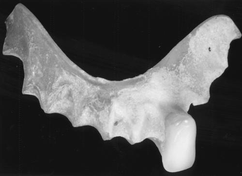
A man aged 60, attending casualty after taking a drug overdose, complained of sore throat with mild discomfort on swallowing. There was no clear history of foreign body ingestion and he was able to drink. The lateral soft tissue neck X-ray and chest X-ray were unremarkable. An initial fibreoptic laryngopharyngoscopy was normal, but on later review the patient indicated that he might have swallowed his denture. Fibreoptic nasendoscopy again showed a normal hypopharynx with no pooling of saliva. The scope was passed into the oesophagus where the denture was found to be lying transversely. The patient then underwent a rigid oesophagoscopy with removal of the denture (Figure 1) and recovered without incident.
Figure 1.
The denture recovered in case 2
Case 3
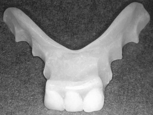
A man of 57 was referred for an opinion in relation to a legal claim against his dentist. He had been fitted with an acrylic immediate replacement lower partial denture carrying three incisor teeth that had soon become very loose and uncomfortable, particularly when the patient was eating. He was advised by the dentist to persist in eating with the denture since he would become accustomed to it and it would 'tighten up' with use. A specific reassurance was also given that the denture was too large to swallow (it was 4 cm wide with a maximum depth of 3 cm). However, the denture did lodge in the patient's throat while he was swallowing a drink and by the time he reached casualty it had entered the oesophagus. As the prosthesis did not show on a radiograph and could not be retrieved with the aid of an oesophagoscope, laparotomy and gastrotomy had to be undertaken before the it could be retrieved (Figure 2).
Figure 2.
The denture recovered in case 3, showing similar deficiencies in design to that in Figure 1
PREVIOUS CASES
An electronic search under the term 'swallowed partial denture' yielded 34 references for the period 1973–2001, including 3 that dealt with inhaled dentures.1–3 The instances reported in each publication ranged between one and five and these publications cited further references not found by our original criteria. The phenomenon of the swallowed or inhaled denture is thus well documented. Nor is the hazard confined to partial dentures; there were also reports relating to the ingestion of complete (full) lower dentures.4,5
At the time of case 3, telephone enquiries to dental advisers at UK protection societies suggested that any medicolegal consequences of swallowed dentures were infrequent. In relation to other categories of claim, this may well be so. However, one adviser subsequently reviewed his society's database to find that, in the period 1977–2003, a total of 26 claims were made against dental practitioners in respect of swallowed dentures (Phillips MW, personal communication). Although the database was not always specific as to whether the denture was partial or complete, the published work suggests that most would have been partial. In addition to the swallowed dentures, 3 complaints relating to inhaled dentures had also been received. It was of particular note that 47% of the swallowed-denture complaints specifically cited failure in diagnosis. Of the 13 cases that were now closed, 6 had resulted in payment to the claimant.
FEATURES AT PRESENTATION
Two of our patients were unable to give an initial history of having swallowed their dentures when presenting with secondary symptoms. This seems by no means unusual, especially in the case of patients with learning and mental health disorders.6–8 An early challenge is thus posed for the casualty officer who has restricted information to guide the diagnostic process. If it is observed that there are some natural teeth missing, the possibility of a swallowed denture should be included in the differential diagnosis. Parker and co-workers9 concluded that denture wearers were no more likely than similarly aged non-denture wearers to present with swallowed foreign bodies, but this could be misleading. Their investigation specifically excluded neurologically compromised patients and other disadvantaged categories. The most likely presenting symptom after swallowing of a denture is dysphagia, with other complaints related to how far the denture has progressed and time since swallowing. Thus further reports may also be anticipated of sore throat, choking sensation, retrosternal pain, sweating and a raised temperature and coughing up blood.
Early diagnosis and treatment will avoid the oedematous reaction and mucosal infection and necrosis that heighten the risk of rigid oesophagoscopy.10 Reported late complications of the undiagnosed swallowed denture include extraluminal migration from the oesophagus causing either a diverticulum11 or perforation12 (once a perforation has occurred, further severe sequelae may be anticipated, e.g. tracheo-oesophageal fistula13), the need to resect 18 cm of ileum,14 enterocolonic fistula15 and sigmoid colon perforation16,17.
IMAGING AND LOCATION
Poly(methylmethacrylate), the plastic from which most dentures are made, is radiolucent. Porcelain teeth produce light shadows on a plain radiograph but it is the metal pins attaching the teeth to the denture base that make them readily discernible. However, with the improvement in appearance and wear resistance of the better quality plastic and composite artificial teeth, porcelain teeth are seldom used on dentures in the UK.
Although a plain X-ray may well not identify a swallowed denture, the investigation has been recommended to exclude pneumomediastinum or gas within the soft tissues.18 A soft-tissue exposure is more likely to suggest the presence of a plastic denture than a standard exposure but, as with our experience in case 2, cannot be relied upon. Similarly a barium contrast medium before radiography is seldom helpful since it will coat all sides of a radiolucent object. Also, barium swallows can make subsequent endoscopy more difficult.19
Despite the many calls for use of radio-opaque denture base materials20–27 no such product seems available in the UK. The abridged published version of the report prepared by Brauer for the American Dental Association28 is an excellent source of information on the subject. At the time it was written, 1981, no plastic radio-opaque material was commercially available with physical properties, appearance and ease of handling to match those in radiolucent products. The amount of heavy metal salts and glass fillers that needed to be incorporated was sufficient to weaken the material, thereby increasing the possibility of fracture and the risk of swallowing a denture fragment. These inclusions also affected the appearance of the material.
Tsoa et al.29 conducted a clinical evaluation which, they claimed, validated the acceptability of a radioopaque acrylic that was available at the time. Their favourable conclusion was based on the finding of continuing radio-opacity of dentures after 5 years and that those patients who responded to the recall found their dentures 'reasonably satisfying'. However, only 22 of the original 102 patients attended for the recall, so we know nothing about the fate of the remaining eighty sets of dentures. Later work29,30 investigated the use of 40% poly(2,3-dibromopropylmethacrylate), introduced into the poly(methylmethacrylate) to render the denture base plastic radio-opaque. Because bromine was incorporated into the polymeric structure rather than present as a filler, the strength of the material was less affected. The material does not seem to have been marketed, possibly because of concerns that the halide might have cancer-inducing potential.
All metal partial dentures are readily detected on standard radiographs, but this fact did not prevent one epileptic patient from carrying such a prosthesis in the pyriform sinus for eleven months while a series of doctors tried to resolve his complaints of choking, dyspnoea and dysphagia.3 Metal components in a plastic denture, such as wire retainers or clasps, will also aid location on a radiograph. Clasps should render a partial denture less likely to dislodge but require regular review and maintenance for continuing efficiency. If swallowed, such components are likely to damage the gut lining.6,12
Denture labelling, to prevent the prosthesis entering the wrong mouth, is of value especially in a care home.31 Although this is a separate issue from the radiological location of swallowed dentures, both purposes could be served by the use of an embedded metal foil identity tag system in plastic denture bases.32 However, more than one tag may be required to counter the possibility that the denture may fracture in use and only one part be swallowed.
Acrylic dentures are more likely to be discernible by CT, since the process is more sensitive to small changes in X-ray attenuation, than by plain radiography.33,34 They can also be shown by MRI, the difficulty being access to MRI equipment in an emergency.
Direct visualization of a swallowed denture with a flexible or rigid endoscope is possible while the prosthesis remains in the hypopharynx or oesophagus. Examination with a fibre-optic instrument is more readily undertaken but, as our experience shows, not totally reliable. Although early oesophagoscopy has been recommended, the procedure is still not without the risk of perforation.10,35. Oesophagoscopy may miss the denture and Youngs et al.36 described a case in which rigid oesophagoscopy failed to identify a prosthesis that had passed into an extraluminal position.
THE PROVISION OF SMALL DENTURES
The major lesson from the published work is that a denture does not have to be small to be swallowed. Configuration as well as overall dimensions is important. Thus a horseshoe shaped denture swallowed 'end-on' and vertically may well rotate into the hypopharynx and oesophagus, though its width would make it difficult to swallow flat and transversely. Such a consideration would explain why swallowing complete (full) lower dentures has been reported.
The hazard of the small 'side plate' has long been recognized.37 Alternatives include conventional bridgework, resin-bonded bridgework (where it is desirable to restrict the preparation of abutment teeth) and implant-borne bridges.38 There remains the possibility that such fixed prostheses will become detached and swallowed39 but, because they are smaller, have metal components and lack features liable to engage and traumatize the gut wall, such an event is less likely to cause the complications of a swallowed denture.
Where a removable denture has to be provided, it should be designed in such a manner as to render it retentive and stable. This consideration is of particular importance in treatment planning for the epileptic patient and those with learning difficulties. The minimal complete lower denture base reduces both the size and the stability of the denture. For the partial denture, the principles of retention (direct and indirect) and cross-arch bracing are particularly important. Checking over the dentures and undertaking necessary maintenance should be part of the regular dental recall/review process for the patient. Apart from the denture swallowing risk (and for reasons of overall dental health), patients should be advised not to wear dentures at night.
RECOMMENDATIONS
Any planned dental prosthesis demands careful thought. Some form of embedded radio-opaque marker(s) should be part of the design considerations.
The patient should be advised on the wearing and care of their dentures and the need to return regularly for maintenance.
Care workers need to know that certain individuals will be unable to perceive or report the disappearance of a denture; they should be alert to the possibility of swallowing or inhalation.
Medical personnel, especially those called upon to manage emergencies, should likewise be aware of the multiple hazards.
Acknowledgments
We thank Mr P R Prinsley and Mr D J Premachandra for permission to report the patients under their care.
References
- 1.Knowles JE. Inhalation of dental plates—a hazard of radiolucent materials. J Laryngol Otol 1991;105: 681-2 [DOI] [PubMed] [Google Scholar]
- 2.Douzinas EE, Goumas P, Bilalis D. Respiratory distress following the inhalation of a non radio-opaque dental prosthesis. Ann Med Interne (Paris) 1989;140: 413-14 [PubMed] [Google Scholar]
- 3.Gionvannitti JA. Aspiration of a partial denture during an epileptic seizure. J Am Dent Assoc 1981;103: 895. [DOI] [PubMed] [Google Scholar]
- 4.Hazelrigg CO. Ingestion of mandibular complete denture. J Am Dent Assoc 1984;108: 208. [DOI] [PubMed] [Google Scholar]
- 5.Perenack DM. Ingestion of mandibular complete denture. J Am Dent Assoc 1980;101: 802. [DOI] [PubMed] [Google Scholar]
- 6.Jacobs LI. Ingestion of partial denture. J Am Dent Assoc 1980;101: 801. [DOI] [PubMed] [Google Scholar]
- 7.Carbery A, Provencal M. A case of swallowing a partial denture. J Can Dent Assoc 1993;59: 841-4 [PubMed] [Google Scholar]
- 8.Green JG, Durham TM, King TA. Management of patients with swallowed dental objects. Am J Dent 1988;1: 147-50 [PubMed] [Google Scholar]
- 9.Parker AJ, Yardley PJ, Owen GO. Dental prostheses and the impacted swallowed foreign body. J R Coll Surg Edin 1993;38: 337-9 [PubMed] [Google Scholar]
- 10.Nandi P, Ong GB. Foreign body in the oesophagus: review of 2394 cases. Br J Surg 1978;75: 5-9 [DOI] [PubMed] [Google Scholar]
- 11.Olak J, Jeyasingham K. Cervical oesophageal diverticulum associated with an impacted denture. Can J Surg 1991;34: 614-17 [PubMed] [Google Scholar]
- 12.Treska TP, Smith CC. Swallowed partial denture. Oral Surg Med Oral Pathol 1991;72: 756-7 [DOI] [PubMed] [Google Scholar]
- 13.Rajesh PB, Goiti JJ. Late onset tracheo-oesophageal fistula following a swallowed dental plate. Eur J Cardiothorac Surg 1993;7: 661-2 [DOI] [PubMed] [Google Scholar]
- 14.Goodacre CJ. A dislodged and swallowed unilateral removable partial denture. J Prosthet Dent 1987;58: 124-5 [DOI] [PubMed] [Google Scholar]
- 15.Sejdinaj I, Powers RC. Enterocolonic fistula from swallowed denture. JAMA 1973;225: 994. [PubMed] [Google Scholar]
- 16.Ghori A, Dorricott NJ, Sanders DS. A lethal ectopic denture: an unusual case of sigmoid perforation due to unnoticed swallowed dental plate. J R Coll Surg Edin 1999;44: 203-4 [PubMed] [Google Scholar]
- 17.Cleator IG, Christie J. An unusual case of swallowed dental plate and perforation of the sigmoid colon. Br J Surg 1973;60: 163-5 [DOI] [PubMed] [Google Scholar]
- 18.Nageris B, Feinmesser R. Dentures in the oesophagus complicated by pneumomediastinum. Ear Nose Throat J 1990;69: 737-8 [PubMed] [Google Scholar]
- 19.Willshire PC, Clarke CP, Daniel FJ. Dentures: difficult oesophageal foreign bodies. Aust N Z J Surg 1993;63: 736-8 [DOI] [PubMed] [Google Scholar]
- 20.Phillips WL. Impaction of dentures in the oesophagus. J Prosthet Dent 1971;26: 222-4 [DOI] [PubMed] [Google Scholar]
- 21.Chandler HH, Bowen RL, Paffenbarger GC. The need for radio-opaque denture base materials: a review of the literature. J Biomed Mater Res 1971;5: 245-52 [DOI] [PubMed] [Google Scholar]
- 22.Kerr AG. Swallowed dentures. Br Dent J 1966;120: 595-8 [PubMed] [Google Scholar]
- 23.Banham TM. Dangers of radio-translucent dental plates. BMJ 1965;2: 302. [DOI] [PMC free article] [PubMed] [Google Scholar]
- 24.McCabe JF, Wilson HJ. A radio-opaque denture material. J Dent 1976;4: 254-64 [DOI] [PubMed] [Google Scholar]
- 25.Combe EC. Further studies on radio-opaque denture material. J Dent 1972;1: 93-7 [DOI] [PubMed] [Google Scholar]
- 26.Chandler HH, Bowen RL, Paffenbarger GC. Development of a radio-opaque denture base material. J Biomed Mater Res 1971;5: 253-265 [DOI] [PubMed] [Google Scholar]
- 27.Stafford GD, MacCulloch WT. Radio-opaque denture base materials. Br Dent J 1971;131: 22-4 [DOI] [PubMed] [Google Scholar]
- 28.Brauer GM. The desirability of using radio-opaque plastics in dentistry—a status report. J Am Dent Assoc 1981;102: 347-9 [DOI] [PubMed] [Google Scholar]
- 29.Tsao DH, Guilford HJ, Kazanoglu A, Bell DH. Clinical evaluation of a radiopaque denture base resin. J Prosthet Dent 1984;51: 456-8 [DOI] [PubMed] [Google Scholar]
- 30.Davy KW, Causton BE. Radio-opaque denture base: a new acrylic co-polymer. J Dent 1982;10: 254-64 [DOI] [PubMed] [Google Scholar]
- 31.McNally L, Gosney MA, Doherty U, Field EA. The orodental status of a group of elderly in-patients: a preliminary assessment. Gerodont 1999;16: 81-4 [DOI] [PubMed] [Google Scholar]
- 32.Reeson MG. A simple and inexpensive inclusion technique for denture identification. J Prosthet Dent 2001;86: 441-2 [DOI] [PubMed] [Google Scholar]
- 33.Newton JP, Abel RW, Lloyd CH, Yemm R. The use of computed tomography in the detection of radiolucent denture base material in the chest. J Oral Rehabil 1987;14: 193-202 [DOI] [PubMed] [Google Scholar]
- 34.McLaughlin MG, Dwayne LC, Garuana V. Computed tomographic detection of swallowed dentures. Comput Med Imaging Graph 1989;13: 161-3 [DOI] [PubMed] [Google Scholar]
- 35.Cooke LD, Baxter PW. Accidental impaction of partial dental prostheses in the upper gastrointestinal tract. Br Dent J 1992;172: 451-2 [DOI] [PubMed] [Google Scholar]
- 36.Youngs RP, Gatland D, Brookes J. Swallowed radiolucent dental prostheses: risk of extraluminal oesophageal perforation. J Laryngol Otol 1988;63: 736-8 [DOI] [PubMed] [Google Scholar]
- 37.Goodacre CJ. A dislodged and swallowed unilateral removable partial denture. J Prosthet Dent 58: 124-5 [DOI] [PubMed]
- 38.Walmsley AD, Walsh TF, Burke FJT, Shortall ACC, Lumley PJ, Hayes-Hall R. Restorative Dentistry. Edinburgh: Churchill-Livingstone, 2002
- 39.Beaumont RH. Retrieval of a swallowed casting six weeks after ingestion. Oral Surg Oral Med Oral Pathol 1987;64: 287-8 [DOI] [PubMed] [Google Scholar]


