Dysphagia and odynophagia are sometimes associated with cervical osteophytes, but a causal connection can seldom be proved.1
CASE HISTORY
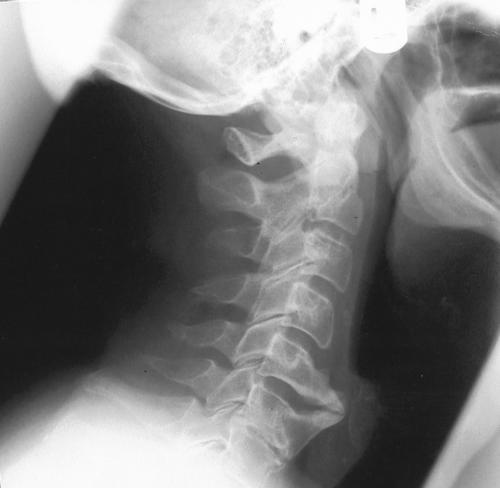
A man of 60 sought medical attention when he developed numbness in the back of the right arm after carrying luggage on holiday. Lateral X-rays of the neck showed a solitary massive cervical osteophyte complex at the level of the fifth and sixth cervical vertebrae (Figure 1). For the past 10 years he had been treated repeatedly with antibiotics for sore throat, odynophagia and mild dysphagia but had not been investigated for these. He was now referred for an ENT opinion. He was a non-smoker and there had been no weight loss, voice change or dyspnoea.
Figure 1.
Preoperative lateral neck X-ray showing the osteophytes at the level of C5/6
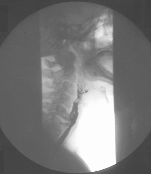
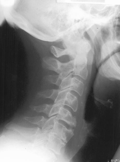
Pernasal flexible fibreoptic examination of the pharynx showed bulging of the posterior pharyngeal wall at the level of the larynx, covered by normal smooth pharyngeal mucosa. Nothing further was revealed by a barium swallow (Figure 2). The osteophyte was therefore considered the cause of his throat symptoms and was excised with a chisel, via an anterolateral transcervical approach, by the ENT consultant (APJ). Three months later the patient was completely symptom-free and a lateral neck X-ray (see Figure 3) and barium swallow showed that the external compression had been eradicated.
Figure 2.
Preoperative barium swallow
Figure 3.
Postoperative lateral neck X-ray showing excision site of osteophytes
COMMENT
Osteophytes are common, and occur mainly at the sites of maximal cervical movement—namely, C6/5 and C6/7.2 Surprisingly, they do not often cause dysphagia. Le Roux, in his study of 1200 cases of dysphagia, did not report any as being caused by osteophytes alone.3 The coexistence of the two can therefore be a clinical trap for the unwary,4 but in the present case there can be little doubt about the causation. In view of the 10-year history of symptoms, hypopharyngeal or upper oesophageal malignancy was highly unlikely. The arm numbness seems to have been an incidental symptom which fortunately prompted the taking of a lateral neck X-ray; with the use of a non-steroidal anti-inflammatory it disappeared spontaneously.
How do cervical osteophytes cause dysphagia? A large osteophyte may do so by simple mechanical obstruction or by impinging on mobile structures such as the cricoid cartilage. Local inflammation or associated muscular spasm may also be implicated,5 in which case conservative management with soft diet, anti-inflammatory medications and antibiotics may be sufficient.6 Surgery may be the best option in a well-nourished patient with severe obstructive symptoms and can be done either per-orally or transcervically. The per-oral approach carries a higher theoretical risk of deep neck space infection and cervical spine osteomyelitis, because pharyngotomy is required. A transcervical approach may be posterolateral or anterolateral. In the posterolateral approach the contents of the carotid sheath are retracted anteriorly with the larynx and pharynx. This offers wide exposure to the prevertebral space but requires more retraction of the carotid sheath and carries a much higher risk of injury to the sympathetic chain.7 In the anterolateral approach the contents of the carotid sheath are retracted posteriorly. Special care is needed to preserve the recurrent laryngeal nerve, injury to which can result in temporary or permanent voice change.7 Either technique can be complicated by bleeding pharyngocutaneous fistula, oesophageal perforation, airway obstruction due to pharyngeal oedema, transient aspiration and trauma to the vagus, spinal accessory and hypoglossal nerves. With cautious technique, secondary instability of the cervical spine leading to neurological damage should be wholly avoidable.
References
- 1.Crowther JA, Ardran GM. Dysphagia due to cervical spondylosis. J Laryngol Otol 1985;99: 1167-9 [DOI] [PubMed] [Google Scholar]
- 2.Saunders WH. Cervical osteophytes and dysphagia. Ann Otol Rhinol Laryngol 1970;79: 1091-7 [DOI] [PubMed] [Google Scholar]
- 3.Le Roux BT. Dysphagia and its causes. Geriatrics 1962;17: 560-4 [PubMed] [Google Scholar]
- 4.Ladenheim SE, Marlowe FI. Dysphagia secondary to cervical osteophytes. Am J Otolaryngol 1999;20: 184-9 [DOI] [PubMed] [Google Scholar]
- 5.Eviatar E, Harell M. Diffuse idiopathic skeletal hyperostosis with dysphagia [review]. J Laryngol Otol 1987;101: 627-32 [DOI] [PubMed] [Google Scholar]
- 6.Ozgocmen S, Kiris A, Kocakoc E, Ardicoglu O. Osteophyte-induced dysphagia: report of three cases. Joint Bone Spine 2002;69: 226-9 [DOI] [PubMed] [Google Scholar]
- 7.Sobol SM, Rigual NR. Anterolateral extrapharyngeal approach for the cervical osteophyte-induced dysphagia. Ann Otol Rhinol Laryngol 1984;93: 498-504 [DOI] [PubMed] [Google Scholar]


