Abstract
The V protein expressed by pathogenic Yersinia pestis is an important virulence factor and protective immunogen. The presence of linear B-cell epitopes in the V protein was investigated by using a series of 17 overlapping linear peptides. Groups of 10 mice were immunized intraperitoneally with 30 μg of each peptide on days 0, 30, and 60. Although the V protein-specific antibody response to the peptides varied, most of the peptides elicited high antibody titers. The immunized mice were challenged subcutaneously with 60 50% lethal doses (LD50) (1 LD50 = 1.9 CFU) of a virulent Y. pestis strain, CO92. None of the peptide-immunized mice survived challenge. The animals immunized with the V protein were completely protected against challenge. The immunogenicity of some of the V peptides was increased by conjugating them to keyhole limpet hemocyanin. Only one peptide (encompassing amino acids 1 to 30) conjugate demonstrated some protection; the others were not protective. In additional experiments, V peptides that reacted well with sera from mice surviving Y. pestis infection were combined and used to immunize mice. Although the combined peptides appeared to be very immunogenic, they were not protective. Therefore, the protective B-lymphocyte epitope(s) in the V protein is most likely to be conformational.
Yersinia pestis, a gram-negative bacillus, is the causative agent of plague and is one of the most virulent bacteria presently known (7, 10, 11). The extreme virulence of this bacterium can be attributed to its ability to efficiently invade and subvert the mammalian innate immune system, resulting in an overwhelming infection. The capacity of Y. pestis to disarm the innate immune system is determined by numerous virulence factors encoded on its chromosome and three plasmids (7, 10, 11).
One of the factors with a dominant role in promoting the virulence of Y. pestis is the V protein (8, 37). V is a secreted protein of approximately 39 kDa which is encoded by the 75-kb low-calcium-response plasmid (4, 8, 9, 30, 31). There is experimental evidence suggesting that the V protein acts to suppress the innate immune response (8, 26, 27, 29). Attenuated bacterial strains demonstrated increased virulence in mice given repeated doses of purified V protein (26). Additionally, V protein alters cytokine profiles during Yersinia infections, which may contribute to immune system subversion (27, 29). In addition to its effect on the host, the V protein is involved in the regulation of the low calcium response of Y. pestis (4, 30, 31, 37).
Previous experiments performed with mice illustrated the efficacy of the V protein as a vaccine against lethal subcutaneous (s.c.) and aerosol infection with both F1-positive and F1-negative Y. pestis strains (1, 18, 23, 24, 41, 42). Wild-type (F1-positive) organisms form a capsule composed of the Y. pestis specific F1 protein, while the F1-negative strains have lost the ability to produce this capsule. The licensed Plague Vaccine USP does not elicit antibodies to the V antigen but relies on inducing antibodies to the F1 capsular protein. Mice immunized with the current licensed vaccine are therefore not protected against the F1-negative organisms. The ability of candidate V protein vaccines to protect mice from fatal disease caused by Y. pestis appears to result from the generation of protective V-specific antibodies. The passive transfer of both V-specific polyclonal and monoclonal antisera protects animals from challenge with virulent Y. pestis (22, 25, 36, 38). In mice immunized with the V protein, there appeared to be a correlation between the quantity and isotype of V-specific antibody induced and protection against disease (1, 23, 42).
To gain a more detailed understanding of which regions of the V protein are responsible for eliciting the protective immunity, studies have been conducted to epitope map the V antigen. These studies were initially conducted by Motin et al. (25). Using a series of genetically engineered truncated V proteins fused to protein A, they concluded that the protective epitopes were located between amino acid residues 168 and 275 of the V protein. However, they did not test this fragment directly for its ability to remove the protective activity of sera generated against the entire V protein. More recently, Hill et al. (19) actively immunized mice with both N-terminal and C-terminal truncations of the V protein fused to glutathione S-transferase. Based on the pattern of protection generated by immunization with the various V fusion proteins, they speculated that the protective epitopes were located between amino acid residues 135 and 275, again without directly testing this fragment in an active immunization protocol. A more recent study demonstrated that Yersinia spp. appear to express one of two major forms of the V antigen and that antibodies generated against one form are unable to protect against the other (33). Interestingly, the major difference in the two forms occurs between amino acids 225 and 232 (33). Therefore, three separate studies with very different approaches suggested that this region of the V protein contains protective epitopes.
In an effort to determine if a protective linear epitope existed in this region, we studied the presence of linear B-cell epitopes in this region (amino acids 130 to 280), as well as the rest of the V protein, by using a series of 17 overlapping linear peptides. These V peptides were used to determine whether a linear region of protein had the capacity to elicit a protective immune response in mice. This information would assist efforts to develop an in vitro correlate of immunity for new plague vaccines containing the V protein and to develop V peptide vaccines. Additionally, we have examined the reactivity of linear V peptides with sera from mice exposed to native V protein through infection with Y. pestis in an attempt to develop possible V diagnostic reagents. We report here the results of our attempts to determine if a protective linear epitope exists in the V antigen and to determine which V peptides react with sera from Y. pestis-infected animals.
MATERIALS AND METHODS
Peptide synthesis and design.
Peptides that encompassed the entire V protein were designed from the published sequence of the Y. pestis KIM 5 strain V gene as determine by Price et al. (31). The peptides that were used in an unconjugated form were designed to be 30 amino acids long. Generally, the peptides overlapped by 10 amino acids, with the exception of the peptide pairs V9/V10 and V14/V15, which overlapped by 23 and 19 amino acids, respectively. The sequences of these peptides are shown in Table 1. The V peptides were synthesized at Macromolecular Resources (Fort Collins, Colo.), purified by reversed-phase high-pressure liquid chromatography, and sequenced by mass spectroscopy.
TABLE 1.
Sequences of V peptides used in this study
| Peptide | Residues | Sequence |
|---|---|---|
| V1 | 1–30 | MIRAYEQNPQHFIEDLEKVRVEQLTGHGSS |
| V2 | 21–50 | VEQLTGHGSSVLEELVQLVKDKNIDISIKY |
| V3 | 41–70 | DKNIDISIKYDPRKDSEVFANRVITDDIEL |
| V4 | 61–90 | NRVITDDIELLKKILAYFLPEDAILKGGHY |
| V5 | 81–110 | EDAILKGGHYDNQLQNGIKRVKEFLESSPN |
| V6 | 101–130 | VKEFLESSPNTQWELRAFMAVMHFSLTADR |
| V7 | 121–150 | VMHFSLTADRIDDDILKVIVDSMNHHGDAR |
| V8 | 141–170 | DSMNHHGDARSKLREELAELTAELKIYSVI |
| V9 | 161–190 | TAELKIYSVIQAEINKHLSSSGTINIHDKS |
| V10 | 168–197 | SVIQAEINKHLSSSGTINIHDKSINLMDKN |
| V11 | 188–217 | DKSINLMDKNLYGYTDEEIFKASAEYKILE |
| V12 | 208–237 | KASEYKILEKMPQTTIQVDGSEKKIVSIK |
| V13 | 228–257 | GSEKKIVSIKDFLGSENKRTGALGNLKNSY |
| V14 | 248–275 | GALGNLKNSYSYNKDNNELSHFATTCSD |
| V15 | 257–286 | YSYNKDNNELSHFATTCSDKSRPLNDLVSQ |
| V16 | 277–306 | SRPLNDLVSQKTTQLSDITSRFNSAIEALN |
| V17 | 297–326 | RFNSAIEALNRFIQKYDSVMQRLLDDTSGK |
Peptide-carrier conjugation.
Peptides that appeared poorly immunogenic or nonimmunogenic were resynthesized to contain a C-terminal cysteine and conjugated to keyhole limpet hemocyanin (KLH) through this additional residue (Chiron Minotopes Peptide Systems, San Diego, Calif.). These peptide-carrier conjugates were purified by gel filtration. The sequences of these peptides are shown in Table 2.
TABLE 2.
Sequence of conjugated V peptides used in this study
| Peptide | Residues | Sequence |
|---|---|---|
| V18 | 1–30 | MIRAYEQNPQHFIEDLEKVRVEQLTGHGSS(C) |
| V19 | 101–120 | VKEFLESSPNTQWELRAFMA(C) |
| V20 | 116–135 | RAFMAVMHFSLTADRIDDDI(C) |
| V21 | 168–197 | SVIQAEINKHLSSSGTINIHDKSINLMDKN(C) |
| V22 | 277–296 | SRPLNDLVSQKTTQLSDITS(C) |
| V23 | 287–306 | KTTQLSDITSRFNSAIEALN(C) |
Immunization of animals.
Groups of 10 female, 8- to 9-week-old, outbred Swiss Webster mice (Hsd:ND4) obtained from Harlan Sprague-Dawley (Indianapolis, Ind.) were given three intraperitoneal (i.p.) immunizations of either peptide in free form or conjugated to KLH. The initial immunization consisted of either 30 μg of free peptide in complete Freund’s adjuvant (CFA) (Sigma Chemical, St. Louis, Mo.) or 50 μg of peptide-carrier conjugate in CFA. The manufacturer of the peptide-carrier conjugate estimated that approximately 30% of the weight of the conjugates was peptide; therefore, the mice received about 15 μg of peptide. The use of a higher concentration of peptide was not attempted. The amount of conjugate given to the mice was the amount suggested by the manufacturer for giving optimal antibody responses in mice. The subsequent immunizations after 30 and 60 days consisted of either 30 μg of free peptide or 50 μg of conjugated peptide in incomplete Freund’s adjuvant (IFA). Separate groups of mice were also immunized s.c. with 0.2 ml of Plague Vaccine USP, lot 1128X1 (Greer Laboratories, Lenior, N.C.) or with 10 μg of recombinant histidine-tagged V protein (a gift from Matthew Mauro, Naval Research Center, Washington, D.C.). CFA and IFA were used with the recombinant V antigen to immunize mice as described for the peptides. Anesthetized mice were bled from the retro-orbital sinus 10 to 14 days after the second and third immunizations to assess the immunoglobulin G (IgG) titer to the V antigen in serum. The response of individual mice was monitored by implanting transponders (BioMedic Data System, Seaford, Del.) s.c. (3). The transponders provided a positive identification system. All experiments involving animals were conducted in accordance with the regulations described in the Guide for the Care and Use of Laboratory Animals (28).
Analysis of sera by ELISA.
The analysis of the V antigen-specific and V peptide IgG-specific serum antibody response was conducted by a standard indirect enzyme-linked immunosorbent assay (ELISA) (1, 5, 6). Briefly, microtiter plates (Nunc, Naperville, Ill.) were coated with 50 μl of either V peptide (10 μg/ml) or recombinant V protein (1 μg/ml) in 15 mM Na2CO3–35 mM NaHCO3 (pH 9.6) for 16 to 20 h at room temperature. The plates were then blocked with 0.1% (wt/vol) bovine serum albumin in phosphate-buffered saline (PBS) (pH 7.4)–0.1% (vol/vol) Tween 20 by adding 300 μl to each well and then incubating the plates for 2 h at room temperature. After being blocked, the plates were washed five times with PBS–0.1% Tween 20 by using an automated plate washer (Skatron Instrument, Sterling, Va.). To the blocked plates, sera obtained from immunized animals (100 μl) were serially diluted across the wells of the microtiter plates, and the plates were incubated for 1 h at 37°C. The sera were removed, and the plates were washed five times with 300 μl of PBS–0.1% Tween 20. Goat anti-mouse IgG (heavy plus light chains) conjugated to horseradish peroxidase (Kirkegaard & Perry Laboratories, Gaithersburg, Md.) was diluted to 1:5,000 in PBS–0.1% Tween 20, and 100 μl of this was added to each of the microtiter wells. After 1 h of incubation at 37°C, the plates were again washed five times. The plates were then developed with 100 μl of the chromogenic substrate 2.2′-azino-di-[3-ethylbenzthiazoline sulfonate (6)] solution (Kirkegaard & Perry Laboratories). The reaction was allowed to proceed for 30 min and then stopped by the addition of 100 μl of 10% sodium dodecyl sulfate per well. The optical density was measured at 405 to 417 nm with an ELISA plate reader (Dynatech Laboratories, Chantilly, Va.). The end-point titer was defined as the highest serum dilution with a value of at least 0.1 optical density unit.
Challenge studies.
Mice were challenged approximately 30 days after the last immunization as previously described (1). This postimmunization time point was chosen to minimize any nonspecific resistance which may have been induced by the adjuvant. The mice were challenged s.c. with 50 to 100 50% lethal doses (LD50) (1 LD50 = 1.9 CFU) of Y. pestis CO92 (15). All manipulations were performed in a Biological Safety Level-3 containment facility. The mice were monitored for 30 days, after which the remaining mice were exsanguinated and their spleens were examined for residual viable Y. pestis infection.
Statistical analysis.
The Fisher exact test and log-rank test were used to compare the results from the various vaccinated groups of animals (35).
RESULTS
Capacity of the overlapping V peptides to induce a heterologous response to the V protein.
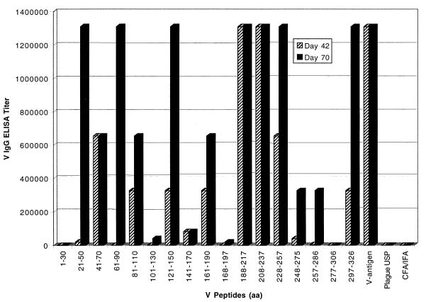
In initial experiments, groups of 10 mice were immunized with the 17 overlapping V peptides (Table 1) as well as the intact recombinant V protein, the current whole-cell Plague Vaccine USP, and the adjuvant alone. As illustrated in Fig. 1, the heterologous immune response to the V antigen elicited by the individual V peptides after the second immunization was variable. Seven of the V peptides elicited responses that were similar to that for the V antigen response, six of the V peptides elicited responses that were below that for the recombinant V protein, and four of the peptides elicited no observable response. Sera from mice immunized with peptides encompassing amino acids 1 to 30 and 277 to 306 did not react with the recombinant V protein used in the ELISA, and peptides spanning amino acids 101 to 130 and 168 to 197 elicited only a weak heterologous response.
FIG. 1.
Heterologous immune response to the V antigen elicited by the individual V peptides. Groups of Hsd:ND4 mice (10 mice/group) were immunized with 30 μg of peptide (30-mer) on days 0, 30, and 60. After the second (day 42) and third (day 72) immunizations, mice were bled, the sera from the individual animals were pooled, and the presence of V-specific antibodies was determined by ELISA. aa, amino acids.
Homologous V peptide specific immune response.
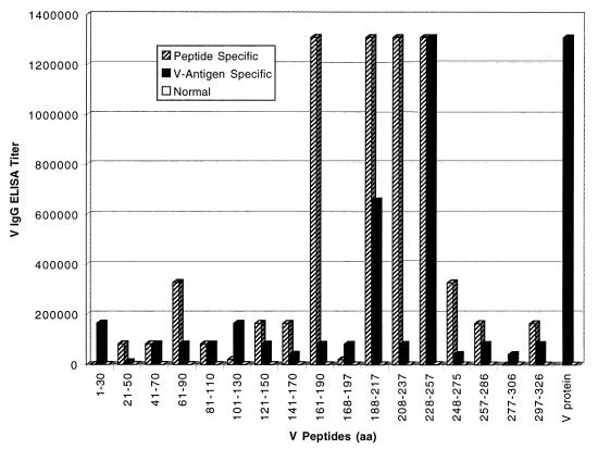
The analysis described in the preceding section was specifically designed to evaluate the ability of the peptides to elicit a response capable of reacting in a heterologous fashion with the V antigen. The analysis described below was designed to determine the peptide-specific antibody response generated by the immunized animals. Each peptide was tested with sera from the peptide-immunized mice, sera from naive mice, and sera from mice immunized with the whole V protein. As illustrated in Fig. 2, the peptide-specific response was detected for each peptide except for the peptides encompassing V amino acids 1 to 30 and 277 to 306, the same peptides for which no heterologous response was detected. The ability of these two peptides to bind to the wells of the microtiter plate was confirmed by their reactivity with V antigen-specific sera (Fig. 2). The peptides encompassing amino acids 101 to 130 and 168 to 197 generated a weak but detectable peptide-specific response. This analysis demonstrated that the peptides encompassing amino acids 1 to 30, 101 to 130, 168 to 197, and 277 to 306 were either weakly immunogenic or nonimmunogenic and that the problem was not due to a lack of cross-reactivity with the recombinant V antigen.
FIG. 2.
Homologous peptide-specific response to each V peptide as measured by a peptide ELISA. Each peptide was adsorbed to the wells of a microtiter plate at 10 μg/ml, and the pooled sera from peptide-immunized, V-immunized, and Hsd:ND4 mice given adjuvant alone were analyzed for reactivity with the V peptide panel. aa, amino acids.
Immunogenicity of the conjugated V peptides.
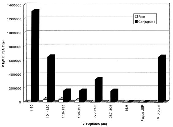
There were four V peptides for which very little heterologous or homologous antibody response was generated. In some instances, the low immunogenicity of a small peptide can be attributed to the lack of an appropriate T-helper lymphocyte epitope contained within the peptide (5, 16). To enhance the immunogenicity of the V peptides shown to be incapable of generating an adequate antibody response, the identical peptides or shorter derivatives of these peptides were synthesized with a cysteine residue at the C terminus and were conjugated to the carrier molecule KLH (Table 2). As shown in Fig. 3, the immunogenicity of each nonimmunogenic peptide was increased by conjugation to KLH, as indicated by a significant, albeit variable, increase in the heterologous antibody response to the V protein compared to that for the free V peptide.
FIG. 3.
Effect of conjugation to KLH on the immunogenicity of nonimmunogenic peptides. Hsd:ND4 mice were immunized with either 30 μg of free peptide or 50 μg of conjugated peptide on days 0, 30, and 60. Additional animals served as controls and were immunized with the carrier molecule KLH, Plague Vaccine USP, or the recombinant V protein. The mice were bled on day 70, and the sera were analyzed by ELISA for the presence of V-specific antibodies. aa, amino acids.
Protection against fatal plague after peptide immunization.
To determine whether the V peptides were capable of inducing a protective response, mice were challenged with 50 to 100 LD50 of virulent Y. pestis CO92. It has previously been shown that immunization with the full-length V protein can protect mice against greater than 106 LD50; therefore, 50 to 100 LD50 is a minimal challenge (1). The challenge results, illustrated in Table 3, indicated that none of the V peptides in free form were able to induce a protective immune response. There was no increase in the mean time to death, which provided additional evidence that even high-titer antibody responses to the linear V peptides did not induce a protective immune response. The recombinant V protein, as well as the Plague Vaccine USP, protected the mice, as previously reported (1).
TABLE 3.
Summary of the V peptide challenge experimentsa
| Immunogen | ELISA titerb | MTD (days) ± SDc | No. of mice surviving/ total no.d |
|---|---|---|---|
| V peptide 1–30 | <320 | 3.7 ± 0.67 | 0/10 |
| V peptide 21–50 | 1,310,720 | 5.0 ± 1.56 | 0/10 |
| V peptide 41–70 | 655,360 | 4.0 ± 0 | 0/10 |
| V peptide 61–90 | 1,310,720 | 4.0 ± 0.70 | 0/9 |
| V peptide 81–110 | 655,360 | 4.6 ± 1.07 | 0/10 |
| V peptide 101–130 | 5,120 | 4.0 ± 0.94 | 0/10 |
| V peptide 121–150 | 1,310,720 | 4.6 ± 0.97 | 0/10 |
| V peptide 141–170 | 81,920 | 4.4 ± 1.26 | 0/10 |
| V peptide 161–190 | 655,360 | 4.5 ± 0.97 | 0/10 |
| V peptide 168–197 | 20,480 | 3.9 ± 0.88 | 0/10 |
| V peptide 188–217 | 1,310,720 | 4.6 ± 1.43 | 0/10 |
| V peptide 208–237 | 1,310,720 | 3.9 ± 1.20 | 0/10 |
| V peptide 228–257 | 1,310,720 | 4.9 ± 0.99 | 0/10 |
| V peptide 248–275 | 327,680 | 4.5 ± 1.71 | 0/10 |
| V peptide 257–286 | 327,680 | 4.6 ± 1.17 | 0/10 |
| V peptide 277–306 | <320 | 4.1 ± 0.99 | 0/10 |
| V peptide 297–327 | 1,310,720 | 4.9 ± 1.17 | 0/9 |
| V protein in CFA | 1,310,720 | 10/10 | |
| Plague Vaccine USP | <320 | 15.3 ± 8.08 | 7/10 |
| CFA/IFA alone | 640 | 4.8 ± 1.12 | 0/10 |
Hsd:ND4 mice were immunized three times i.p. with either peptide, V protein, or Plague Vaccine USP.
Sera obtained from immunized animals after the third immunization were pooled and analyzed for the presence of antibodies to the V antigen.
Mean time to death ± standard deviation.
Mice were challenged s.c. with 60 LD50 of Y. pestis CO92 approximately 30 days after the last immunization.
Mice given the peptide conjugates developed greater antibody titers to the heterologous V protein than did mice immunized with the free peptide. However, except for the conjugated peptide representing the region from amino acids 1 to 30, protection was not conferred (Table 4). The conjugated peptide representing the region from amino acids 1 to 30 appeared to be capable of inducing low-level protection, since four of nine mice survived challenge (P = 0.033) and there was a corresponding delay in the mean time to death (P = 0.0001) for the mice not surviving challenge (Table 4) compared to the results for the groups of mice immunized with KLH carrier alone.
TABLE 4.
Summary of the V peptide conjugate challenge experimenta
| Immunogen | ELISA titerb | MTD (days) ± SDc | No. of mice surviving/ total no.d |
|---|---|---|---|
| V peptide 1–30 plus KLH | 1,310,720 | 7.8 ± 1.3 | 4/9 |
| V peptide 101–120 plus KLH | 655,360 | 4.7 ± 0.9 | 0/9 |
| V peptide 116–136 plus KLH | 163,840 | 4.7 ± 1.1 | 0/10 |
| V peptide 168–197 plus KLH | 163,840 | 4.7 ± 0.9 | 0/10 |
| V peptide 277–296 plus KLH | 327,680 | 4.6 ± 0.5 | 0/10 |
| V peptide 287–306 plus KLH | 163,840 | 5.1 ± 0.7 | 0/10 |
| V protein in CFA | 1,310,720 | 9/9 | |
| Plague Vaccine USP | <640 | 21 ± 6.4 | 8/10 |
| KLH alone | <640 | 5.1 ± 0.9 | 0/10 |
Hsd:ND4 mice were immunized three times i.p. with either peptide, V protein, or Plague Vaccine USP.
Serum obtained from immunized animals was pooled and analyzed for the response to recombinant V antigen.
MTD ± SD, mean time to death ± standard deviation.
Mice were challenged s.c. with 100 LD50 of Y. pestis CO92 approximately 30 days after the last immunization.
Immunogenicity of peptide combinations.
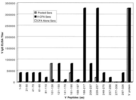
To determine whether the presence of antibodies to more than one linear V region might be protective, mice were immunized with combinations of linear V peptides. To avoid combinations of peptides that were simply random, the entire V peptide panel was screen for reactivity with sera from Y. pestis-infected animals surviving infection (2, 34). As shown in Fig. 4, peptides encompassing amino acids 101 to 130, 161 to 190, and 228 to 257 reacted well with sera pooled from infected animals, while a second group, encompassing amino acids 141 to 170, 208 to 237, and 248 to 275, were less reactive. Based on these results, the immunodominant peptides (amino acids 101 to 130, 161 to 190, and 228 to 257) were combined and mice were immunized with this mixture. In a second peptide combination, these three peptides were mixed with those encompassing amino acids 141 to 170, 208 to 237, and 248 to 275 and this mixture, containing six peptides in equal molar amounts, was used to immunize mice. These peptide combinations were very immunogenic and reacted well with the heterologous V protein in ELISA. However, as with the individual peptides, protection was not conferred and there was no delay in the mean time to death (data not shown).
FIG. 4.
Analysis of sera, pooled from representative infected Hsd:ND4 mice, for antibodies capable of reacting with each of 17 overlapping peptides. Microtiter plates were coated with 500 ng of each peptide, and then each peptide was analyzed for reactivity with pooled sera from mice surviving infection with wild-type Y. pestis, mice immunized with the V protein, and normal mice. aa, amino acids.
DISCUSSION
In an effort to extend previous V epitope-mapping studies (19, 25), the occurrence of linear B-lymphocyte epitopes in the Y. pestis V antigen were analyzed with a panel of 17 overlapping V peptides that encompassed the entire V antigen. The immunogenicity of the peptides was initially assessed by immunizing separate groups of mice with each individual V peptide formulated in adjuvant. The V peptides were generally immunogenic, with the exception of four peptides, which were subsequently conjugated to the carrier molecule KLH, thereby increasing their immunogenicity. With the exception of the V peptide conjugate spanning the region from amino acids 1 to 30, mice immunized with the individual peptides in either free or conjugated form failed to survive challenge and there was no increase in the mean time to death. However, the V peptide spanning the region from amino acids 1 to 30 induced some protection when conjugated to KLH. Individual V peptides spanning the region from amino acids 135 to 275, defined as a region containing protective epitopes (19, 25, 33), were incapable of inducing a protective response, indicating that there were no protective linear epitopes in this region.
In addition to individual peptides, animals were immunized with combinations of peptides selected for their capacity to react with the sera from mice with antibodies to native V antigen. We reasoned that the protection induced by the V antigen might be due to antibodies produced to more than one linear region. Mice immunized with these peptides responded with high anti-V antibody titers; however, these peptide combinations were unable to induce a protective response.
In addition to providing information about which peptides could be combined, the analysis of the V peptide panel with sera from plague-infected mice identified V peptides that could be used as diagnostic reagents. Some of the V peptides (amino acids 101 to 130, 161 to 190, and 228 to 257) reacted with sera from animals surviving Y. pestis infection at significant levels. Therefore, these peptides may be useful as diagnostic reagents in an ELISA designed to detect the presence of V-specific antibodies after immunization or infection as reported for other pathogens (2, 34).
The capacity of linear peptides to neutralize or protect against pathogens has been reported previously (5, 16, 17, 20, 21, 32, 39, 40). A classic example of linear peptides inducing a protective response against an infectious disease is that of the virus causing foot-and-mouth disease (5, 16). Furthermore, Chong et al. (14) demonstrated that peptides derived from the S1 subunit of the pertussis toxin were capable of inducing antibodies with the ability to neutralize the enzymatic active of this protein. However, failures to protect with linear peptides have been reported (12–14). Linear peptides in either a free, conjugated, or combined form derived from the P1 and P2 proteins of Haemophilus influenzae were immunogenic in animals but failed to confer protection (12, 13).
In conclusion, our results demonstrated that while the V peptides were immunogenic in either a free, conjugated, or combined form, only the conjugated form of the peptide encompassing amino acids 1 to 30 induced any protection. Thus, we suggest that the significant protective epitope(s) in the V antigen results from the combination of noncontiguous amino acid residues to create a conformational epitope(s). Data from our experiments demonstrated that the use of linear V peptides given to mice in three 30-μg immunizations failed to confer protection against a relatively small challenge dose of Y. pestis. It has previously been demonstrated that recombinant V protein adsorbed to the adjuvant, aluminum hydroxide, can confer protection against much higher LD50s (1, 18, 23, 42). Interestingly, it was demonstrated that one of the four nonimmunogenic V peptides, when conjugated to KLH, conferred partial protection against a small challenge dose, but, again, this does not match the protective effect demonstrate by immunization with the completed V protein (1, 18, 22, 42). Whether other peptide conjugates not synthesized for the experiments reported here would have had the capacity to elicit a protective immune response against Y. pestis will have to be determined before the use of a V peptide vaccine can be totally excluded. However, the response to most of the free V peptides consisted of a mature IgG antibody response at relatively high titer, indicating the involvement of CD4-positive T lymphocytes. Although unlikely, conjugation might allow the production of a more functional antibody to the peptide; therefore, this will have to be explored in future experiments. In addition, the results of our experiments indicate that some of the V peptides appear to be potential candidates for inclusion in a V-diagnostic ELISA, a finding which will be examined in more detail in future experiments.
ACKNOWLEDGMENTS
We thank Chris Bolt, Jill Trotter, Steve Tobery, and Robert Hill for technical assistance.
REFERENCES
- 1.Anderson G W, Jr, Leary S C, Williamson E D, Titball R W, Welkos S L, Worsham P L, Friedlander A M. Recombinant V antigen protects mice against pneumonic and bubonic plague caused by F1-capsule-positive and -negative strains of Yersinia pestis. Infect Immun. 1996;64:4580–4585. doi: 10.1128/iai.64.11.4580-4585.1996. [DOI] [PMC free article] [PubMed] [Google Scholar]
- 2.Antoni G, Maso G D, Berti B, Soldatini C, Cocola F. Detection of antigenic determinants in the Treponema pallidum membrane protein TmpA using overlapping synthetic peptides. J Immunol Methods. 1996;189:137–140. doi: 10.1016/0022-1759(95)00254-5. [DOI] [PubMed] [Google Scholar]
- 3.Ball D J, Argentieri G, Krause R, Lipinski M, Robison R L, Stoll R E, Visscher G E. Evaluation of a microchip implant system used for animal identification in rats. Lab Anim Sci. 1991;41:185–186. [PubMed] [Google Scholar]
- 4.Bergman T, Hakansson S, Forsberg A, Norlander L, Macellaro A, Backman A, Bolin I, Wolf-Watz H. Analysis of the V antigen IcrGVH-yopBD operon of Yersinia pseudotuberculosis: evidence for a regulatory role of LcrH and LcrV. J Bacteriol. 1991;173:1607–1616. doi: 10.1128/jb.173.5.1607-1616.1991. [DOI] [PMC free article] [PubMed] [Google Scholar]
- 5.Boersma W J A, Haaijman J J, Claassen E. Use of synthetic peptide determinants for the production of antibodies. In: Cullo A C, editor. Immunohistochemistry. II. New York, N.Y: John Wiley & Sons, Inc.; 1993. pp. 1–77. [Google Scholar]
- 6.Boudet F, Theze J, Zouali M. UV-treated polystyrene microtitre plates for use in an ELISA to measure antibodies against synthetic peptides. J Immunol Methods. 1991;142:73–82. doi: 10.1016/0022-1759(91)90294-p. [DOI] [PubMed] [Google Scholar]
- 7.Brubaker R R. Factors promoting acute and chronic diseases caused by yersiniae. Clin Microbiol Rev. 1991;4:309–324. doi: 10.1128/cmr.4.3.309. [DOI] [PMC free article] [PubMed] [Google Scholar]
- 8.Brubaker R R. The V antigen of Yersiniae: an overview. Contrib Microbiol Immunol. 1991;12:127–133. [PubMed] [Google Scholar]
- 9.Brubaker R R, Sample A K, Yu D Z, Zahorchak R J, Hu P C, Fowler J M. Proteolysis of V antigen from Yersinia pestis. Microb Pathog. 1987;2:49–62. doi: 10.1016/0882-4010(87)90114-8. [DOI] [PubMed] [Google Scholar]
- 10.Burrows T W. Virulence of Pasteurella pestis. Nature (London) 1957;179:1246–1247. doi: 10.1038/1791246a0. [DOI] [PubMed] [Google Scholar]
- 11.Burrows T W, Bacon G A. The effects of loss of different virulence determinants on the virulence and immunogenicity of strains of Pasteurella pestis. Br J Exp Pathol. 1958;39:278–291. [PMC free article] [PubMed] [Google Scholar]
- 12.Chong P, Yang Y-P, Persaud D, Haer M, Tripet B, Tam E, Sia C, Klein M. Immunogenicity of synthetic peptides of Haemophilus influenzae type b outer membrane protein P1. Infect Immun. 1995;63:3751–3758. doi: 10.1128/iai.63.10.3751-3758.1995. [DOI] [PMC free article] [PubMed] [Google Scholar]
- 13.Chong P, Yang Y-P, Fahim R, McVerry P, Sia C, Klein M. Immunogenicity of overlapping synthetic peptides covering the entire sequence of Haemophilus influenzae type B outer membrane protein P2. Infect Immun. 1993;61:2653–2661. doi: 10.1128/iai.61.6.2653-2661.1993. [DOI] [PMC free article] [PubMed] [Google Scholar]
- 14.Chong P, Zobrist G, Sia C, Loosmore S, Klein M. Identification of T- and B-cell epitopes of the S2 and S3 subunits of pertussis toxin by use of synthetic peptides. Infect Immun. 1992;60:4640–4647. doi: 10.1128/iai.60.11.4640-4647.1992. [DOI] [PMC free article] [PubMed] [Google Scholar]
- 15.Doll J M, Zeitz P S, Ettestad P, Bucholtz A L, Davis T, Gage K. Cat-transmitted fatal pneumonic plague in a person who traveled from Colorado to Arizona. Am J Trop Med Hyg. 1994;51:109–114. doi: 10.4269/ajtmh.1994.51.109. [DOI] [PubMed] [Google Scholar]
- 16.Francis M J, Hastings G Z, Syred A D, McGinn B, Brown F, Rowlands D. Non-responsiveness to a foot and mouth disease peptide overcome by addition of foreign helper T-cell determinants. Nature. 1987;330:168–171. doi: 10.1038/330168a0. [DOI] [PubMed] [Google Scholar]
- 17.Gilleland L B, Gilleland H E., Jr Synthetic peptides representing two protective, linear B-cell epitopes of outer membrane protein of Pseudomonas aeruginosa elicit whole-cell-reactive antibodies that are functionally pseudomonad specific. Infect Immun. 1995;63:2347–2351. doi: 10.1128/iai.63.6.2347-2351.1995. [DOI] [PMC free article] [PubMed] [Google Scholar]
- 18.Heath D G, Anderson G W, Jr, Mauro J M, Welkos S L, Andrews G P, Friedlander A M. A recombinant capsular F1-V antigen fusion protein vaccine protects against experimental bubonic and pneumonic plague. Vaccines (Cold Spring Harbor) 1997;97:197–200. doi: 10.1016/s0264-410x(98)80110-2. [DOI] [PubMed] [Google Scholar]
- 19.Hill J, Leary S E C, Griffin K F, Williamson E D, Titball R W. Abstracts of the 96th General Meeting of the American Society of Microbiology 1996. Washington, D.C: American Society for Microbiology; 1996. Localisation of immunologically important regions of Yersinia pestis V antigen, abstr. B-222; p. 193. [Google Scholar]
- 20.Hughes E E, Gilleland H E., Jr Ability of synthetic peptides representing epitopes of outer membrane protein F of Pseudomonas aeruginosa to afford protection against P. aeruginosa infection in a murine acute pneumonia model. Vaccine. 1995;13:1750–1753. doi: 10.1016/0264-410x(95)00166-x. [DOI] [PubMed] [Google Scholar]
- 21.Langeveld J P M, Casal J I, Cortes E, van de Wetering G, Boshuizen R S, Schaaper W M M, Dalsgaard K, Meloen R H. Effective induction of neutralizing antibodies with the amino terminus of VP2 of canine parvovirus as a synthetic peptide. Vaccine. 1994;12:1473–1480. doi: 10.1016/0264-410x(94)90158-9. [DOI] [PubMed] [Google Scholar]
- 22.Lawton W D, Erdman R L, Surgalla M J. Biosynthesis and purification of V and W antigen in Pasteurella pestis. J Immunol. 1963;91:179–184. doi: 10.21236/ad0299868. [DOI] [PubMed] [Google Scholar]
- 23.Leary S E C, Williamson E D, Griffin K F, Russell P, Eley S M, Titball R W. Active immunization with recombinant V antigen from Yersinia pestis protects mice against plague. Infect Immun. 1995;63:2854–2858. doi: 10.1128/iai.63.8.2854-2858.1995. [DOI] [PMC free article] [PubMed] [Google Scholar]
- 24.Motin V L, Nedialkov Y A, Brubaker R R. V antigen-polyhistidine fusion peptide: binding to LcrH and active immunity against plague. Infect Immun. 1996;64:4313–4318. doi: 10.1128/iai.64.10.4313-4318.1996. [DOI] [PMC free article] [PubMed] [Google Scholar]
- 25.Motin V L, Nakajima R, Smirnov G B, Brubaker R R. Passive immunity to yersiniae mediated by anti-recombinant V antigen and protein A-V antigen fusion peptide. Infect Immun. 1994;62:4192–4201. doi: 10.1128/iai.62.10.4192-4201.1994. [DOI] [PMC free article] [PubMed] [Google Scholar]
- 26.Nakajima R, Motin V L, Brubaker R R. Suppression of cytokines in mice by protein A-V antigen fusion peptide and restoration of synthesis by active immunization. Infect Immun. 1995;63:3021–3029. doi: 10.1128/iai.63.8.3021-3029.1995. [DOI] [PMC free article] [PubMed] [Google Scholar]
- 27.Nakajuima R, Brubaker R R. Association between virulence of Yersinia pestis and suppression of gamma interferon and tumor necrosis factor alpha. Infect Immun. 1993;61:23–31. doi: 10.1128/iai.61.1.23-31.1993. [DOI] [PMC free article] [PubMed] [Google Scholar]
- 28.National Research Council, Institute of Laboratory Animal Resources. Guide for the care of and use of laboratory animals. U.S. Department of Health and Human Services publication (NIH) 86-23. U.S. Bethesda, Md: Department of Health and Human Services; 1986. [Google Scholar]
- 29.Nedialkov Y A, Motin V L, Brubaker R R. Resistance to lipopolysaccharide mediated by the Yersinia pestis V antigen-polyhistidine fusion peptide: amplification of interleukin-10. Infect Immun. 1997;65:1196–1203. doi: 10.1128/iai.65.4.1196-1203.1997. [DOI] [PMC free article] [PubMed] [Google Scholar]
- 30.Price S B, Cowan C, Perry R D, Straley S C. The Yersinia pestis V antigen is a regulatory protein necessary for Ca2+-dependent growth and maximal expression of low-Ca2+ response virulence genes. J Bacteriol. 1991;173:2649–2657. doi: 10.1128/jb.173.8.2649-2657.1991. [DOI] [PMC free article] [PubMed] [Google Scholar]
- 31.Price S B, Leung K Y, Barve S S, Straley S C. Molecular analysis of IcrGVH, the V antigen operon of Yersinia pestis. J Bacteriol. 1989;171:5646–5663. doi: 10.1128/jb.171.10.5646-5653.1989. [DOI] [PMC free article] [PubMed] [Google Scholar]
- 32.Robinson K, Bellaby T, Chan W C, Wakelin D. High levels of protection induced by a 40-mer synthetic peptide vaccine against the intestinal nematode parasite Trichinella spiralis. Immunology. 1995;86:495–498. [PMC free article] [PubMed] [Google Scholar]
- 33.Roggenkamp A, Geiger A M, Leitritz L, Kessler A, Heesemann J. Passive immunity to infection with Yersinia spp. mediated by anti-recombinant V antigen is dependent on polymorphism of V antigen. Infect Immun. 1997;65:446–451. doi: 10.1128/iai.65.2.446-451.1997. [DOI] [PMC free article] [PubMed] [Google Scholar]
- 34.Salo P, Narvanen A, Leinonen M. Mapping of immunoreactive sites of pneumococcal pneumolysin by use of synthetic peptides. Infect Immun. 1993;61:2822–2826. doi: 10.1128/iai.61.7.2822-2826.1993. [DOI] [PMC free article] [PubMed] [Google Scholar]
- 35.SAS Institute, Inc. SAS/STAT user’s guide, version 6. 4th ed. Vol. 2. Cary, N.C: SAS Institute, Inc.; 1989. [Google Scholar]
- 36.Sato K, Nakajima R, Hara F, Une T, Osada Y. Preparation of monoclonal antibody to V antigen from Yersinia pestis. Contrib Microbiol Immunol. 1991;12:225–229. [PubMed] [Google Scholar]
- 37.Skrzypek E, Straley S C. Differential effects of deletions in the lcrV on secretion of V antigen, regulation of the low-Ca2+ response, and virulence of Yersinia pestis. J Bacteriol. 1995;177:2530–2542. doi: 10.1128/jb.177.9.2530-2542.1995. [DOI] [PMC free article] [PubMed] [Google Scholar]
- 38.Une T, Brubaker R R. Roles of V antigen in promoting virulence and immunity in Yersiniae. J Immunol. 1984;133:2226–2230. [PubMed] [Google Scholar]
- 39.Tabatabai L B, Pugh G W., Jr Modulation of immune responses in Balb/c mice vaccinated with Brucella abortus Cu-Zn superoxide dismutase synthetic peptide vaccine. Vaccine. 1994;12:919–924. doi: 10.1016/0264-410x(94)90035-3. [DOI] [PubMed] [Google Scholar]
- 40.Taubman M A, Holmberg C J, Smith D J. Immunization of rats with synthetic peptide constructs from the glucan-binding or catalytic region of mutants streptococcal glucosyltransferase protects against dental caries. Infect Immun. 1995;63:3088–3093. doi: 10.1128/iai.63.8.3088-3093.1995. [DOI] [PMC free article] [PubMed] [Google Scholar]
- 41.Wake A, Maruyama T, Akiyama K, Yamamoto M. The role of virulence antigens (VW) in the protection of mice against Yersinia pestis infection. Curr Microbiol. 1983;8:73–77. [Google Scholar]
- 42.Williamson E D, Eley S M, Griffin K F, Green M, Russell P, Leary S E C, Oyston P C F, Easterbrook T, Reddin K M, Robinson A, Titball R W. A new improved sub-unit vaccine for plague: the basis of protection. FEMS Immunol Med Microbiol. 1995;12:223–230. doi: 10.1111/j.1574-695X.1995.tb00196.x. [DOI] [PubMed] [Google Scholar]


