

Abstract
OBJECTIVES:
Three-dimensional evaluation of maxillary sinus volume using VR mesh and correlate it with the volume of maxillary canine, lateral incisor, and dentoalveolar and skeletal dimensions in both impacted and non-impacted sites.
MATERIALS AND METHODS:
In a retrospective cross-sectional study, cone beam computed tomography scans of 100 patients with unilateral palatally impacted maxillary canine were analyzed. Canine, lateral incisor, and maxillary sinus volumes on the impaction and non-impaction sides were measured using DICOM files, with three-dimensional softwares (MIMICS) and VR mesh to accomplish this assessment.
RESULTS:
There were no significant differences in canine and lateral incisors volumes between male and female patients in the impacted side at (p > 0.05) while the maxillary sinuses volume revealed a statistical difference (P = 0.022) between male (12642 ± 810) and female (12481 ± 650) patients in the impacted side. A strong positive correlation was found between canine and lateral incisor volume for male patients in the non-impacted (r = 0.420**, P = 0.008) and impacted sides (r = 0.334*, P = 0.038).
CONCLUSIONS:
There were no significant differences in the canine volume, the height of the anterior alveolar ridge (AARHMLI), and anterior dentoalveolar (ADAHMLI) of maxillary lateral incisors in the impacted/non-impacted side, but it showed a statistically significant difference for the lateral incisors volume in the impacted and non-impacted sides for male and female patients. The maxillary sinus volumes revealed a statistical difference between males and females in impacted and non-impacted sides.
Keywords: Canine impaction, cone beam computed tomography, MIMICS, palatal canine displacement, VR mesh
Introduction
Maxillary canine impaction was somewhat relatively common. So when not taking into consideration of the third molar, the maxillary canine is the tooth that becomes impacted the most frequently.[1] The prevalence of the impacted maxillary canines is described to be in the range between 0.9% and 3.3%.[1] Reports stated that the majority of maxillary canine impaction occurs unilaterally at which most of the cases (50%–85%) are palatally displaced.[1] The etiology of these sorts of impactions has been correlated to one of two theories; the genetic theory and the guidance theory. According to the genetic viewpoint, palatally displaced maxillary canines are mostly the result of genetic causes.[2] Additionally, Becker[3] demonstrated a 2.4 × increase in impacted canines next to missing laterals. Both genetic and local environmental factors may be to blame for this, which is consistent with both views.[3] However, the precise cause of an impacted canine condition is still unknown.
Among the four paranasal sinuses, the maxillary sinus is considered to be the largest, and it is a bilateral air-filled chamber that existed in the maxilla.[4] Oishi et al.[5] used the conventional coronal and sagittal planes to measure the distance between the canine and posterior teeth and the maxillary sinus floor, and the results revealed considerable discrepancies between the measurements in these two planes. The canine teeth showed a significant disparity. This can be due to the maxilla's curved anatomy and the canine tooth's unique position in the jaw. The maxillary sinus volume is statistically remarkably smaller in the side of impaction (11377.17 ± 1386.87 mm3) compared to the contralateral side (12770.71 ± 1621.24 mm3).[6] A decrease in the maxillary sinus volume can be predicted when the maxillary canines are impacted.[6]
Modern two-dimensional (2D) radiographs, such as periapical, occlusal, and panoramic radiographs, continue to be the most popular utilized method for the initial diagnosis, treatment planning, and localization of un-erupted teeth despite the recent introduction of novel diagnostic imaging techniques.[7] However, 2D radiography pictures are severely limited in their ability to show the precise location of these teeth, the effect they will have on adjacent teeth, the anatomy of roots, and other nearby structures which is absolutely relevant for treatment planning.[8]
By eliminating image superimposition, computed tomography (CT) enables the rebuilding of scanned-in structures in several planes in addition to three-dimensional (3D) reconstructions.[8] Cone-beam computed tomography (CBCT), a more contemporary and widely accessible modality in dentistry, generates diagnostic 3D in high-quality pictures with the lowest distortion at a reasonably low cost and with a much lower radiation dose than other CT modalities.[9] A recent CBCT study shows that the maxillary sinus extends to the canine area in 68.9% of the cases and the incisor region in 15.5%.[10] The high frequency of the sinus extension to the canine tooth area necessitates a thorough evaluation of the region regarding the relationship between the maxillary sinus and the dentition.
Materials and Methods
This study depends on retrospective cross-sectional samples of a total of 100 patients with unilateral palatally maxillary impacted canines and non-orthodontically treated patients (out of 1000 cases). The samples were collected from (x). Ethical approval was gotten from the Research Ethics Committee, reference no. UoM.Dent/H.48/22. The patient's ages ranged from 14 to 20 years old (M: 16.30 2.27), (F: 15.23 1.33). The inclusion criteria were: excluding the third molar, full permanent dentition in both jaws with unilaterally palatally impacted canines on the maxilla, no history of orthodontic treatment or orthognathic surgery, or severe distorting of the dental arches brought on by a cleft lip or palate, absence of agenesis, absence of any buccally or bilateral impacted canines, normal breathing pattern, absence of maxillary hypoplasia, and absence of any upper respiratory tract infection or pathology. Scanning parameters with a cone-beam computed tomography (CBCT) machine (Carestream 8100) were 90 kVp, 15 s, and 2.5 mA, with a field of view 16 × 12 cm2 and voxel size (150 × 150 × 150 µm3).
The measurements were made using CBCT-derived anteroposterior radiographs. These measurements were anterior alveolar ridge maxillary lateral incisor (AARHMLI) through drawing a straight line extended from the floor of the nostrils till the upper lateral incisors bony ridge on the side of impacted and non-impacted canine, which is parallel to the mid-sagittal plane as shown in Figure 1a and anterior dentoalveolar maxillary lateral incisor (ADAHMLI) through drawing a straight line from the floor of the nostrils till the incisal edge of maxillary lateral incisors on the side of impacted and non-impacted canine, which is parallel to the mid-sagittal plane) as shown in Figure 1b. These two parameters were measured in millimeters.
Figure 1.

(a) The anterior alveolar ridge height. (b) The anterior dentoalveolar height
Software 3D modeling and design, the Materialise interactive medical image control system (MIMICS) program 20.0, was used to import the digital imaging and communication in medicine (DICOM) files. MIMICS program 20.0 is typically used to generate a 3D surface model from stacks of 2D image data. The segmentation of the images was a delicate operation that required the collaboration of two observers. Thereafter, to remove any chance of reading bias, inter-examiner calibration was carried out. Additionally, the researcher performed the procedure of segmentation blindly one week later.
The segmentation process of the maxillary canines and lateral incisors was done; semi-automatically we cropped the area of interest and then we used the Mimics Research software's “automatic threshold” feature to create the segmentation mask. To automatically recognize all of the teeth's Hounsfield values and limits, scan after scan the threshold was adjusted, and to decrease the mistakes in distinguishing pulpal tissue from dentin, we included the volumes of the pulpal tissue for each tooth. Single tooth mask was then improved in terms of quality and accuracy by first manually removing, slice by slice in the axial and sagittal views using Edit Mask, additional portions of the segmentation mask that occur outside the tooth contour, such as some of the surrounding bone that shares the same H.U. with the tooth to be segmented and portions of the adjacent teeth that are involved in the mask during the action of cropping. When the 3D tooth model was accomplished, the volume of the tooth models was determined by the MIMICS program, which is the same program we did segmentation in it in Figures 2-4.
Figure 2.

Creation of the three-dimensional tooth model by MIMICS from CBCT
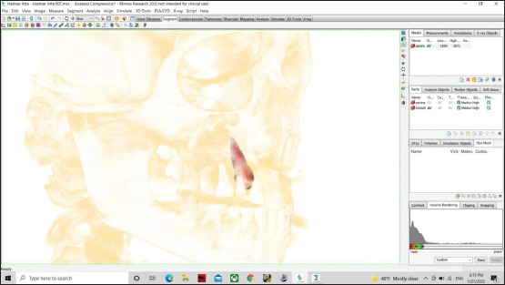
Figure 4.

Measuring the volume of the Lateral incisor in the impacted side using MIMICS
Figure 3.
Using the volume rendering to show the 3D impacted canine tooth model
Automatic-segmentation of the maxillary sinuses (200 MS, F = 122, M = 78) was also done to see if there is a difference in its volume in the impacted and non-impacted sides. First, we used the dynamic region grow (which allows growing a mask from a selected point without having to threshold) in MIMICS, followed by some steps until we obtained accurate borders of the maxillary sinuses as shown in Figure 5. The Materialise interactive medical image control system (MIMICS) tool was used to determine the volume of the sinus models after the three-dimensional sinus model was finished. Since manual segmentation takes a lot of time and requires a lot of operator skill, it is not a procedure that can be done in a routine clinical setting. Manual threshold selection is still needed for semi-automatic segmentation algorithms. Furthermore, the manual changes of segmented structures take a lot of time and could result in operator errors.[11]
Figure 5.

The three-dimensional Maxillary sinus model via MIMICS from CBCT
Virtual Grid was founded in 2003 and developed VR Mesh, an advanced 3D point cloud, and mesh processing software tool. The MIMICS software was used to convert the DICOM file into stereo lithographic or standard tessellation language (STL), because the DICOM files are not recognized by the VR Mesh application as in Figure 6. V R Mesh covers point cloud classification, feature extraction, and point cloud meshing. In the present study, VR Mesh software was being used to calculate the tooth and sinus volumes; the development of this software was carried out by an American company (Bellevue, Washington), and it is used in many fields involving dentistry as in Figure 7.
Figure 6.

Converting DICOM files into STL using MIMICS
Figure 7.

Measuring sinus 3D models volume using VR mesh
Results
The male represents (39%) (of 39 patients) while female patients signify (61%) (of 61 patients), the mean age for male patients was 16.30 ± 2.27 years while for female patients 15.23 ± 1.33 years, 47 (47%) of unilateral palatally impacted canine in the left side of the patient while 53 (53%) in the right side as shown in [Tables 1 and 2].
Table 1.
Descriptive statistics of right- and left-impacted maxillary canine
| Side | n (%). | Impacted canine |
P | ||||||
|---|---|---|---|---|---|---|---|---|---|
| Age | Mean | SE | SD | Minimum | Median | Maximum | |||
| Left side | 47 (47%) | 20.000 | 15.000 | 14.00 | 1.486 | 0.217 | 15.553 | >0.05 | |
| Right side | 53 (53%) | 25.000 | 16.000 | 14.00 | 2.368 | 0.325 | 16.170 | ||
Table 2.
Representing the gender and age of the patients
| Parameters | Male No. (%) | Female No. (%) | P |
|---|---|---|---|
| Gender | 39 (39) | 61 (61) | P<0.01 |
| Age (year) (Mean ± SD) | 16.30±2.27 | 15.23±1.33 | P<0.01 |
Regarding the ADAHMLI and the AARHMLI, there were no statistically significant differences for both males and females (p > 0.05) in the impacted side with a Mean ± SD (M: 26.456 ± 1.067, F: 26.523 ± 1.127) and (M: 21.687 ± 0.950, F: 21.703 ± 0.970). But in the non-impacted side, ADAHMLI (M: 26.630 ± 1.093, F: 26.697 ± 1.151) and the AARHMLI (M: 21.830 ± 0.951, F: 21.857 ± 0.970) showed no statistical significance (p > 0.05) between male and female patients as shown in [Table 3].
Table 3.
Comparing ADAHMLI and AARHMLI between male and female patients
| Sex | Mean | SE | SD | Minimum | Median | Maximum | P | |
|---|---|---|---|---|---|---|---|---|
| Impacted | ||||||||
| ADAHMLI | Male | 26.456 | 0.107 | 1.067 | 24.600 | 26.300 | 29.100 | >0.05 |
| female | 26.523 | 0.144 | 1.127 | 24.600 | 26.400 | 29.100 | >0.05 | |
| AARHMLI | Male | 21.687 | 0.0950 | 0.950 | 20.000 | 21.700 | 24.000 | >0.05 |
| Female | 21.703 | 0.124 | 0.970 | 20.000 | 21.900 | 24.000 | ||
| Non impacted | ||||||||
| ADAHMLI | Male | 26.630 | 0.110 | 1.093 | 24.300 | 26.450 | 29.300 | >0.05 |
| Female | 26.697 | 0.149 | 1.151 | 24.300 | 26.550 | 29.300 | ||
| AARHMLI | Male | 21.830 | 0.0951 | 0.951 | 20.100 | 21.900 | 24.100 | >0.05 |
| Female | 21.857 | 0.124 | 0.970 | 20.100 | 22.000 | 24.100 | ||
There were no significant differences in canine and lateral incisor volumes between male and female patients in the impacted side at P > 0.05, while the maxillary sinus volumes revealed a statistical difference (P = 0.022) between male (12642 ± 810) and female (12481 ± 650) patients in the impacted side. While in the non-impacted side, there was a statistical difference in the volume of lateral incisors and maxillary sinuses P = (0.003), (0.001), respectively, between male and female patients as shown in [Table 4].
Table 4.
Comparing the canine, lateral incisors, and maxillary sinus volume between male and female patients
| Sex | Mean | SE | SD | Minimum | Median | Maximum | P | |
|---|---|---|---|---|---|---|---|---|
| Impacted | ||||||||
| Canine volume | Male | 537.12 | 4.54 | 45.42 | 460.85 | 534.39 | 665.66 | >0.05 |
| Female | 538.63 | 5.14 | 40.16 | 467.61 | 540.20 | 611.20 | ||
| Lateral incisor volume | Male | 312.44 | 2.33 | 23.28 | 244.12 | 309.35 | 404.09 | >0.05 |
| Female | 314.23 | 2.46 | 19.19 | 286.16 | 310.20 | 385.09 | ||
| Maxillary sinus volume | Male | 12642 | 81.4 | 810 | 11029 | 12477 | 15212 | 0.022 |
| Female | 12481 | 83.3 | 650 | 11393 | 12289 | 14198 | ||
| Non impacted | ||||||||
| Canine volume | Male | 542.32 | 4.61 | 45.84 | 465.72 | 542.73 | 660.61 | >0.05 |
| Female | 543.47 | 5.27 | 40.83 | 472.30 | 545.71 | 615.70 | ||
| Lateral incisor volume | Male | 363.81 | 3.94 | 39.41 | 282.14 | 366.30 | 578.20 | 0.003 |
| Female | 372.35 | 5.57 | 43.46 | 297.35 | 370.50 | 578.20 | ||
| Maxillary sinus volume | Male | 12914 | 79.4 | 790 | 11393 | 12743 | 15452 | 0.001 |
| Female | 12698 | 82.7 | 646 | 11656 | 12487 | 14422 | ||
When comparing the canine volume in impacted and non-impacted sides for male patients (P > 0.05), there was no significant difference, while it showed a statistically significant difference for the lateral incisors volume (P = 0.002), maxillary sinuses volume (P = 0.002) in impacted and non-impacted sides. For the female patients, the canine and maxillary sinus volumes showed no statistical significance (P > 0.05) between the impacted and non-impacted sides. The P value was (0.003) for the lateral incisors volume in the female patients, which means there was a statistical difference as shown in [Table 5].
Table 5.
Comparing the canine, lateral incisors, and maxillary sinus volume between impacted and non-impacted sides for male and female patients separately
| Sex variables | Impacted/non-impacted | Mean | SE | SD | Minimum | Median | Maximum | P |
|---|---|---|---|---|---|---|---|---|
| Male | Impacted | 537.12 | 4.54 | 45.42 | 460.85 | 534.39 | 665.66 | >0.05 |
| Canine volume | Non-impacted | 542.32 | 4.61 | 45.84 | 465.72 | 542.73 | 660.61 | |
| Male | Impacted | 312.44 | 2.33 | 23.28 | 244.12 | 309.35 | 404.09 | 0.002 |
| Lateral incisor volume | Non-impacted | 363.81 | 3.94 | 39.41 | 282.14 | 366.30 | 578.20 | |
| Male | Impacted | 12642 | 81.4 | 810 | 11029 | 12477 | 15212 | 0.003 |
| Maxillary sinus volume | Non-impacted | 12914 | 79.4 | 790 | 11393 | 12743 | 15452 | |
| Female | Impacted | 538.63 | 5.14 | 40.16 | 467.61 | 540.20 | 611.20 | >0.05 |
| Canine volume | Non-impacted | 543.47 | 5.27 | 40.83 | 472.30 | 545.71 | 615.70 | |
| Female | Impacted | 314.23 | 2.46 | 19.19 | 286.16 | 310.20 | 385.09 | 0.003 |
| Lateral incisor volume | Non-impacted | 372.35 | 5.57 | 43.46 | 297.35 | 370.50 | 578.20 | |
| Female | Impacted | 12481 | 83.3 | 650 | 11393 | 12289 | 14198 | >0.05 |
| Maxillary sinus volume | Non-impacted | 12698 | 82.7 | 646 | 11656 | 12487 | 14422 |
The correlation coefficient among parameters (AARHMLI, ADAHMLI, the canine, the lateral incisors, and the maxillary sinuses volume) using Spearman's correlation coefficient as shown in [Tables 6-9]. A strong positive correlation was found between canine and lateral incisor volume for male patients in the non-impacted (r = 0.420**, P = 0.008) and impacted sides (r = 0.334*, P = 0.038). Also, a positive correlation was found between ADAHMLI and lateral incisor volume for female patients in the impacted side (r = 0.296*, P = 0.021) as shown in [Table 10].
Table 6.
Spearman's correlation coefficient among these parameters (ADAHMLI, AARHMLI, the canine and lateral incisors, and the maxillary sinuses volume) in non-impacted male
| ADAHMLI (non-impacted side) | AARHMLI (non-impacted) | (Non-impacted) canine volume | Lateral incisor volume (non-impacted) | Maxillary sinus volume (non-impacted) | |
|---|---|---|---|---|---|
| ADAHMLI (non-impacted side) | |||||
| r | 1.000 | −0.273 | −0.115 | 0.085 | 0.077 |
| P | 0.093 | 0.488 | 0.608 | 0.639 | |
| n | 39 | 39 | 39 | 39 | 39 |
| AARHMLI (non-impacted | |||||
| r | −0.273 | 1.000 | 0.076 | −0.232 | 0.109 |
| P | 0.093 | 0.646 | 0.155 | 0.509 | |
| n | 39 | 39 | 39 | 39 | 39 |
| (Non-impacted) canine volume | |||||
| r | −0.115 | 0.076 | 1.000 | 0.420** | −0.149 |
| P | 0.488 | 0.646 | 0.008 | 0.365 | |
| n | 39 | 39 | 39 | 39 | 39 |
| Lateral incisor volume (non-impacted) | |||||
| r | 0.085 | −0.232 | 0.420** | 1.000 | −0.073 |
| P | 0.608 | 0.155 | 0.008 | 0.658 | |
| n | 39 | 39 | 39 | 39 | 39 |
| Maxillary sinus volume (non-impacted) | |||||
| r | 0.077 | 0.109 | −0.149 | −0.073 | 1.000 |
| P | 0.639 | 0.509 | 0.365 | 0.658 | |
| n | 39 | 39 | 39 | 39 | 39 |
Table 9.
Spearman's correlation coefficient among these parameters (ADAHMLI, AARHMLI, the canine, and lateral incisors, and the maxillary sinuses volume) in impacted females
| ADAHMLI (impacted) | AARHMLI (impacted) | (Impacted) canine volume | Lateral incisor volume (impacted) | Maxillary sinus volume (impacted) | |
|---|---|---|---|---|---|
| ADAHMLI (impacted) | |||||
| r | 1.000 | −0.065 | −0.006 | 0.296* | 0.051 |
| P | 0.617 | 0.964 | 0.021 | 0.696 | |
| n | 61 | 61 | 61 | 61 | 61 |
| AARHMLI (impacted) | |||||
| r | −0.065 | 1.000 | 0.040 | 0.133 | −0.038 |
| P | 0.617 | 0.762 | 0.306 | 0.771 | |
| n | 61 | 61 | 61 | 61 | 61 |
| (Impacted) canine volume | |||||
| r | −0.006 | 0.040 | 1.000 | −0.212 | −0.004 |
| P | 0.964 | 0.762 | 0.101 | 0.975 | |
| n | 61 | 61 | 61 | 61 | 61 |
| Lateral incisor volume (impacted) | |||||
| r | 0.296* | 0.133 | −0.212 | 1.000 | −0.109 |
| P | 0.021 | 0.306 | 0.101 | 0.404 | |
| n | 61 | 61 | 61 | 61 | 61 |
| Maxillary sinus volume (impacted) | |||||
| r | 0.051 | −0.038 | −0.004 | −0.109 | 1.000 |
| P | 0.696 | 0.771 | 0.975 | 0.404 | |
| n | 61 | 61 | 61 | 61 | 61 |
Table 10.
Positive Spearman's correlation coefficient was found between these parameters
| Parameter A | Parameter B | R | P |
|---|---|---|---|
| (Non-impacted) canine volume/male | Lateral incisor volume (non-impacted)/male | 0.420** | 0.008 |
| AARHMLI (non-impacted)/female | Lateral incisor volume (non-impacted)/female | 0.319* | 0.012 |
| (Impacted) canine volume/male | Lateral incisor volume (impacted)/male | 0.334* | 0.038 |
| ADAHMLI (impacted)/female | Lateral incisor volume (impacted)/female | 0.296* | 0.021 |
Table 7.
Spearman's correlation coefficient among these parameters (ADAHMLI, AARHMLI, the canine and the lateral incisors, and the maxillary sinuses volume) in non-impacted female
| ADAHMLI (non-impacted side) | AARHMLI (non-impacted) | (Non-impacted) canine volume | Lateral incisor volume (non-impacted) | Maxillary sinus volume (non-impacted) | |
|---|---|---|---|---|---|
| ADAHMLI (non-impacted side) | |||||
| r | 1.000 | −0.050 | −0.021 | 0.185 | 0.066 |
| P | 0.701 | 0.870 | 0.154 | 0.612 | |
| n | 61 | 61 | 61 | 61 | 61 |
| AARHMLI (non-impacted) | |||||
| r | −0.050 | 1.000 | 0.035 | 0.319* | −0.033 |
| P | 0.701 | 0.792 | 0.012 | 0.803 | |
| n | 61 | 61 | 61 | 61 | 61 |
| (Non-impacted) canine volume | |||||
| r | −0.021 | 0.035 | 1.000 | 0.011 | 0.006 |
| P | 0.870 | 0.792 | 0.933 | 0.965 | |
| n | 61 | 61 | 61 | 61 | 61 |
| Lateral incisor volume (non-impacted | |||||
| r | 0.185 | 0.319* | 0.011 | 1.000 | −0.140 |
| P | 0.154 | 0.012 | 0.933 | 0.283 | |
| n | 61 | 61 | 61 | 61 | 61 |
| Maxillary sinus volume (non-impacted) | |||||
| r | 0.066 | −0.033 | 0.006 | −0.140 | 1.000 |
| P | 0.612 | 0.803 | 0.965 | 0.283 | |
| n | 61 | 61 | 61 | 61 | 61 |
Table 8.
Spearman's correlation coefficient among these parameters (ADAHMLI, AARHMLI, the canine, and lateral incisors, and the maxillary sinuses volume) in impacted males
| ADAHMLI (impacted) | AARHMLI (impacted) | (Impacted) canine volume | Lateral incisor volume (impacted) | Maxillary sinus volume (impacted) | |
|---|---|---|---|---|---|
| ADAHMLI (impacted) | |||||
| r | 1.000 | −0.265 | −0.079 | 0.129 | 0.075 |
| P | 0.102 | 0.633 | 0.435 | 0.649 | |
| n | 39 | 39 | 39 | 39 | 39 |
| AARHMLI (impacted) | |||||
| r | −0.265 | 1.000 | 0.044 | −0.093 | 0.092 |
| P | 0.102 | 0.791 | 0.572 | 0.580 | |
| n | 39 | 39 | 39 | 39 | 39 |
| (Impacted) canine volume | |||||
| r | −0.079 | 0.044 | 1.000 | 0.334* | −0.060 |
| P | 0.633 | 0.791 | 0.038 | 0.716 | |
| n | 39 | 39 | 39 | 39 | 39 |
| Lateral incisor volume (impacted) | |||||
| r | 0.129 | −0.093 | 0.334* | 1.000 | 0.059 |
| P | 0.435 | 0.572 | 0.038 | 0.723 | |
| n | 39 | 39 | 39 | 39 | 39 |
| Maxillary sinus volume (impacted) | |||||
| r | 0.075 | 0.092 | −0.060 | 0.059 | 1.000 |
| P | 0.649 | 0.580 | 0.716 | 0.723 | |
| n | 39 | 39 | 39 | 39 | 39 |
The calibration of the inter-examiner between the two observers revealed a statistically no significant difference as shown in [Tables 11 and 12].
Table 11.
Test of normality comparing between the first and after a week of reading among these parameters (the canine, lateral incisors, and the maxillary sinuses volume) in the impacted and non-impacted sides
| Kolmogorov-Smirnova |
Shapiro-Wilk |
|||||
|---|---|---|---|---|---|---|
| Statistic | df | Sig. | Statistic | df | Sig. | |
| (Impacted) canine volume | 0.067 | 99 | 0.200* | 0.971 | 99 | 0.030 |
| Impacted canine after week | 0.067 | 99 | 0.200* | 0.971 | 99 | 0.030 |
| (Non-impacted) canine volume | 0.081 | 99 | 0.110 | 0.974 | 99 | 0.045 |
| (Non-impacted) canine volume after a week | 0.081 | 99 | 0.110 | 0.974 | 99 | 0.045 |
| Lateral incisor volume (impacted) | 0.157 | 99 | 0.000 | 0.905 | 99 | 0.000 |
| Lateral incisor volume (impacted) after a week | 0.157 | 99 | 0.000 | 0.905 | 99 | 0.000 |
| Lateral incisor volume (non-impacted) | 0.204 | 99 | 0.000 | 0.698 | 99 | 0.000 |
| Lateral incisor volume (non-impacted) after a week | 0.203 | 99 | 0.000 | 0.698 | 99 | 0.000 |
| Maxillary sinus volume (non-impacted) | 0.103 | 99 | 0.012 | 0.952 | 99 | 0.001 |
| Maxillary sinus volume (non-impacted) after a week | 0.103 | 99 | 0.012 | 0.952 | 99 | 0.001 |
| Maxillary sinus volume (impacted) | 0.522 | 99 | 0.000 | 0.079 | 99 | 0.000 |
| Maxillary sinus volume (impacted) after a week | 0.522 | 99 | 0.000 | 0.079 | 99 | 0.000 |
Table 12.
Descriptive statistics between the first and after a week of reading among these parameters (the canine, lateral incisors, and the maxillary sinuses volume) in the impacted and non-impacted sides
| Mean | n | Std. Deviation | Std. Error Mean | |
|---|---|---|---|---|
| Pair 1 | ||||
| (Impacted) canine volume | 536.9105 | 100 | 45.12070 | 4.51207 |
| Impacted canine after a week | 536.9229 | 100 | 45.11895 | 4.51189 |
| Pair 2 | ||||
| (Non-impacted) canine volume | 542.5497 | 100 | 45.66561 | 4.56656 |
| (Non-impacted) canine volume after a week | 542.5516 | 100 | 45.66868 | 4.56687 |
| Pair 3 | ||||
| Lateral incisor volume (impacted) | 312.4374 | 100 | 23.28548 | 2.32855 |
| Lateral incisor volume (impacted) after a week | 312.4401 | 100 | 23.28524 | 2.32852 |
| Pair 4 | ||||
| Lateral incisor volume (non-impacted) | 363.8054 | 100 | 39.40814 | 3.94081 |
| Lateral incisor volume (non-impacted) after a week | 363.8090 | 100 | 39.41099 | 3.94110 |
| Pair 5 | ||||
| Maxillary sinus volume (non-impacted) | 12913.9639 | 100 | 790.23589 | 79.42170 |
| Maxillary sinus volume (non-impacted) after a week | 12913.9695 | 100 | 790.23592 | 79.42170 |
| Pair 6 | ||||
| Maxillary sinus volume (impacted) | 12649.8578 | 100 | 809.22079 | 80.92208 |
| Maxillary sinus volume (impacted) after a week | 12649.8636 | 100 | 809.22052 | 80.92205 |
When the tooth and sinus volumes were calculated using the VR Mesh program, it was discovered that, with the exception of a few tiny decimals, they nearly had the same value.
Discussion
In the current study, 1:1.5 male-to-female ratios of palatally impacted canines coincide with the 1:2 male-to-female ratios (out of 1333 cases) reported by Moreira Telmo et al.[12] The prevalence of maxillary canine impaction in (x) population was (6.78%), and in terms of the investigated sample's gender, the number of impacted canines was higher in females than in males and also reported that the impaction occurred more unilaterally than on both sides that agrees with our findings.[13] The intensity and direction (positive or negative) of a relationship between two variables can be summarized using Spearman's rank correlation coefficient. The answer will always be between 1.0 (a perfect positive correlation) and −1.0 (a perfect negative correlation). Spearman's correlation coefficient was used instead of Pearman's correlation coefficient, because most of the parameters included in our study were not normally disturbed. A strong correlation was found between canine and lateral incisor volume in the impacted side, which coincides with the correlation that was done[14], and they found that the lateral incisor volume, buccolingual and mesiodistal width of the lateral incisor crown, root and the total lateral incisor length, the angulation of the lateral incisor to the midline, and the axis of neighboring canine were considered to be strong predictors for the impaction of the maxillary canine.
The findings of this study indicate favor for smaller volumes and shorter root lengths of maxillary lateral incisors in palatally displaced canines since it is able to have a significant local impact.[15] These findings support the guidance theory by showing that maxillary canine eruption is hampered by smaller maxillary lateral incisors, and it may be concluded that individuals who have smaller maxillary lateral incisor crowns, roots, and volumes are more susceptible to a deviated eruption of the maxillary canine.[14]) For the maxillary first premolars, canines, and central incisors, except for the length of the first premolar, there were no statistically significant differences in root lengths or volumes among the three groups,[15] which agree with our results in that there was no significant difference in the canine volume in the (impacted/non-impacted sides) for male and female patients.
CBCT is an effective and noninvasive diagnostic imaging modality in clinical dentistry due to its limited radiation exposure and ability of producing highly accurate life-size images. The constant overestimation of the volume may be explained by a number of factors that have an impact on the images produced by the used CBCT scanning. The voxel size, the field of view (FOV), scattered x-rays, and subjectivity in the segmentation process can be recognized as some of them.[16] It was determined that the CBCT is strong and accurate for measuring the tooth volume in vivo and was statistically comparable to measurements made in vitro.[17] Additionally, the manual segmentation steps have a certain amount of subjectivity built into them that can continuously skew the results in favor of a decrease or an increase in volume.[16] In conclusion, CBCT can be used to accurately reconstruct 3D tooth models for linear, volumetric, and geometric measurements.[18]
The volume of teeth depends on the threshold interval and segmentation methods, which are variable for each software as well as the operator.[19] Mimics software has more options for segmentation and has a slight learning curve.[19] In the present study, the segmentation was done semi-automatically and picked with the whole tooth 3D volume examination rather than merely dental roots that are apical to the cementoenamel junction (CEJ), because it is regarded as being more definitive.[20]
The purpose of the current study was to compare the dentoalveolar and skeletal dimensions of the maxillae in a sample with unilaterally palatally impacted canines to the contralateral normal erupted side in order to assess the effectiveness of CBCT in volume measured using the (MIMICS) program and VR Mesh. Volumetric measurements can be made using a variety of computer applications, including Mimics, Dolphin, and ITK-Snap, among others. No previous studies measured the volume of the maxillary sinus in patients with unilateral palatally maxillary impacted canines using VR Mesh. The maxillary canine eruption occurs between 11 and 12 years of age, while the maxillary sinus reaches its maximum size between 12 and 15 years of age and it does not significantly increase in its volume after 12 years of age.[21] Therefore, in the current study, the maxillary sinus volume was evaluated in patients aged above 12 years with unilateral palatally maxillary canine impactions. They discovered that male patients had the considerably greater mean right, left, and total maxillary sinus volumes than female patients. The fact that the males have typically a larger physical form than the females in most dimensions can be ascribed to the disparity in maxillary sinus volumes between the sexes.[22] In contrast, the present study found that thesinus volume was greater in males than in females. Additionally, the results will not be altered by the existence of an impacted canine on either the right or left side because prior research found no discernible difference in the volumes of the maxillary sinuses on the right and left sides.[23] Dental impaction may be connected to a decrease in the maxillary sinus volume.[23] The maxillary sinus volumes increased statistically significantly in the non-impaction sides 12770.71 ± 1621.24 mm3 compared to the impaction side 11377.17 ± 1386.87 mm3,[6] which was similar to our results. In the current study, smaller maxillary sinus volume was observed on the impacted canine side than on the opposing side. This result agreed with that of Zeynep et al.[24] who stated that when the canine was impacted on the right side, the volume of the right maxillary sinus was substantially lower than that of the left side. On the other hand, Kalabalık and Tarım Ertas[23] observed no discernible variation between the right and left sides, leading them to conclude that unilaterally impacted teeth have no effect on the maxillary sinus volume. Zeynep A et al.[24] reported that a deeply impacted canine has an impact on maxillary sinus volume by measuring the distance from the canine cusp tip to the palatal plane in their research.
The results of the current study show that non-significant differences when the AARHMLI and ADAHMLI were measured (p > 0.05), which agrees with the D'Oleo-Aracena et al. study;[25] in that there were statistically negligible variations between the impacted and non-impacted sides in all of these dimensions. In the study of Tadinada et al.,[26] however, the results were different; the impacted side's alveolar bone dimensions (from the nasal floor to the alveolar ridge) were substantially smaller than those on the non-impacted side. In this study, measurements were done in the canine region depending on the fact that incisor eruption occurs before canines, so we believe that the height of the incisors should not be affected. VR Mesh Reverse, which was used in the current study, is the unique technology that we have that helps you convert large point clouds to meshes quickly and accurately. And a comprehensive toolset is provided, including point cloud cleanup, mesh repair/editing, registration, inspection, and measurement.
Outlining the precise location of the impacted canine in three dimensions is critical in the planning of the treatment and offers good assessment-making that may permit uninterrupted access or traction and less time-consuming treatment and less invasive.[27] Furthermore, the treating orthodontist was able to quickly reposition the impacted canine into normal occlusion due to the precise 3D image of the affected tooth.[27]
The number of samples with a unilaterally impacted maxillary canine was relatively low, which makes it difficult to accurately quantify prevalence without using many samples. We think that more enquiries with bigger sample numbers are necessary to define the relationship between impacted canine and maxillary sinus volume in the general population.
Conclusion
The results of the current study show non-significant differences in the AARHMLI and ADAHMLI measured (p > 0.05). There is no significant difference in the canine volume in the (impacted/non-impacted side), but it showed a statistically significant difference for the Lateral incisor volume in the impacted and non-impacted side for male and female patients. The Maxillary sinuses volume revealed statistical differences between males and females in impacted and non-impacted sides.
Ethical approval
Dear Dr, On behalf of Research Ethics Committee, I can confirm that the committee has reviewed the above project and permission to commence has been granted on the basis described in the application form as well as on the understanding that the study is conducted in accordance with the research guidelines followed in the University of Mosul/College of Dentistry. REC reference no. is Uom.Dent/H.48/22.
Financial support and sponsorship
Nil.
Conflicts of interest
There are no conflicts of interest.
Acknowledgements
I would like to thank the kindest and most gracious Allah for giving me the strength, serenity, faith, and disposition to complete this study. Special thanks to everyone who supported and helped me in any way during the preparation of this research.
References
- 1.Ericson S, Kurol J. Radiographic assessment of maxillary canine eruption in children with clinical signs of eruption disturbance. Eur J Orthod. 1986;8:133–40. doi: 10.1093/ejo/8.3.133. [DOI] [PubMed] [Google Scholar]
- 2.Litsas G, Acar A. A review of early displaced maxillary canines: Etiology, diagnosis and interceptive treatment. Open Dent J. 2011:39–47. doi: 10.2174/1874210601105010039. [DOI] [PMC free article] [PubMed] [Google Scholar]
- 3.Becker A. 2nd. United Kingdom: Informa Healthcare; 2007. The Orthodontic Treatment of Impacted Teeth. [Google Scholar]
- 4.Endo T, Abe R, Kuroki H, Kojima K, Oka K, Shimooka S. Cephalometric evaluation of maxillary sinus sizes in different malocclusion classes. Odontology. 2010;98:65–72. doi: 10.1007/s10266-009-0108-5. [DOI] [PubMed] [Google Scholar]
- 5.Oishi S, Ishida Y, Matsumura T, Kita S, Sakaguchi-Kuma T, Imamura T. A cone-beam computed tomographic assessment of the proximity of the maxillary canine and posterior teeth to the maxillary sinus floor: Lessons from 4778 roots. Am J Orthod Dentofacial Orthop. 2020;157:792–802. doi: 10.1016/j.ajodo.2019.06.018. [DOI] [PubMed] [Google Scholar]
- 6.Elmarhoumy SM, Safwat W, Ellaithy M. Cone-beam computed tomography evaluation of maxillary sinus volume in patients with unilaterally maxillary impacted canines. Egypt Dent J. 2022;68:1165–70. [Google Scholar]
- 7.Chalakkal P, Thomas AM, Chopra S. Displacement, location, and angulation of unerupted permanent maxillary canines and absence of canine bulge in children. Am J Orthod Dentofacial Orthop. 2011;139:34–50. doi: 10.1016/j.ajodo.2009.03.044. [DOI] [PubMed] [Google Scholar]
- 8.Mah JK, Alexandroni S. Cone-beam computed tomography in the management of impacted canines. Semin Orthod. 2010;16:199–204. [Google Scholar]
- 9.Scarfe WC, Farman AG, Sukovic P. Clinical applications of cone-beam computed tomography in dental practice. J Can Dent Assoc. 2006;72:75–80. [PubMed] [Google Scholar]
- 10.Zhang YQ, Yan XB, Meng Y, Zhao YN, Liu DG. Morphologic analysis of maxillary sinus floor and its correlation to molar roots using cone beam computed tomography. Chin J Dent Res. 2019;22:29–36. doi: 10.3290/j.cjdr.a41772. [DOI] [PubMed] [Google Scholar]
- 11.Cellina M, Gibelli D, Cappella A, Toluian T, Pittino CV, Carlo M, et al. Segmentation procedures for the assessment of paranasal sinuses volumes. Neuroradiol J. 2021;34:13–20. doi: 10.1177/1971400920946635. [DOI] [PMC free article] [PubMed] [Google Scholar]
- 12.Moreira T, Braga A, Ferreira A. Prevalence of palatally impacted canines. Int J Dent Sci Res [Google Scholar]
- 13.Al-Turaihi BA, Ali IH, Alhamadani GM, Alam MK. Patterns of maxillary canine impaction in Iraqi population. Pesqui Bras Odontopediatria Clín Integr. 2020;20:e5266. [Google Scholar]
- 14.Koral S, Arman Özçırpıcı A, Tunçer Nİ. Association between Impacted maxillary canines and adjacent lateral incisors: A retrospective study with cone beam computed tomography. Turk J Orthod. 2021;34:207–13. doi: 10.5152/TurkJOrthod.2021.20148. [DOI] [PMC free article] [PubMed] [Google Scholar]
- 15.Leonardi R, Muraglie S, Crimi S, Pirroni M, Musumeci G, Perrotta R. Morphology of palatally displaced canines and adjacent teeth, a 3-D evaluation from cone-beam computed tomographic images. BMC Oral Health. 2018;18:156. doi: 10.1186/s12903-018-0617-0. [DOI] [PMC free article] [PubMed] [Google Scholar]
- 16.Liu Y, Olszewski R, Alexandroni ES, Enciso R, Xu T, Mah JK. The validity of in vivo tooth volume determinations from cone-beam computed tomography. Angle Orthod. 2010;80:160–6. doi: 10.2319/121608-639.1. [DOI] [PMC free article] [PubMed] [Google Scholar]
- 17.Li W, Chen F, Zhang F, Ding W, Ye Q, Shi J, et al. Volumetric measurement of root resorption following molar mini-screw implant intrusion using cone beam computed tomography. PLoS One. 2013:e60962. doi: 10.1371/journal.pone.0060962. [DOI] [PMC free article] [PubMed] [Google Scholar]
- 18.Sang YH, Hu HC, Lu SH, Wu YW, Li WR, Tang ZH. Accuracy assessment of three-dimensional surface reconstructions of in vivo teeth from cone-beam computed tomography. Chin Med J (Engl) 2016;129:1464–70. doi: 10.4103/0366-6999.183430. [DOI] [PMC free article] [PubMed] [Google Scholar]
- 19.Nimbalkar S. Accuracy of volumetric analysis software packages in the assessment of tooth volume using CBCT. Loma Linda University Electronic Theses, Dissertations & Projects. 2016;400 [Google Scholar]
- 20.Forst D, Nijjar S, Flores-Mir C, Carey J, Secanell M, Lagravere M. Comparison of in vivo 3D cone-beam computed tomography tooth volume measurement protocols. Prog Orthod. 2014;15:69. doi: 10.1186/s40510-014-0069-2. [DOI] [PMC free article] [PubMed] [Google Scholar]
- 21.Saccucci M, Cipriani F, Carderi S, Di Carlo G, D-Attilio M, Rodolfino D. Gender assessment through three-dimensional analysis of maxillary sinuses by means of cone beam computed tomography. Eur Rev Med Pharmacol Sci. 2015;19:185–93. [PubMed] [Google Scholar]
- 22.Kalabalıkn F, Tarım Ertaş E. Investigation of maxillary sinus volume relationships with nasal septal deviation, concha bullosa, and impacted or missing teeth using cone-beam computed tomography. Jpn Soc Oral Maxillofac Radiol. 2018 doi: 10.1007/s11282-018-0360-x. [DOI] [PubMed] [Google Scholar]
- 23.Kalabalık F, Tarım Ertas E. Investigation of maxillary sinus volume relationships with nasal septal deviation, concha bullosa, and impacted or missing teeth using cone beam computed tomography. Oral Radiol. 2019;35:287–95. doi: 10.1007/s11282-018-0360-x. [DOI] [PubMed] [Google Scholar]
- 24.Zeynep A, Alper A, Hakan E, Palomo J. Maxillary sinus volume in patients with impacted canines. Angle Orthod. 2017;87:25–32. doi: 10.2319/122915-895.1. [DOI] [PMC free article] [PubMed] [Google Scholar]
- 25.D Oleo-Aracena MF, Arriola-Guillén LE, Rodríguez-Cárdenas YA, Ruíz-Mora GA. Skeletal and dentoalveolar bilateral dimensions in unilateral palatally impacted Canine using cone beam computed tomography. Prog Orthod. 2017;18:7. doi: 10.1186/s40510-017-0160-6. [DOI] [PMC free article] [PubMed] [Google Scholar]
- 26.Tadinada A, Mahdian M, Vishwanath M, Allareddy V, Upadhyay M, Yadav S. Evaluation of alveolar bone dimensions in unilateral palatally impacted canine: A cone beam computed tomographic analyses. Eur J Orthod. 2015;37:596–602. doi: 10.1093/ejo/cju098. [DOI] [PubMed] [Google Scholar]
- 27.Alqerban A, Jacobs R, van Keirsbilck PJ, Aly M, Swinnen S, Fieuws S, et al. The effect of using CBCT in the diagnosis of canine impaction and its impact on the orthodontic treatment outcome. J Orthod Sci. 2014;3:34–40. doi: 10.4103/2278-0203.132911. [DOI] [PMC free article] [PubMed] [Google Scholar]