

Abstract
Extractions are commonly used to alleviate moderate to severe crowding, retract protrusive incisors, or correct anteroposterior inconsistencies in the maxillomandibular area. The choice of which teeth to be extracted requires a thorough assessment of the dentition of the patient, taking into account treatment objectives, dental and periodontal properties as well as ease of mechanics with minimum iatrogenic effects. This case report discusses the successful treatment with myofunctional appliance of a growing patient with skeletal Class II malocclusion followed by fixed mechanotherapy involving atypical teeth extraction.
Keywords: Atypical extraction, skeletal malocclusion, transposition
Introduction
The primary goal of orthodontics as a discipline is to maintain the functional, structural, and esthetic integrity of teeth and their supporting structures. Every dental unit, according to Dr. Edward H. Angle, must provide optimal skeletal, soft tissue, and dental health, as well as facial esthetic harmony. Different malocclusions, asymmetries, transposed teeth complexities, midline anomalies, and extreme arch length differences, however, necessitate the sacrificing of certain dental units—mainly premolars, but also atypical teeth such as lower incisors or even the cornerstone canines.[1,2] While premolars are most commonly extracted in case of tooth material arch length discrepancy as it is usefully sited to relieve anterior crowding and to correct molar relationship, canines are rarely extracted.
The mechanical, esthetic, and practical aspects of canine extraction have been explored by Saa dé and O Ghougassian.[3] Canine rise in lateral excursive movements has received a lot of attention in the past. Thornton[4] discovered, however, that there is no scientific evidence for the practical superiority of one occlusal scheme over another. The evidence-based literature does not support the need to develop a canine-protected occlusion in orthodontic patients. As a result, group function or premolar guidance will safely replace canine guidance.
Orthodontic treatment aims to restore proper occlusion and facial aesthetics, while also preserving joint and periodontal stability, as well as the health of tooth support systems. However, in a number of situations, a multidisciplinary clinical approach is needed in order to achieve all objectives.[5]
The aim of this case report is to discuss a comprehensive treatment of a growing patient with skeletal Class II malocclusion along with transposition between the canine and lateral incisor in the lower left quadrant.
Case Report
Male patient 13 years old, sought orthodontic treatment with chief complaint of having forwardly positioned upper front teeth and irregularly placed lower front teeth.
The clinical examination revealed the patient in good general health. In the frontal view, the patient presented a symmetrical face, increased lower facial third dimension, dolicofacial pattern, and absent lip seal with increased incisor display at rest.
In the lateral view, he presented a convex profile, a recessive chin due to retro-positioned mandible, acute nasolabial angle, and obtuse cervico-mandibular angle. The smile was asymmetric due to the crowding and was non-consonant.
The functional pattern analysis evidenced mixed breathing, despite being predominantly oral, in addition to phonation and deglutition with anterior interposition of tongue. The tonsils, adenoids, and temporomandibular joint were normal, with hyperactivity of both upper and lower lips.
The intraoral examination revealed mild upper arch crowding, severe lower arch crowding, lack of adequate overbite, 8 mm of overjet and increased axial inclination of maxillary incisors. There was a Class I molar relationship on the left side, half unit Class II on the right side with Class I canines and the left lower quadrant tooth #32 was partially transposed with #33; which was totally blocked out of the arch buccally. [Figure 1]
Figure 1.
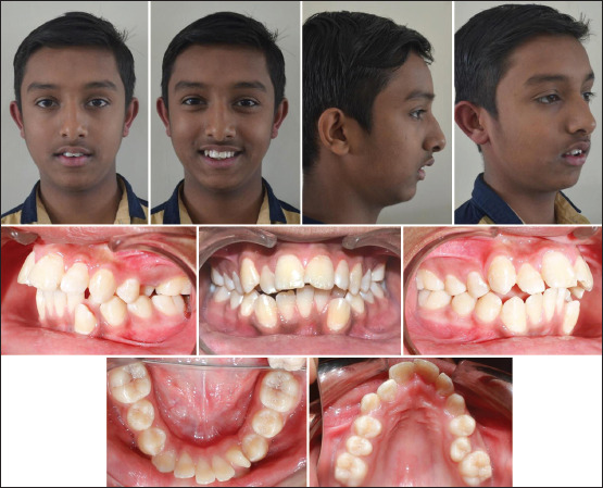
Pretreatment extra-oral and intra-oral photographs
The dental cast assessments revealed a tooth material excess in both maxillary and mandibular arches. The panoramic radiograph showed overlapping images of tooth #32 and #33. The trabeculae contour was normal and the lamina dura was intact for all teeth with developing third molars also evident.
The skeletal pattern was assessed by means of lateral cephalogram and revealed a combination of orthognathic maxilla and a retrognathic mandible thus evidencing a Class II skeletal pattern (SNA = 81°, SNB = 75°, and ANB = 6°), with facial vertical growth (SN.GoGn = 33° and Y-axis = 64°). The dental pattern revealed increased axial inclination and protrusion of maxillary incisors (1-NA = 7.5 mm and 39.5°). Lastly, a convex profile was found, with the upper lower lip positioned 7 mm forward relative to the S line [Figure 2].
Figure 2.

Pretreatment Lateral Cephalograms & Orthopantomogram (a & d), Mid-treatment Lateral Cephalogram (b) and Post-treatment Lateral Cephalograms & Orthopantomogram (c & e)
Treatment objectives
Address the chief complaint, i.e., to correct the inclination and positioning of upper anteriors and proper alignment of lower teeth.
Correction of increased overjet and reduced overbite.
Correct the molar relation on the right side and obtain proper canine relationships bilaterally.
Correct the skeletal Class II relation.
Improve the smile and soft tissue profile.
Treatment alternatives
With the list of objectives defined for this case, multiple treatment plans were taken into consideration:
To address the skeletal malocclusion with myofunctional appliance in phase 1 followed by extractions of all first premolars during phase 2, that is fixed mechanotherapy.
To address the skeletal malocclusion with myofunctional appliance in phase 1 followed by asymmetric extractions during phase 2, which would be tooth #14, #24, #33, and #44.
To treat this case by camouflaging the skeletal malocclusion via extractions and fixed mechanotherapy.
The second treatment plan was chosen for this particular case because we wanted to take advantage of the growth left in the patient to correct the skeletal disproportion as well as to correct the position of the teeth by fixed mechanotherapy. Extraction of tooth #33 would have aided us in creating simpler mechanics with minimal side effects and some anchorage preservation in the lower arch.
TREATMENT PLAN AND MECHANICS OF CHOICE: The orthodontic planning consisted of addressing the skeletal Class II by myofunctional appliance; twin block in this case, with an expansion screw so as to correct the posterior cross-bite that would develop after mandibular advancement. An occipital-pull headgear was used to control sagittal and vertical growth. Following the active and supporting stage of the twin block appliance, the patient underwent fixed mechanotherapy with MBT 0.022” X 0.028”. Based on a comprehensive space analysis, extraction of all first premolars was decided, but to correct the partial transposition and for the ease of mechanics, tooth #33 was chosen for extraction instead of #34. Following a phase of leveling and alignment, retraction of anterior teeth was carried out on 19 × 25 SS wires keeping maximum anchorage in both arches. The occipital headgear was modified to fit into the headgear tube and continued for anchorage preservation. Following space closure, Intermaxillary elastics were used for settling the occlusion.
Results
The treatment objectives were fulfilled, as shown by the assessment of results achieved after the orthodontic treatment was carried out over a period of 24 months. The facial profile improved due to the mandibular advancement and decreasing protrusion of the maxillary incisors. In terms of tooth positioning, there were significant uprighting and retraction of maxillary incisors, in addition to the correction of the anterior open bite. Proper occlusion was achieved from a functional perspective, with incisal guidance during protrusive movement of the mandible and disocclusion of canines on the right side (working occlusion), without balancing interference during lateral guidance. On the left side, group disocclusion was aimed due to tooth #34 replacing tooth #33. Therefore, functional occlusion and esthetic outcomes were achieved. Subsequently, the patient was subjected to a minor restorative procedure to develop the anatomy of the canine on the premolar in the third quadrant [Figures 3 and 4].
Figure 3.

Mid-treatment extra-oral and intra-oral photographs after treatment with twin block appliance
Figure 4.

Post-debond extra-oral and intra-oral photographs
The skeletal pattern assessment revealed, although the patient no longer presented facial growth, the ANB angle decreased to 3°. This occurred not just because of mandibular advancement but also due to retraction of incisors and considerable decrease in axial inclination [Table 1].
Table 1.
Pre-treatment, post functional appliance therapy and post debond Cephalometric analysis
| Parameter | Normal Value/Range | Pre-treatment | Post-functional | Post-treatment |
|---|---|---|---|---|
| SNA | 82° | 81° | 81° | 81° |
| SNB | 80° | 75° | 78° | 78° |
| ANB | 2° | 6° | 3° | 3° |
| Upper 1 to NA | 22°, 4 mm | 39.5°, 7.5 mm | 38.5°, 7 mm | 25°, 4.5 mm |
| Lower 1 to NB | 25°, 4 mm | 25°, 4.5 mm | 26°, 5 mm | 24°, 4 mm |
| Upper 1 to SN | 102±2° | 119° | 118 | 104° |
| IMPA | 90° | 92° | 94° | 92° |
| Inter-incisal Angle | 135° | 109° | 110° | 131° |
| Upper 1 to A-Pog | 2.7 mm, 1-5 mm | +10 mm | +9.5 mm | +3.5 mm |
| Lower 1 to A-Pog | 1+3 mm | +2 mm | +3 mm | +1 mm |
| SN-MP | 32° | 33° | 33° | 33° |
| PP-MP | 25° | 26° | 26° | 26° |
| Jaraback Ratio | 62-65% | 62% | 61% | 60% |
| Maxillary length | 80 mm | 80 mm | 80 mm | |
| Mandibular length | 95 mm | 96.5 mm | 96.5 mm | |
| LAFH | 59 mm | 60 mm | 61 mm | |
| Naso-labial Angle | 102±4° | 93° | 93° | 103° |
| N Perpendicular to point A | 0±3.7 mm | +3 mm | +3 mm | +3 mm |
| N Perpendicular to pogonion | 4-6 mm | -11 mm | -5 mm | -6 mm |
| Wits Appraisal | 0 mm | 4.3 mm | 0.5 mm | 0.5 mm |
| E Line | -2 mm | +3 mm | +3 mm | -1 mm |
IMPA: Incisor mandibular plane angle, SN-MP: SN-Mandibular plane angle, PP-MP: Palatal plane-mandibular plane angle, LAFH: Lower anterior face height
The total cephalometric superimposition revealed slight clockwise rotation of the mandible, very slight opening of mandibular plane, decrease in protrusion of maxillary and mandibular incisors, and improved lip position. The partial maxillary superimposition revealed mild extrusion and decreased protrusion of maxillary incisors, with a more significant palatal movement of the crown. The partial mandibular superimposition revealed mild extrusion and decreased protrusion of mandibular incisors, with less significant movement of the crown lingually, probably because it was a case of mandibular anterior crowding. By the end of fixed mechanotherapy, the patient had well-aligned arches, Angle's Class I molars, Class I canines, and proper overjet and overbite with significant improvement in the overall facial appearance [Figure 5].
Figure 5.

Treatment Superimposition (Black: Pre-treatment, Blue: Mid-treatment/Post-functional & Red: Post-treatment)
Discussion
The adult dentition is prone to asymmetrical and awry complications, necessitating unusual extractions and treatment preparation. The decision to extract should be made in order to achieve equilibrium between the upper and lower arches, with no deficiency or excess space remaining. Soft-tissue breakdown, which manifests clinically as gingival recession, is often associated with an ectopically placed tooth. When teeth are relocated orthodontically into a new location, they lose their attachment and more bone is lost.[2]
Although these extractions may seem to be incongruous, they were necessary for this case to achieve an acceptable esthetic and structural target. When done correctly, atypical extractions can produce very satisfactory results. Before preparing any extraction, it is important to consider the midline of the dentition, periodontal health of the teeth in question, mechanics, and overall benefit to the patient with minimum iatrogenic damage. No dental unit should be compromised unless the need for extraction is clearly justified.[6,7]
Whatever the treatment plan may be, the only goal that it should abide by should be to provide a stable occlusion along with a pleasing facial profile.
Treatment options are influenced by a number of factors, including the dental arch, affected teeth, crown and root position, degree of resorption, malocclusion, clinician experience, and patient motivation.[8,9] As for the patient reported herein, malocclusion hindered aesthetics which was impaired due to the excessively recessive mandible, incompetent lips, presence of anterior open bite, protruded maxillary incisors, and severe mandibular dental crowding.
In addition to the problems mentioned, the case was further complicated by the partial transposition present between tooth #32 and #33. In view of the treatment options available in the literature, alignment of transposed teeth would not be recommended. This is because the reported case presented with a partial transposition affecting the canine in labial version without enough space for alignment. Despite reaching less favorable outcomes when the order of teeth is not corrected, a number of clinicians opt for such orthodontic therapy, which is rendered simpler. They, thus, recommend correction of pseudo or incomplete transposition only so as to prevent root resorption, recession, and hard-to-control mechanics.[5] In the case reported herein, significant root resorption was absent at treatment completion, with only generalized rounding of root apices being found.
Additionally, the approach demands longer treatment time as well as meticulous torque and direction of force control, so as to move the transposed teeth while preserving the buccal bone cortex. Therefore, lack of space and unfavorable position of canine and preservation of anchorage in the lower arch were decisive in opting for extraction of one transposed tooth. Thiruvenkatachari[10] did a study to assess the esthetic perceptions of patient smiles among dental professionals and laypeople with respect to maxillary canine extractions. It was concluded that no statistically significant difference was found in the smile attractiveness between canine extraction and premolar extraction patients as assessed by general dentists, laypeople, and orthodontists.
A positive visual treatment objective, increased overjet, and patients's age were the key factors that propelled us to treat this case initially with a twin block appliance with an expansion screw; although certain factors like vertical growth pattern and lower anterior crowding were not the most favorable points. But the final decision was based on the idea of taking advantage of the growth that was left; even if we could obtain mild skeletal and dental changes, it would help in reducing the overjet and give some definition to the chin area.
Some amount of intrusion of the first premolar was also performed in order to achieve optimal marginal gingiva levels along with a conservative buildup of premolar. This was the treatment of choice aimed at enhancing aesthetics and function, in addition to allowing shorter treatment time.
Conclusion
When done correctly, atypical extractions can produce very satisfactory results. Factors like treatment time, ease of mechanics, avoidance of any iatrogenic damages as well as the periodontal health of the teeth in question are key points in determining which tooth to extract. As shown in this case report, however, satisfactory esthetic and functional outcomes can be achieved by doing atypical extractions like canine in selected cases where the benefits outweigh the risks.
Declaration of patient consent
The authors certify that they have obtained all appropriate patient consent forms. In the form, the legal guardian has given his consent for images and other clinical information to be reported in the journal. The guardian understands that names and initials will not be published and due efforts will be made to conceal identity, but anonymity cannot be guaranteed.
Financial support and sponsorship
Nil.
Conflicts of interest
There are no conflicts of interest.
References
- 1.Tayer BH. The asymmetric extraction decision. Angle Orthod. 1992;62:291–7. doi: 10.1043/0003-3219(1992)062<0291:TAED>2.0.CO;2. [DOI] [PubMed] [Google Scholar]
- 2.Kumar C, Garg K, Vaidik PK, Mangal A, Parmar P. The aberrant extraction of a maxillary canine and two lower incisors. Natl J Maxillofac Surg. 2018;9:86–9. doi: 10.4103/njms.NJMS_11_18. [DOI] [PMC free article] [PubMed] [Google Scholar]
- 3.Saadé Ces A, Ghougassian S. Canine extractions: Functional, esthetic, and practical considerations. J Clin Orthod. 2021;55:210301–10. [PubMed] [Google Scholar]
- 4.Thornton L. Anterior guidance: Group function/canine guidance: A literature review. J Prosthet Dent. 1990;64:479–82. doi: 10.1016/0022-3913(90)90048-h. [DOI] [PubMed] [Google Scholar]
- 5.Matsumoto MAN, Stuani MBS. Tooth transposition: A multidisciplinary approach. Dental Press J Orthod. 2018;23:97–107. doi: 10.1590/2177-6709.23.1.097-107.bbo. [DOI] [PMC free article] [PubMed] [Google Scholar]
- 6.Shastri D, Tandon P, Nagar A. Atypical extractions in adult treatment. J Clin Orthod. 2015;49:312–8. [PubMed] [Google Scholar]
- 7.Farret MM, Farret MM. Practical and esthetic considerations in adult canine-extraction treatment. J Clin Orthod. 2013;47:353–60. [PubMed] [Google Scholar]
- 8.Sarver DM, Ackerman MB. Dynamic smile visualization and quantification: Part 1. Evolution of the concept and dynamic records for smile capture. Am J Orthod Dentofacial Orthop. 2003;124:4–12. doi: 10.1016/s0889-5406(03)00306-8. [DOI] [PubMed] [Google Scholar]
- 9.Bahreman AA. Lower incisor extraction in orthodontic treatment. Am J Orthod. 1977;72:560–7. doi: 10.1016/0002-9416(77)90024-0. [DOI] [PubMed] [Google Scholar]
- 10.Thiruvenkatachari B, Javidi H, Griffiths SE, Shah AA, Sandler J. Extraction of maxillary canines: Esthetic perceptions of patient smiles among dental professionals and laypeople. Am J Orthod Dentofacial Orthop. 2017;152:509–15. doi: 10.1016/j.ajodo.2017.02.015. [DOI] [PubMed] [Google Scholar]