In adults but not children, small-bowel intussusception is usually traceable to a physical lesion. We report a causation that does not seem to have been described before.
CASE HISTORY
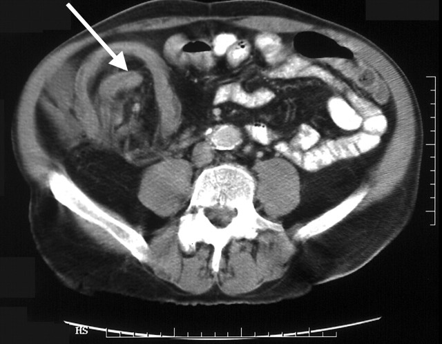
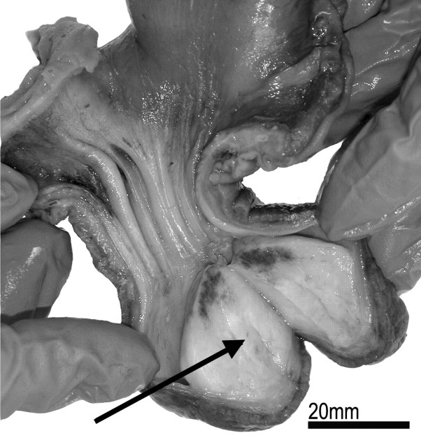
A man aged 28 came to hospital after 24 hours of profuse rectal bleeding (bright red blood) associated with colicky central abdominal pain, vomiting and diarrhoea. Over the preceding twelve months he had been troubled by intermittent crampy abdominal pains about which he had not sought medical advice. There was no medical or family history of note. On examination he was pale, sweaty and in obvious discomfort, and an ovoid mass 10 cm in maximum diameter was palpable in the peri-umbilical region. The abdomen was generally tender but clinical peritonism was absent. His haemoglobin was 7.5 g/dL. Plain radiographs of the chest and abdomen were unremarkable but CT revealed a complex mass, consistent with the palpable lesion, involving small bowel and proximal large bowel. Radiological features were suggestive of an ileocolic intussusception, although no definite lead-point or causal lesion was identifiable (Figure 1). At laparotomy the presence of an enterocolic intussusception was confirmed, and when it was reduced by gentle manual traction the lead-point was identified as a small jejunal lesion that had travelled via the ileocaecal valve into the ascending colon. A standard resection with primary anastomosis was performed to remove the abnormal and intussuscepted region of small bowel. Pathological examination of the resected specimen revealed an inverted Meckel's diverticulum that contained a lipomatous lesion about 30 mm in diameter, arising from its tip (Figure 2).
Figure 1.
CT of abdomen showing egg-in-cup sign
Figure 2.
Resected specimen showing Meckel's diverticulum with lipoma in tip
COMMENT
Intussusception is seen mainly in childhood, when it is idiopathic in 95% of cases. In the rare adult cases1 a causal lesion is identified in as many as 90%. Symptoms of intussusception in the adult are usually protracted, over weeks or months, and include abdominal pain, nausea, vomiting, rectal bleeding, altered bowel habit and weight loss. The paediatric triad of acute abdominal pain, a palpable sausage-shaped abdominal mass and ‘redcurrant jelly’ stools is seldom observed in adults though it can arise in acute-on-chronic cases such as we report here. On CT, the bowel-in-bowel or egg-in-cup sign is diagnostic, and occasionally the underlying lesion is identifiable. Usually this proves to be benign—a neoplasm, lymphoid hyperplasia, or an adhesion. Although an inverted Meckel's diverticulum as a causal lesion has been described several times,3-6 we have found no previous report of an association with a lipoma as the lead-point.
References
- 1.Azar T, Berger DL. Adult intussusception. Ann Surg 1997;226: 134-8 [DOI] [PMC free article] [PubMed] [Google Scholar]
- 2.Begos DG, Sandor A, Modlin IM. The diagnosis and management of adult intussusception. Am J Surg 1997;173: 88-94 [DOI] [PubMed] [Google Scholar]
- 3.James GK, Berean KW, Nagy AG, Owen DA. Inverted Meckel's diverticulum: an entity simulating an ileal polyp. Am J Gastroenterol 1998;93: 1554-5 [DOI] [PubMed] [Google Scholar]
- 4.Lu CL, Chen CY, Chiu ST, Chang FY, Lee SD. Adult intussuscepted Meckel's diverticulum presenting mainly lower gastrointestinal bleeding. J Gastroenterol Hepatol 2001;16: 478-80 [DOI] [PubMed] [Google Scholar]
- 5.Dujardin M, de Beeck BO, Osteaux M. Inverted Meckel's diverticulum as a leading point for ileoileal intussusception in an adult: case report. Abdom Imaging 2002;27: 563-5 [DOI] [PubMed] [Google Scholar]
- 6.Formejster I, Mattern R. Ileus due to ileo-cecal intussusception caused by lipoma coexistent with Meckel's diverticulum. Wiad Lek 1971;24: 1101. [PubMed] [Google Scholar]


