

Highlights
-
•
Accurate and rapid diagnosis of amyotrophic lateral sclerosis (ALS) is important to prevent erroneous interventions.
-
•
The recent Gold Coast criteria are easily applicable and have high sensitivity and specificity.
-
•
Future developments will help to distinguish ALS as a specific clinical-pathologic entity.
Keywords: Amyotrophic Lateral Sclerosis, Diagnostic criteria, Differential diagnosis, Electromyography, Motor neuron disease
Abstract
Accurate and rapid diagnosis of amyotrophic lateral sclerosis (ALS) is essential in order to provide accurate information for patient and family, to avoid time-consuming investigations and to permit an appropriate management plan. ALS is variable regarding presentation, disease progression, genetic profile and patient reaction to the diagnosis. It is obviously important to exclude treatable conditions but, in most patients, for experienced neurologists the diagnosis is clear-cut, depending on the presence of progressive upper and lower motor neuron signs. Patients with signs of restricted lower motor neuron (LMN) or upper motor neuron (UMN) dysfunction may present diagnostic difficulty, but electromyography (EMG) is often a determinant diagnostic test since it may exclude other disorders. Transcranial magnetic stimulation may aid detection of UMN dysfunction, and brain and spinal cord MRI, ultrasound and blood neurofilament measurements, have begun to have clinical impact, although none are themselves diagnostic tests. Several sets of diagnostic criteria have been proposed in the past; all rely on clinical LMN and UMN signs in different anatomic territories, EMG changes, exclusion of other disorders, and disease progression, in particular evidence of spreading to other anatomic territories. Fasciculations are a characteristic clinical feature and increased importance is now attached to fasciculation potentials detected by EMG, when associated with classical signs of denervation and reinnervation. The Gold Coast diagnostic criteria rely on the presence of UMN and LMN signs in one (or more) anatomic territory, or LMN signs in two (or more) anatomic territories, recognizing the fundamental clinical requirements of disease progression and exclusion of other diseases. Recent studies confirm a high sensitivity without loss of specificity using these Gold Coast criteria. In considering the diagnosis of ALS a critical question for future understanding is whether ALS should be considered a syndrome or a specific clinico-pathologic entity; this can only be addressed in the light of more complete knowledge.
1. Introduction
The group of neuromuscular diseases associated with progressive weakness caused by motor neuron degeneration are designated motor neuron diseases (MND). The most common and most severe of these is amyotrophic lateral sclerosis (ALS), characterized by rapid progression, respiratory involvement and short survival.
1.1. Diagnosis of ALS
Accurate and rapid diagnosis of ALS is relevant for many reasons. A definite diagnosis will relieve anxiety for the patient and their family associated with multiple, ultimately non-relevant investigations and persistent uncertainties about diagnosis and management of a disorder without effective therapy (Gwathmey et al., 2023). Diagnosis also allows initiation of earlier clinical management including possible entry into a clinical trial, and permits earlier life planning and adaptations, as well requesting the necessary healthy system support (Househam and Swash, 2000). Importantly, early definite diagnosis avoids unnecessary surgical intervention, which can itself accelerate disease progression (Pinto et al., 2014).
1.2. Spinal muscular atrophy syndromes
The diagnosis of ALS relies on the exclusion of mimic conditions that can create diagnostic uncertainty. Two such conditions are considered below: the different forms of Spinal Muscular Atrophy and Kennedy's disease.
The designation spinal muscular atrophy (SMA) is used to categorize a range of conditions characterized by spinal lower motor neuron degeneration, rarely involving bulbar motoneurons, causing symmetric weakness, frequently of juvenile-onset and related to genetic abnormalities in the SMN genes. The absence of UMN signs serves to distinguish SMA syndromes from ALS. Progression is slower in adult-onset SMA and respiratory muscles tend not to be affected (Kolb and Kissel, 2011).
SMA may present at birth or in infancy, in adolescence or in adulthood, tending to be most severe in infantile onset cases. Several specific SMA phenotypes have been recognized. Hirayama disease is a benign motor myelopathy characterized by asymmetric or unilateral upper limb weakness, of juvenile onset, with stabilization after initial progression over a few years. It is more common in Japan and other Asian countries (Hirayama, 2000). It has been associated with forward displacement of the posterior dural sac of the cervical spinal cord, due to instability of the posterior longitudinal ligament, causing traumatic or perhaps vascular degeneration of the anterior horns (Hirayama et al., 1987, Hirayama and Tokumaru, 2000).
Another relevant specific condition is Kennedy's disease (X-linked recessive bulbospinal neuronopathy) that affects men and is associated with a mutation in the androgen receptor gene. It is one of the uncommon neurological disorders caused by a CAG repeat expansion (polyglutamine diseases). Typically its onset is in the 5th decade, with slowly progressive bulbar involvement, an atrophic tongue, perioral fasciculation-myokymia, limb weakness and fasciculations, cramps, and postural hand tremor. It is frequently associated with testicular atrophy and gynecomastia. No clinical or neurophysiological sign of corticospinal tract lesion can be detected, and respiratory function is preserved. Sensory nerve action potentials (SNAPs) are of low amplitude or absent (sensory ganglionopathy), a finding that is a marker distinct from ALS. Kennedy's disease is considered a multisystem disorder with features of sexual dysfunction, dysmetabolism (dyslipidemia and diabetes), urinary tract symptoms and muscle fiber involvement (Breza and Koutsis, 2019).
In this chapter, we will provide a scheme for prompt diagnosis in suspected ALS.
2. Clinical presentation
2.1. Clinical features
Presentation of ALS is variable, and this can impose diagnostic difficulties. Most cases start with limb weakness (spinal-onset), but about 25–30 % of patients present with dysarthria, dysphagia or, more rarely, dysphonia and these are categorized as bulbar-onset (Masrori and Van Damme, 2020). The phenotypic presentation is influenced by the patients’ geographical origin, or perhaps their racial origin. The percentage of bulbar-onset patients is about 15 % in China (Chen et al., 2015) where it is strongly associated with sex and age. In Europe, bulbar-onset ALS is more frequent in elderly women (Masrori and Van Damme, 2020). About 3–4 % of ALS patients present with respiratory insufficiency. In general, this is a presentation found in older men, with marked weight loss; a syndrome with a poor prognosis in spite of prompt ventilatory support (Pinto et al., 2023). A small number of patients present with other, related symptoms, such as axial weakness (sometimes associated with drop-neck), fasciculations, cramps, emotional lability (“pathological” laughter and/or crying, provoked by a minor stimulus) and weight-loss. Cognitive changes are reported in about half of all ALS patients, but only 10–15 % show clinical signs of fronto-temporal dementia. Cognitive involvement is considered part of the disease spectrum (Lomen-Hoerth et al., 2002). In one study about 3 % of patients presented with cognitive changes, even without any motor symptoms, rapidly progressing to predominantly upper motor neuron (UMN) involvement of the bulbar region (Gromicho et al., 2021). These older patients have a poor prognosis and there is often a longer delay in achieving a definite diagnosis (Alves et al., 2023).
2.2. Phenotypes
Although ALS is recognized as a disease affecting the brain beyond the classical motor system (Masrori and Van Damme, 2020), since it can affect cognition, metabolism, and the autonomic nervous system, its most defining feature is muscular atrophy and weakness, progressive functional disability and fast progression. ALS affects both UMN and the lower motor neurons (LMN) and, for this reason, clinical diagnosis is typically supported by the presence of both UMN and LMN clinical signs. Some patients present with isolated LMN signs (progressive muscular atrophy, PMA) and about 20 % of these will develop overt UMN signs during disease progression (Kim et al., 2009). However, most patients with the PMA form of ALS remain with pure LMN signs, even though at autopsy there is often degeneration of the corticospinal tracts in the cord (Ince et al., 2003). This is perhaps consistent with the frequent clinical difficulty in assessing UMN signs in classic ALS when there is severe LMN muscular atrophy in affected limbs. PMA represents some 10 % of all ALS patients; it is more frequent in older men and shows slower progression and longer survival than typical ALS (Kim et al., 2009). PMA seems to be less common in China (Chen et al., 2015), but because it is associated with older age, it has become more commonly diagnosed in Western countries, as expected as the population ages. For example, it increased from 5.9 % to 17.4 % over a 27 year period in one center (Alves et al., 2023).
The absence of UMN signs often leads to diagnostic uncertainty at clinical presentation, due to the need to consider a number of clinically similar disorders, especially myopathies and motor neuropathies (Cortés-Vicente et al., 2017). However, the diagnostic delay experienced by patients with PMA is similar to typical spinal-onset ALS patients, in whom UMN signs may be difficult to elicit. Bulbar-onset patients have a shorter diagnostic delay than the other groups (Alves et al., 2023). In addition to the autopsy evidence of corticospinal tract degeneration in about 50 % of PMA patients (Ince et al., 2003), both conventional and paired-pulse stimulation transcranial magnetic stimulation (TMS) are abnormal in a large percentage of these patients, depending on the method applied (Triggs et al., 1999, Vucic and Kiernan, 2007, Menon et al., 2016). PMA does not differ from typical ALS regarding inevitable progression, leading to respiratory insufficiency and death. The reasons for the limitations of the clinical method in detecting UMN signs in ALS patients have been discussed elsewhere (Swash, 2012, Swash et al., 2020), but include destruction of the α- and γ-motor neuron and interneuron connexions, and spinal circuitry changes, in addition to dominant muscle atrophy.
Spinal-onset ALS is typically asymmetric, with weakness and wasting beginning insidiously, often in distal segments, associated with fasciculations, generally affecting the right arm more than the left, although both right and left legs have the same probability of being first affected (Swinnen and Robberecht, 2014, Gromicho et al., 2020). Progressive hand weakness without pain or other sensory symptoms, with more severe involvement of the medial hand muscles (first dorsal interosseous and abductor pollicis brevis) and relative preservation of the hypothenar muscles, termed the split-hand phenomenon, is strongly suggestive of ALS, especially if it is associated with UMN signs in the same limb (Corcia et al., 2021a).
Some patients, generally categorized as PMA, present with symmetric proximal upper limb weakness not spreading to other regions for at least 12 months. This syndrome has been somewhat casually termed the flail arm or “man-in-a-barrel syndrome”. (Chiò et al., 2020), although it is doubtful that it represents a specific disorder. It is characterized by male predominance and slower progression. However, as in typical ALS, the presence of UMN signs in these patients is associated with a worse prognosis (Gromicho et al., 2023), a feature that underscores the association between UMN signs and faster disease progression (Chiò et al., 2011, Gromicho et al., 2020). Another typical presentation in spinal-onset patients is a progressive unilateral foot-drop (Eisen, 2009), without pain or other sensory symptoms, in general spreading to the contralateral side, and sometimes resembling a polyneuropathy (Cappellari et al., 2008).
The diagnostic label progressive bulbar palsy is used less nowadays. It was formerly applied to define patients with UMN (previously called pseudobulbar phenotype) and/or LMN involvement limited to the bulbar region, but its distinction from the predominant bulbar phenotype is uncertain. ALS patients with this predominantly bulbar phenotype show facial muscle weakness, in particular lip closure weakness, weakness of jaw closure and opening, palatal, laryngeal and pharyngeal dysfunction, and flexor muscle weakness, in addition to symmetric tongue wasting, weakness and fasciculations, indicating that brainstem regions other than the medulla are affected. Brisk jaw jerk, brisk pharyngeal reflex and spastic tongue are common signs of UMN involvement in this region, and these UMN features coincide with the LMN involvement. Altogether, these changes will cause dysarthria, dysphonia, dysphagia and sialorrhea as the main symptoms (Kühnlein et al., 2008).
A pure UMN syndrome not showing signs of LMN dysfunction defines primary lateral sclerosis (PLS). Clinical criteria for diagnosis of PLS have recently been revised, creating definite disease (no LMN signs for more than 4 years) and probable (no LMN signs for more than 2 years). Clearly, electromyography (EMG) is essential for diagnosis using these criteria (Turner et al., 2020) in order to separate PLS from ALS. PLS is a rare syndrome, representing 3–5 % of ALS cases (Masrori and Van Damme, 2020). The exclusion of other diseases by imaging, serology and other biological tests, and by genetic studies is mandatory (Turner et al., 2020). Bulbar-features help to ensure the clinical diagnosis. Some PLS patients have family members affected with ALS, and some causative mutations are expressed as PLS or ALS (Corcia et al., 2021b). Moreover, some PLS patients progress to ALS even after more than 10 years of disease progression and many PLS patients have minor EMG changes not sufficient to clinically classify them as ALS (de Carvalho et al., 2020). These observed EMG changes can slowly progress over the years (Silva et al., 2021). The classical and historical distinction between PLS and predominant UMN ALS is cloudy and is perhaps of doubtful importance but currently PLS is accepted as a distinct ALS phenotype.
2.3. Facial onset sensory and motor neuronopathy
Facial onset sensory and motor neuronopathy (FOSMN syndrome) was described as a new, presumed MND variant in 2006, mostly affecting men (Vucic et al., 2006). It is characterized by sensory changes in the face, arms and thorax, associated with slowly progressive motor impairment and fasciculations in the bulbar region and neck, and arms. Needle EMG reveals neurogenic changes in bulbar and cervical myotomes, and sometimes in other anatomical regions. Nerve conduction studies disclosed low SNAPs in upper limbs. Its neurodegenerative origin was confirmed by identifying the loss of LMN in the hypoglossal nucleus and anterior horns of the cervical spinal cord, associated with loss of sensory neurons in the trigeminal sensory nucleus and dorsal root ganglia (Vucic et al., 2006). In addition extensive TDP-43-positive neuronal/glial inclusions in the brainstem and hypoglossal nerve nuclei were detected at autopsy (Sonoda et al., 2013). Moreover, a few patients progress to typical ALS and respiratory failure (Fluchere et al., 2011). Interestingly, UMN signs are generally not observed, in accord with normal threshold-tracking and triple-stimulation TMS studies (Fluchere et al., 2011, Vucic et al., 2012) and without abnormal neuropathological findings in the motor cortex (Sonoda et al., 2013, Rossor et al., 2019). However, in some patients genetic mutations typically associated with ALS are detected (such as SOD1 and TARDBP), showing the complexity of this disease (Vázquez-Costa et al., 2019).
3. Differential diagnosis
The differential diagnosis of ALS is potentially wide, but most of these disorders are really of theoretical rather than practical clinical interest; the most relevant are summarized in Table 1. In the past some disorders, such as heavy-metal intoxication, Lyme disease and multifocal motor neuropathies were emphasized, but these are less of a problem in the modern era. Nevertheless, one should be careful to avoid missing a treatable condition. There are a few more relevant disorders, which seem to us to need an update but, in clinical practice, relatively few other conditions merit consideration.
Table 1.
ALS mimicking disorders.
| UMN | LMN | UMN + LMN |
|---|---|---|
| Multiple sclerosis | Post-polio syndrome | Allgrove syndrome |
| Myelopathy | Benign fasciculations or Benign cramps-fasciculations syndrome | Hyperthyroidism |
| Corticobasal degeneration | Motor neuropathies | Hyperparathyroidism |
| Progressive supranuclear palsy | Diabetic amyotrophy | Adrenomyeloneuropathy |
| Lathyrism | Plexopathies | Adult polyglucosan body disease |
| Konzo | Nerve entrapments | Lead intoxication |
| Tropic tropical paraparesis (HTLV-1- myelopathy) | Spinal muscular atrophy | Mercury intoxication |
| Kennedy's disease | HIV infection | |
| Tay-Sachs disease | Amyotrophic spondylolytic myeloradiculopathy | |
| Syringomielia | Spinal dural arteriovenous fistula | |
| Multiple root lesions | ||
| Multifocal myositis | ||
| Inclusion body myopathies | ||
| Distal myopathies | ||
| Myasthenia gravis (bulbar ALS) | ||
| Radiation-induced plexopathy/nerve trunk lesion | ||
| Bulbar-Onset Anti-IgLON5 Disease |
ALS: amyotrophic lateral sclerosis; UMN: upper motor neuron signs; LMN: lower motor neuron signs; HTLV-1- Human T-lymphotropic virus type 1; HIV- human immunodeficiency virus.
3.1. Post-polio syndrome
“Post-polio syndrome” is a term that was given to the development of progressive weakness, increasing atrophy and more severe cramp and fasciculation in patients with a history of polio, even affecting previously clinically spared segments (Dalakas, 1995). While the severity of polio may appear focal, involving restricted region(s), the disease is widespread, involving motor neuron pools outside the obviously affected regions, perhaps predisposing them to premature degeneration. Postpolio syndrome in Western countries is currently uncommon following successful vaccination programmes for many years, but may increase with immigration from world regions where polio has remained a prevalent problem for rather longer (Wändell et al., 2023). The concept that post‐polio syndrome originates from ‘exhaustion’ of anterior horn cells with a high metabolic burden, due to reinnervation of a larger than normal motor unit territory, has been supported by the finding that these patients have high motor unit fibre densities in single fibre EMG studies; and reduced spinal cord gray matter areas in the cervical region on MRI, a finding that correlates with decreased muscle strength in the corresponding myotomes (Wendebourg et al., 2022). Although postpolio syndrome can lead to a suspicion of PMA its very long clinical course and the previous history should help in the differential diagnosis. In addition, there are no widespread signs of active denervation, and typical giant motor unit potentials (>10 mV) are observed, consistent with a pattern of long-standing reinnervation (de Carvalho and Swash, 2016). The motor unit potentials tend to be remarkably stable, considering the previous symptomatic weakness (Fig. 1). Furthermore, fasciculation potentials (FPs), although persistent, are not as frequent as in ALS.
Fig. 1.

A small complex motor unit potential with a satellite potential, from a post-polio patient with recent symptoms of weakness aggravation, we can appreciate its remarkable stability (5 superimposed traces, high-pass filter 500 Hz).
3.2. Thyrotoxicosis
Thyrotoxicosis can rarely cause muscle atrophy, fasciculations, weakness and hyperreflexia (Fisher et al., 1985). A similar pattern can be observed in patients with hyperparathyroidism and low phosphate levels (Patten et al., 1974). However, testing for these hormone levels should be considered in the diagnostic work-up of ALS only when there is significant clinical suspicion of endocrine disorder. In fact, the incidence of thyroid dysfunction is similar in ALS patients and subjects with other neuromuscular disorders (Santos Silva et al., 2022).
3.3. Benign fasciculation syndrome
Fasciculations are prominent in ALS, in particular in strong and proximal muscles, and may be the presenting symptom. In addition, patients with ALS often complain of muscular cramps, in general in lower limbs, but sometimes in trunk muscles, which is unusual in benign conditions. Bilateral tongue fasciculation strongly suggests a diagnosis of ALS (Li et al.,1986). The association of fasciculations, weakness, atrophy and UMN signs strongly indicates ALS. In ALS, fasciculations can antedate symptoms of weakness but in these patients EMG is virtually always abnormal (at least in some muscles), which is important regarding the differential diagnosis of benign fasciculation syndrome.
This syndrome of benign fasciculations was described many years ago (Reed and Kurland, 1963). It particularly affects young men, and is associated with cramps (cramps-fasciculation syndrome) raising concern in this internet age in which people can search for diagnoses online, about ALS. This fear is said to be particularly common among healthcare professionals (Simon and Kiernan, 2013). Typically, calf muscles are the most frequently affected, but symptoms can occur in arms, trunk and tongue. Minor EMG changes can sometimes be detected in this population. However clinical and neurophysiological follow-up – always important - confirm its benign nature, and repeat assessments confirm the absence of progression to ALS (Montalvo et al., 2021). Although the clinical message should be clear, some of these subjects continue to be anxious regarding the risk of progression to ALS and seek multiple additional medical opinions. Referral to a clinical psychologist may then be helpful (Blackman et al., 2019).
3.4. Multifocal motor neuropathies
Multifocal motor neuropathies are generally associated with a chronic and slowly progressive weakness in the territory of peripheral nerves, mostly affecting upper limbs. Conduction block should be investigated, in particular by stimulating proximal nerve segments. However, in some cases conduction block is not detected, a condition designated as axonal multifocal motor neuropathy (Katz et al., 2002). For this reason, signs of LMN loss should be investigated by needle EMG in order to study whether these signs are arranged in a nerve or in a myotome territory, only the latter supporting an ALS diagnosis (de Carvalho and Swash, 2016). Moreover, active denervation and FPs are not so profuse in motor neuropathies as compared with ALS (Eisen, 2009). In these patients, peripheral nerve/plexus MRI and ultrasound can detect focal enlargement and T2 hyperintensity, which can contribute to the diagnosis (Goedee et al., 2017). The sensitivity of anti-ganglioside antibody studies is low. In some patients brisk reflexes and fasciculations are observed with circulating anti-GM1 ganglioside antibodies, associated with axonal loss in anterior spinal roots, damage to anterior horn cells, and corticospinal tract degeneration (Adams et al., 1993, Veugelers et al., 1996). Most of these neuropathic patients improve with IgG therapy and this treatment option should be offered to patients in doubtful cases. In the future, new treatments for autoimmune motor neuropathies, namely monoclonal antibodies capable of inhibiting the complement cascade, may be an option (Allen et al., 2022).
The rare syndrome of intraneural malignancy, especially lymphoma, should be borne in mind since this may present with a diffuse motor neuropathy, although careful examination should disclose a multineuropathic distribution (Walk et al., 1998).
3.5. Allgrove syndrome
Allgrove syndrome, a genetic disorder presenting like ALS with UMN and LMN signs and bulbar involvement (Bentes et al., 2001), was first described in 1978. This syndrome is characterized by absence of tears, esophageal achalasia, and autonomic disturbance associated with adrenal insufficiency (the four A syndrome) (Vallet et al., 2012). Neurophysiological changes include widespread signs of motor unit loss, a predominantly motor peripheral neuropathy, cranial neuropathy and increased central motor conduction time on TMS (Bentes et al., 2001). Family history is frequently unhelpful, since the disorder follows an autosomal recessive pattern. Respiratory symptoms occur due to aspiration pneumonitis related to repetitive microaspiration associated with achalasia. There may be a superficial clinical resemblance to ALS, but slow disease progression and the presence of a neuropathy will suggest an alternative diagnosis.
3.6. Inclusion body myositis
The most prevalent acquired muscle disease in people older than 50 years is inclusion body myositis, with a prevalence of approximately 4/100,000 in this age group (Phillips et al., 2000); although, this varies with the ethnic and genetic background. The most frequent initial symptom is quadriceps muscles weakness, resulting in functional limitation for walking up and down stairs, and rising from a low position. At the same time, or soon after, symptoms related to progressive weakness of the finger flexors (namely deep finger flexors) are reported, in particular gripping, using home handheld tools. Atrophy in these territories tends to be very marked, without fasciculations or pain. During disease progression other territories are affected, such as proximal upper limb and distal lower limb segments, and neck muscles. Dysphagia is frequently mentioned (Needham and Mastaglia, 2007). Its pathogenesis is complex, and probably related to an immunological response (cytotoxic T cells and anti-cN1A antibodies) to a myodegenerative process that is thought to be related to accumulation of amyloid precursor protein and amyloid β in the myocyte cytoplasm, associated with other aggregates, including phosphorylated tau and α-synuclein (Greenberg, 2019). In spite of this histological inflammatory component these patients do not improve with immunosuppression. This slowly progressing disorder can suggest a LMN phenotype of ALS. Serum creatine kinase is normal or only mildly elevated and EMG investigation may detect large polyphasic motor unit potentials and a reduced interference pattern in the more affected muscles, suggesting a neurogenic condition (Eisen et al., 1983), but testing relatively asymptomatic muscle usually reveals a myopathic pattern on EMG. The diagnosis can be confirmed by muscle biopsy (routine histological and histochemical studies, immunohistochemistry, and electron microscopy) and MR muscle imaging (Goyal et al., 2023).
3.7. Multiple sclerosis
Patients presenting with UMN syndromes might be suffering from multiple sclerosis, but this is a diagnosis that is strongly suggested by brain MRI findings, since this technique is exquisitely sensitive in detecting white matter abnormalities. Two MRI lesions in specific locations are sufficient to fulfill the multiple sclerosis diagnostic criteria. (Filippi et al., 2019). In one study, from a cohort of 650 ALS patients 5 were identified with concurrent multiple sclerosis, and 4 of them had a C9orf72 expansion, a genetic anomaly often associated with ALS but also found in some unaffected individuals (Ismail et al., 2013). In another report, a young woman presented with bulbar dysfunction, and MRI revealed bilateral, multiple high signal lesions in juxtacortical and periventricular white matter, with gadolinium-enhancement. Her EMG changes, and rapid disease progression were typical of ALS, and genetic testing confirmed C9orf72 expansion (Santos et al., 2017). It is important to study these borderline cases, which highlight the possibility of imaging markers of brain white-matter inflammation in some ALS-related mutations.
3.8. Bulbar-Onset Anti-IgLON5 disease
This a recently described new entity mimicking bulbar-onset ALS, in which patients suffer from progressive dysphagia and sialorrhea, due to marked damage of the trigeminal motor fibers, sometimes associated with facial pain. Cerebral MRI shows enlargement and bilateral T2 fluid-attenuated inversion recovery (FLAIR) hyperintensity of trigeminal nerves. Anti-IgLON5 autoantibodies are detected in and/or CSF, supporting this diagnosis (Cluse et al., 2023).
4. Background to diagnostic criteria
Neurophysiological criteria supporting ALS diagnosis were proposed first by Lambert in 1969. These included: 1) normal sensory nerve action potentials (SNAPs); 2) normal motor nerve conduction velocities (CVs), or CVs more than 70 % of the age-averaged normal values, even when recording from very atrophic muscles; 3) fibrillation/sharp wave potentials (fibs/sw) and fasciculation potentials (FPs) in muscles of the arms and legs, or in the extremities and the cranial muscles, especially the tongue, associated with neurogenic motor unit potentials (MUPs) in reduced number (Lambert, 1969). This set of EMG criteria was useful but was soon considered too stringent. For example, in one study 38 % of ALS patients did not meet Lambert’s criteria (Behnia and Kelly, 1991), mainly because some patients did not show widespread denervation and others had abnormal sensory action nerve potentials (Pugdahl et al., 2008).
4.1. El Escorial criteria
In 1990, a workshop on ALS was held in El Escorial (Spain) sponsored by the Federation of Neurology Subcommittee on Motor Neuron Diseases, in order to develop clinical diagnostic criteria for ALS; these criteria were extensively discussed before their publication (Brooks, 1994). The EL Escorial criteria were explicitly developed for their intended use in clinical trials, and were not intended for clinical diagnostic use. However, they became widely applied in clinical practice, and this led to confusion and dissatisfaction as many patients, unsurprisingly, did not meet the criteria. At that time a critical framework of 4 body regions was proposed that remains in use: bulbar region; cervical region; thoracic region; lumbo-sacral region. It was considered that in each region signs of UMN and LMN dysfunction could be detected on clinical examination (Table 2). However, the identification of UMN abnormality in the thorax or abdomen is quite difficult.
Table 2.
Clinical motor signs in ALS.
 |
ALS: amyotrophic lateral sclerosis.
Three additional important points have been proposed to support the diagnosis of ALS and these should always be carefully sought. They are disease progression (spreading to other body regions); evidence of LMN dysfunction, valid when provided by clinical examination, EMG or neuropathological changes (e.g., muscle biopsy); and exclusion of similar disorders by neurophysiology, neuroimaging and relevant laboratory tests. EMG changes were defined as normal motor CVs studies and SNAPs that excluded peripheral neuropathy (as in the Lambert criteria), absence of conduction block, and the presence of fibs/sw and neurogenic changes in the affected muscle, including neurogenic MUPs with higher firing rate, and loss of motor units (not requiring observation of FPs). In this way, in defining the cervical or lumbo-sacral region as affected it should be necessary only to observe two different muscles, affected as above, but innervated both by different roots and nerves. In the thoracic and bulbar region just one muscle was considered necessary.
The following degrees of diagnostic certainty were defined: definite (UMN and LMN signs in at least 3 body regions); probable (UMN and LMN signs in 2 body regions, possible (UMN and LMN signs in 1 body region, or UMN in 2 body regions, or LMN rostral to one affected region with UMN signs only); and suspected ALS (LMN signs in at least 2 body regions, meaning patients with PMA). Although these criteria were proposed to standardize diagnosis for clinical studies, especially clinical trials, their value for allowing early clinical trial entry has been questioned, since they proved to have low diagnostic sensitivity (Traynor et al., 2000).
In order to have more sensitive diagnostic criteria revised El Escorial criteria were developed in 2000 (Brooks et al., 2000). This revision upgraded EMG abnormalities as equivalent to clinical signs in order to support the diagnosis. For this a new category was created - Probable laboratory-supported ALS. This category required: [signs of UMN and LMN dysfunction in one region OR UMN in one region] AND [EMG changes in at least 2 regions]. The category of suspected ALS was eliminated. Moreover, a mild sensory neuropathy was not considered a finding necessarily excluding ALS diagnosis (Pugdahl et al., 2008). In spite of this effort the same criticism about its utility for early diagnosis was raised (Traynor et al., 2000, Bresch et al., 2013). Contrary to the Lambert criteria, the absence of any emphasis on FPs in the clinical and EMG studies in the Escorial criteria was critically noted (Wilbourn, 1998).
4.2. Awaji criteria
Indeed, abundant and widely distributed fasciculations are a hallmark of ALS, and FPs are regularly seen on EMG of ALS patients (de Carvalho et al., 2017) and it seems now clear that FPs are the first abnormality noted in muscles of ALS patients, antedating signs of active denervation (fibs/sw), abnormal MUPs and signs of end-plate dysfunction (de Carvalho and Swash, 2013). The meaning of fasciculation potentials is a complex issue, it can translate higher cortical excitability, some are driven by cortical stimulation (de Carvalho et al., 2000), or from hyperexcitable spinal motoneurons, since it is possible to voluntarily activate fasciculation potentials (de Carvalho and Swash, 1998). However, most are probably derived from distal axonal unstable branches originating complex and variable fasciculation potentials (Kleine et al., 2008). It is probable that fasciculation potentials from different sources are present in the same muscle at the same time (Kleine et al., 2008), but the relative frequency will depend on the ongoing reinnervation process stage of the affected muscle, more frequently from distal origin when reinnervation is more active (De Carvalho et al., 2017).
This controversy led to a revision, at the Awaji meeting in 2006, to propose a new diagnostic algorithm supported by EMG changes, but respecting the clinical frame of the Escorial criteria, this report was published in 2008 (de Carvalho et al., 2008). The main conclusions were: 1. FPs are equivalent to fibs/sw, meaning that a muscle is considered abnormal when associated with reduced interference pattern and neurogenic MUPs, in particular in strong limbs muscles and in cranial innervated muscles; 2. EMG changes have the same significance as clinical observation for confirming LMN dysfunction in one body region; and 3. that the probable-laboratory supported category had lost its original role and should be deleted. Again it was stressed that there was always a clinical need to exclude other disorders and, especially, it was important always to confirm disease progression. Soon after these criteria were released many studies were published comparing the Awaji criteria with the revised El Escorial analysis. One meta-analysis including all the studies available at the time concluded that the Awaji criteria were more sensitive, and equally specific compared with the revised El Escorial criteria, and that there was a major impact in diagnosis of bulbar-onset patients (Costa et al., 2012). In another meta-analysis including individual patient data from the many published studies, the conclusion was similar, but suggested once more that including a probable-laboratory supported category using the Awaji algorithm for EMG interpretation would increase its positive sensitivity (Geevasinga et al., 2016). An international multicenter study confirmed that the diagnostic sensitivity of the Awaji criteria was higher than that of the revised El Escorial criteria, in particular in bulbar-onset patients. Although inter-rater agreement was poor for both sets of criteria, the higher agreement was on the critical “ALS/Not-ALS” dichotomy, in particular when applying the Awaji criteria (Johnsen et al., 2019). However, the inevitably complex interpretation of the criteria and the requirement for their full knowledge were considered the main causes for this low inter-rater score. Since inter-rater agreement for diagnosis is important for any group of clinical studies, especially clinical trials, this finding stimulated the need for a simplified set of clinically applicable criteria.
4.3. Gold Coast criteria
In response, in 2019, a group of international experts and patient representatives gathered in the Gold Coast (Australia), and proposed a new set of simpler criteria for diagnosing ALS, which were published in the following year (Shefner et al., 2020). The presence of UMN and LMN dysfunction in at least one body region OR signs of LMN dysfunction in at least two body regions was considered as sufficient to diagnose ALS. FPs, such a characteristic feature of ALS, were accepted as equivalent to classical EMG signs of denervation/reinnervation. However, as previously, evidence of disease progression and the exclusion of other relevant disorders was emphasized as essential in the diagnostic process. Moreover, EMG changes were considered equivalent to clinical signs to define LMN dysfunction, as accepted in the revised El Escorial and Awaji criteria. Regarding EMG changes supporting LMN dysfunction, the same guidelines indicated in the Awaji criteria were replicated.
It is important to underscore that for the first time, patients with PMA were considered as ALS patients. Five studies have since been published regarding comparison of sensitivity and specificity between Awaji criteria, revised El Escorial and Gold Coast criteria. In a retrospective study from Australia, the Gold Coast, Awaji and revised El Escorial criteria showed similar diagnostic sensitivities, if the ‘possible ALS’ category was included, but the Gold Coast criteria showed greater sensitivity in lower limb-onset presentations, although the specificity was slightly lower for the Gold Coast criteria (Hannaford et al., 2021). In the other retrospective study, 5 European centers were included. In this European study Gold Coast criteria were more sensitive than the Awaji and revised El Escorial criteria, a finding that was expected because PMA patients were classified as ALS using Gold Coast criteria. In addition, specificity was similar using the three tested criteria (Pugdahl et al., 2021). The higher diagnostic sensitivity of the Gold Coast criteria was confirmed in two further retrospective studies from China (Shen et al., 2021) and Canada (Jewett et al., 2022). In a more recent retrospective study of patients from The Netherlands and Belgium, the Gold Coast criteria had higher sensitivity than the revised El Escorial criteria. In addition, the authors found that there was a similar ALSFRS-R progression rate (and with comparable variability) and median survival time between both groups of patents diagnosed with different criteria. In this way, the Gold Coast criteria did not impact on population heterogeneity, thus not altering the natural trajectory of clinical endpoints in trials, while allowing a higher number of recruited patients (de Jongh et al., 2022).
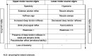
Fig. 2 summarizes the evolution of the diagnostic criteria over the last 30 years.
Fig. 2.

Evolution of the diagnostic criteria to reach the current Gold Coast criteria. Note: we can appreciate the evolution towards simplicity and an upgrade of the EMG role in the diagnosis, as well as the increasing importance of the fasciculation potentials. UMN: upper motor neuron signs; LMN: lower motor neuron signs; EMG: electromyography.
4.4. Final remarks
There are some further important points that need to be addressed. We will briefly discuss them in this section.
One point that should deserve an ongoing evaluation is the value and reliability of the UMN clinical signs in testing ALS patients (Swash, 2012, Swash et al., 2020). These clinical tests are not applied in a standardized way in the many centers following ALS patients, and their interpretation is variable. One example is the role of “retained deep tendon reflexes” in weak and atrophic limbs. These “probable UMN signs” (Younger et al., 1990), are supported as valid markers of UMN lesion by imaging and necropsy (Chan et al., 1999, Ince et al., 2003), but it is uncertain how they are assessed in different centers, even when these centers are involved in clinical trials.
Another point regards the value of a number of methods as markers of UMN and LMN dysfunction for the diagnosis of ALS. We only briefly mention TMS, imaging, EEG and some peripheral markers of LMN loss, as these are developed in other chapters of this book.
TMS is non-invasive, well-tolerated and easily applied method, although conventional measurements have been considered not very sensitive or specific for ALS. A recent contribution shows that central conduction time to lower limbs is sensitive and specific in ALS, allowing twice increased UMN dysfunction detection than clinical observation (Truffert et al., 2023). Markers of increased intra-cortical excitability are sensitive for ALS, but require a more sophisticated methodology, not widely available (Vucic et al., 2023). Following a group of patients, short interval intracortical inhibition (SICI) shows greater abnormality over time (de Carvalho and Swash, 2023), suggesting its potential utility to evaluate treatment response. Additionally, its use to monitor disease onset in presymptomatic carriers is a potentially useful approach in current and future genetic treatments (Vucic et al., 2008).
Electroencephalography (EEG) is an exciting area of research, since its abnormalities are independent of selective LMN or UMN impairment. However, the complexity of the technology and methodology involved limits its use as a method for diagnosis (Nasseroleslami et al., 2019, Dukic et al., 2019). ALS patients show higher cortical connectivity, in particular in frontal and parietal cortices (Blain-Moraes et al., 2013), and these changes increase with disease progression (Nasseroleslami et al., 2019), showing correlation with structural damage, clinical scores (Dukic et al., 2019), and phenotypes (Dukic et al., 2022). A recent study reinforces the potential role of EEG to measure disease progression, showing a higher beta-band power in motor/frontal regions in more severely affected patients (Notturno et al., 2023).
There is a very extensive literature of brain imaging in ALS. However, brain MRI is not used to diagnose ALS but, rather, to exclude other disorders. Some changes can indicate ALS, in particular abnormal signal of the corticospinal tract and corpus callosum (Anand et al., 2023), and diffusion tensor tractography (Fukui et al., 2018). However, other techniques may prove useful, in particular cortical and brain volumetry (Agosta et al., 2007, Verstraete et al., 2012). The role of PET brain imaging is probably limited taking into account its lack of general availability and high cost, although imaging of microglial activation may be a more promising application (Corcia et al., 2012). MRI of the spinal cord assessing atrophy and diffusion tensor imaging metrics is a promising technique with a probable future relevant impact in ALS (Querin et al., 2017), in particular considering its potential to capture changes in pre-symptomatic carriers of ALS-associated genetic mutations (Querin et al., 2019).
Neurofilaments are proteins located inside the axons in all neuronal cells, they form a structural matrix maintaining axonal morphological integrity and promoting axoplasm cargo transportation jointly with microtubules (Norgren et al., 2003). Neurofilament levels are important in ALS, in particular due to its quantitative characteristic, permitting its use to monitor disease activity and perhaps to detect new drugs capable of decreasing its usually rather constant level across disease progression, and with good sensitivity for early diagnosis (Brousse et al., 2023). However, reviewing different contributions it was found that its sensitivity ranges from 0.71 to 0.91; specificity from 0.64 to 0.95; and diagnostic accuracy (area under curve) from 0.77 to 0.98, depending on the patient phenotype, type of neurofilament and laboratory technique used (Xu et al., 2016). Venous blood neurofilament levels are raised months before detectable motor weakness in carriers of ALS-associated mutations, indicating the subclinical neuronal and axonal degeneration that precedes clinical presentation. This could be a critical observation for timely initiation of treatments with effective drugs in susceptible populations (Benatar et al., 2018). It is important to recognize that raised blood or CSF neurofilament levels are not specific tests for ALS, since they are raised in many different central neurodegenerative disorders, including brain ischaemia, multiple sclerosis and other neurodegenerations; but they are useful when considered in the clinical context.
Regarding LMN dysfunction EMG is a sensitive method for diagnosis, and it has been shown to be the essential investigation in the early diagnosis of ALS (Falcão de Campos et al., 2023). In patients with a rapid loss of motor units, the motor unit number estimate (MUNE) is probably more sensitive than MUP analysis in detecting abnormality. MUNE is a non-invasive method easily used in distal limb muscles (Jacobsen et al., 2018). Many different techniques have been investigated over the last 50 years or more (de Carvalho et al., 2018). Currently the most investigated techniques are the Motor Unit Number Index - MUNIX (Neuwirth et al., 2011) and the MScanFit MUNE (Sørensen et al., 2023), both are reliable and easy to apply, although they require a proper trainning. Ultrasound is a sensitive and non-invasive method to detect fasciculations, in particular in cranial-innervated muscles. In addition it can detect decreased muscle thickness and increased muscle echo intensity, as well as peripheral nerve trunk atrophy (Hobson-Webb and Simmons, 2019). Muscle MRI can also be used to detect fasciculations, but the sensitivity of this technique is as yet incompletely understood and the technique is cumbersome and expensive (Whittaker et al., 2019). The added value of muscle ultrasound for reinforcing diagnosis in different centers should be investigated. In the same way, muscle MRI, in particular whole-body muscle MRI, is a new method that might prove effective to confirm early involvement in different anatomical territories and also to monitor disease spreading (Jenkins et al., 2018).
4.5. What is ALS?
A fascinating problem is how to interpret ALS diagnosis. An important aspect to consider is whether ALS should be considered a clinical syndrome, like heart failure, associated with a number of risk factors, including genetic abnormalities, environmental factors (e.g., some toxins, smoking, intense exercise), a partially unknown abnormal immunological response and other factors; or whether it is a discrete disease defined by certain pathological and molecular markers (cytoplasmic aggregates of TDP-43, SOD1, or FUS)?
If ALS is a clinical syndrome the point of interest is the presumed commonality of the pathway leading to motor neuron degeneration, and clinical diagnosis becomes a process of syndrome identification, adjusted for the different stages of the disordered process. This seems to us the likely scenario.
If we accept the classical clinical concept of ALS as a single pathophysiological entity, then a highly sensitive and specific marker might be expected to be found for diagnosis, thus excluding patients in borderline areas of the disease spectrum. Strikingly, this has not been achieved, despite many attempts. Further, it is possible that artificial intelligence tools (for example, machine learning) might help clinicians to achieve a more confident diagnosis, and help us to understand if ALS should be considered a clinical syndrome or a discrete entity (Das et al., 2022, Tavazzi et al., 2023). There is much more work to be done.
The future will reveal our errors today regarding ALS diagnosis, but it is inappropriate to anticipate the future because it will demand new tools that require time for testing their utility.
Declaration of competing interest
The authors declare that they have no known competing financial interests or personal relationships that could have appeared to influence the work reported in this paper.
References
- Adams D., Kuntzer T., Steck A.J., Lobrinus A., Janzer R.C., Regli F. Motor conduction block and high titres of anti-GM1 ganglioside antibodies: pathological evidence of a motor neuropathy in a patient with lower motor neuron syndrome. J. Neurol. Neurosurg. Psychiatry. 1993;56(9):982–987. doi: 10.1136/jnnp.56.9.982. [DOI] [PMC free article] [PubMed] [Google Scholar]
- Agosta F., Pagani E., Rocca M.A., Caputo D., Perini M., Salvi F., et al. Voxel-based morphometry study of brain volumetry and diffusivity in amyotrophic lateral sclerosis patients with mild disability. Hum. Brain Mapp. 2007;28(12):1430–1438. doi: 10.1002/hbm.20364. [DOI] [PMC free article] [PubMed] [Google Scholar]
- Allen J., Nobile-Orazio E., Peric S., Katzberg H., Cadour S., Van de Walle I., et al. Safety, Efficacy, and pharmacokinetics of Argx-117 in adults with multifocal motor neuropathy: a global, multicenter, placebo controlled phase 2 study (Arda) Neurology. 2022;99(23 Supplement 2):S40–S41. [Google Scholar]
- Alves I., Gromicho M., Oliveira Santos M., Pinto S., Pronto-Laborinho A., Swash M., et al. Demographic changes in a large motor neuron disease cohort in Portugal: a 27 year experience. Amyotroph. Lateral Scler. Frontotemporal. Degener. 2023 doi: 10.1080/21678421.2023.2220747. [DOI] [PubMed] [Google Scholar]
- Anand T., Ishaque A., Ta D., Khan M.U., Bharti K., Wu A., et al. Characterization of white matter alterations using diffusion kurtosis imaging in patients with amyotrophic lateral sclerosis. Brain Behav. 2023;6:e3102. doi: 10.1002/brb3.3102. [DOI] [PMC free article] [PubMed] [Google Scholar]
- Behnia M., Kelly J.J. Role of electromyography in amyotrophic lateral sclerosis. Muscle Nerve. 1991;14(12):1236–1241. doi: 10.1002/mus.880141217. [DOI] [PubMed] [Google Scholar]
- Benatar M., Wuu J., Andersen P.M., Lombardi V., Malaspina A. Neurofilament light: a candidate biomarker of presymptomatic amyotrophic lateral sclerosis and phenoconversion. Ann. Neurol. 2018;84(1):130–139. doi: 10.1002/ana.25276. [DOI] [PMC free article] [PubMed] [Google Scholar]
- Bentes C., Santos-Bento M., de Sá J., de Lurdes Sales Luís M., de Carvalho M. Allgrove syndrome in adulthood. Muscle Nerve. 2001;24(2):292–296. doi: 10.1002/1097-4598(200102)24:2<292::aid-mus160>3.0.co;2-x. [DOI] [PubMed] [Google Scholar]
- Blackman G., Cherfi Y., Morrin H., Ellis C.M., Bashford J., Ruths F., et al. The association between benign fasciculations and health anxiety: A report of two cases and a systematic review of the literature. Psychosomatics. 2019;60(5):499–507. doi: 10.1016/j.psym.2019.04.001. [DOI] [PubMed] [Google Scholar]
- Blain-Moraes S., Mashour G.A., Lee H., Huggins J.E., Lee U. Altered cortical communication in amyotrophic lateral sclerosis. Neurosci Lett. 2013;543:172–176. doi: 10.1016/j.neulet.2013.03.028. [DOI] [PMC free article] [PubMed] [Google Scholar]
- Bresch S., Delmont E., Soriani M.H., Desnuelle C. Electrodiagnostic criteria for early diagnosis of bulbar-onset ALS: a comparison of El Escorial, revised El Escorial and Awaji algorithm. Rev. Neurol. 2013;170(2):134–139. doi: 10.1016/j.neurol.2013.10.004. [DOI] [PubMed] [Google Scholar]
- Breza M., Koutsis G. Kennedy's disease (spinal and bulbar muscular atrophy): a clinically oriented review of a rare disease. J. Neurol. 2019;266(3):565–573. doi: 10.1007/s00415-018-8968-7. [DOI] [PubMed] [Google Scholar]
- Brooks B.R. El Escorial World Federation of Neurology criteria for the diagnosis of amyotrophic lateral sclerosis. Subcommittee on Motor Neuron Diseases/Amyotrophic Lateral Sclerosis of the World Federation of Neurology Research Group on Neuromuscular Diseases and the El Escorial“ Clinical limits of amyotrophic lateral sclerosis” workshop contributors. J. Neurol. Sci. 1994;124:96–107. doi: 10.1016/0022-510x(94)90191-0. [DOI] [PubMed] [Google Scholar]
- Brooks B.R., Miller R.G., Swash M., Munsat T.L. El Escorial revisited: revised criteria for the diagnosis of amyotrophic lateral sclerosis. Amyotroph. Lateral Scler. Other Motor Neuron Disord. 2000;1(5):293–299. doi: 10.1080/146608200300079536. [DOI] [PubMed] [Google Scholar]
- Brousse M., Delaby C., De La Cruz E., Kuhle J., Benkert P., Mondesert E., et al. Serum neurofilament light chain cut-off definition for clinical diagnosis and prognosis of amyotrophic lateral sclerosis. Eur. J. Neurol. 2023 doi: 10.1111/ene.15813. [DOI] [PubMed] [Google Scholar]
- Cappellari A., Ciammola A., Silani V. The pseudopolyneuritic form of amyotrophic lateral sclerosis (Patrikios' disease) Electromyogr. Clin. Neurophysiol. 2008;48(2):75–81. [PubMed] [Google Scholar]
- Chan S., Shungu D.C., Douglas-Akinwande A., Lange D.J., Rowland L.P. Motor neuron diseases: comparison of single-voxel proton MR spectroscopy of the motor cortex with MR imaging of the brain. Radiology. 1999;212(3):763–769. doi: 10.1148/radiology.212.3.r99au35763. [DOI] [PubMed] [Google Scholar]
- Chen L., Zhang B., Chen R., Tang L., Liu R., Yang Y., et al. Natural history and clinical features of sporadic amyotrophic lateral sclerosis in China. J. Neurol. Neurosurg. Psychiatry. 2015;86(10):1075–1781. doi: 10.1136/jnnp-2015-310471. [DOI] [PubMed] [Google Scholar]
- Chiò A., Calvo A., Moglia C., Mazzini L., Mora G. Phenotypic heterogeneity of amyotrophic lateral sclerosis: a population based study. J. Neurol. Neurosurg. Psychiatry. 2011;82(7):740–746. doi: 10.1136/jnnp.2010.235952. [DOI] [PubMed] [Google Scholar]
- Chiò A., Moglia C., Canosa A., Manera U., D'Ovidio F., Vasta R., et al. ALS phenotype is influenced by age, sex, and genetics: a population-based study. Neurology. 2020;94(8):e802–e810. doi: 10.1212/WNL.0000000000008869. [DOI] [PubMed] [Google Scholar]
- Cluse F., Hermier M., Demarquay G., Rogemond V., Mallaret M., Svahn J., et al. Trigeminal Nerve Involvement in Bulbar-Onset Anti-IgLON5 Disease. Neurol, Neuroimmunol, Neuroinflamm. 2023;10(6):e200153. doi: 10.1212/NXI.0000000000200153. [DOI] [PMC free article] [PubMed] [Google Scholar]
- Corcia P., Tauber C., Vercoullie J., Arlicot N., Prunier C., Praline J., et al. Molecular imaging of microglial activation in amyotrophic lateral sclerosis. PLoS One. 2012;7(12):e52941. doi: 10.1371/journal.pone.0052941. [DOI] [PMC free article] [PubMed] [Google Scholar]
- Corcia P., Bede P., Pradat P.F., Couratier P., Vucic S., de Carvalho M. Split-hand and split-limb phenomena in amyotrophic lateral sclerosis: pathophysiology, electrophysiology and clinical manifestations. J. Neurol. Neurosurg. Psychiatry. 2021;92(10):1126–1130. doi: 10.1136/jnnp-2021-326266. [DOI] [PubMed] [Google Scholar]
- Corcia P., Lunetta C., Couratier P., Vourc'h P., Gromicho M., Desnuelle C., et al. Familial clustering of primary lateral sclerosis and amyotrophic lateral sclerosis: Supplementary evidence for a continuum. Eur. J. Neurol. 2021;28(8):2780–2783. doi: 10.1111/ene.14960. [DOI] [PubMed] [Google Scholar]
- Cortés-Vicente E., Pradas J., Marin-Lahoz J., De Luna N., Clarimón J., Turon-Sans J., et al. Early diagnosis of amyotrophic lateral sclerosis mimic syndromes: pros and cons of current clinical diagnostic criteria. Amyotroph. Lateral Scler. Frontotemporal Degener. 2017;18(5–6):333–340. doi: 10.1080/21678421.2017.1316408. [DOI] [PubMed] [Google Scholar]
- Costa J., Swash M., de Carvalho M. Awaji criteria for the diagnosis of amyotrophic lateral sclerosis: a systematic review. Arch. Neurol. 2012;69(11):1410–1416. doi: 10.1001/archneurol.2012.254. [DOI] [PubMed] [Google Scholar]
- Dalakas M.C. The post-polio syndrome as an evolved clinical entity: Definition and clinical description. Ann. N. Y. Acad. Sci. 1995;753(1):68–80. doi: 10.1111/j.1749-6632.1995.tb27532.x. [DOI] [PubMed] [Google Scholar]
- Das T., Kaur H., Gour P., Prasad K., Lynn A.M., Prakash A., et al. Intersection of network medicine and machine learning towards investigating the key biomarkers and pathways underlying amyotrophic lateral sclerosis: a systematic review. Brief Bioinform. 2022;23(6):bbac442. doi: 10.1093/bib/bbac442. [DOI] [PubMed] [Google Scholar]
- de Carvalho M., Dengler R., Eisen A., England J.D., Kaji R., Kimura J., et al. Electrodiagnostic criteria for diagnosis of ALS. Clin. Neurophysiol. 2008;119(3):497–503. doi: 10.1016/j.clinph.2007.09.143. [DOI] [PubMed] [Google Scholar]
- de Carvalho M., Kiernan M.C., Swash M. Fasciculation in amyotrophic lateral sclerosis: origin and pathophysiological relevance. J. Neurol. Neurosurg. Psychiatry. 2017;88(9):773–779. doi: 10.1136/jnnp-2017-315574. [DOI] [PubMed] [Google Scholar]
- de Carvalho M., Swash M. Fasciculation potentials: a study of amyotrophic lateral sclerosis and other neurogenic disorders. Muscle Nerve. 1998;21(3):336–344. doi: 10.1002/(sici)1097-4598(199803)21:3<336::aid-mus7>3.0.co;2-b. [DOI] [PubMed] [Google Scholar]
- de Carvalho M., Swash M. Fasciculation potentials and earliest changes in motor unit physiology in ALS. J. Neurol. Neurosurg. Psychiatry. 2013;84(9):963–968. doi: 10.1136/jnnp-2012-304545. [DOI] [PubMed] [Google Scholar]
- de Carvalho M., Swash M. Lower motor neuron dysfunction in ALS. Clin. Neurophysiol. 2016;127(7):2670–2681. doi: 10.1016/j.clinph.2016.03.024. [DOI] [PubMed] [Google Scholar]
- De Carvalho M., Miranda P.C., de Lourdes Sales Luís M., Ducla-Soares E. Neurophysiological features of fasciculation potentials evoked by transcranial magnetic stimulation in amyotrophic lateral sclerosis. J. Neurol. 2000;247:189–194. doi: 10.1007/s004150050561. [DOI] [PubMed] [Google Scholar]
- de Carvalho M., Barkhaus P.E., Nandedkar S.D., Swash M. Motor unit number estimation (MUNE): Where are we now? Clin. Neurophysiol. 2018;129(8):1507–1516. doi: 10.1016/j.clinph.2018.04.748. [DOI] [PubMed] [Google Scholar]
- De Carvalho M., Swash M. Transcranial magnetic stimulation to monitor disease progression in ALS: a review. Amyotroph. Lateral Scler. Frontotemporal Degener. 2023;24(5–6):362–368. doi: 10.1080/21678421.2022.2160649. [DOI] [PubMed] [Google Scholar]
- de Carvalho M., Kiernan M.C., Pullman S.L., Rezania K., Turner M.R., Simmons Z. Neurophysiological features of primary lateral sclerosis. Amyotroph. Lateral Scler. Frontotemporal Degener. 2020;21(sup1):11–17. doi: 10.1080/21678421.2020.1837174. [DOI] [PubMed] [Google Scholar]
- de Jongh A.D., Braun N., Weber M., van Es M.A., Masrori P., Veldink J.H., et al. Characterising ALS disease progression according to El Escorial and Gold Coast criteria. J. Neurol. Neurosurg. Psychiatry. 2022;93(8):865–870. doi: 10.1136/jnnp-2022-328823. [DOI] [PubMed] [Google Scholar]
- Dukic S., McMackin R., Buxo T., Fasano A., Chipika R., Pinto-Grau M., et al. Patterned functional network disruption in amyotrophic lateral sclerosis. Hum. Brain, Mapp. 2019;40:4827–4842. doi: 10.1002/hbm.24740. [DOI] [PMC free article] [PubMed] [Google Scholar]
- Dukic S., McMackin R., Costello E., Metzger M., Buxo T., Fasano A., et al. Resting-state EEG reveals four subphenotypes of amyotrophic lateral sclerosis. Brain. 2022;145(2):621–631. doi: 10.1093/brain/awab322. [DOI] [PMC free article] [PubMed] [Google Scholar]
- Eisen A. Amyotrophic lateral sclerosis: A 40-year personal perspective. J. Clin. Neurosci. 2009;16(4):505–512. doi: 10.1016/j.jocn.2008.07.072. [DOI] [PubMed] [Google Scholar]
- Eisen A., Berry K., Gibson G. Inclusion body myositis (IBM): myopathy or neuropathy? Neurology. 1983;33(9):1109–1114. doi: 10.1212/wnl.33.9.1109. [DOI] [PubMed] [Google Scholar]
- Falcão de Campos C., Gromicho M., Uysal H., Grosskreutz J., Kuzma-Kozakiewicz M., Oliveira Santos M., et al. Trends in the diagnostic delay and pathway for amyotrophic lateral sclerosis patients across different countries. Front. Neurol. 2023;17(13):1064619. doi: 10.3389/fneur.2022.1064619. [DOI] [PMC free article] [PubMed] [Google Scholar]
- Filippi M., Preziosa P., Banwell B.L., Barkhof F., Ciccarelli O., De Stefano N., et al. Assessment of lesions on magnetic resonance imaging in multiple sclerosis: practical guidelines. Brain. 2019;142(7):1858–1875. doi: 10.1093/brain/awz144. [DOI] [PMC free article] [PubMed] [Google Scholar]
- Fisher M., Mateer J.E., Ullrich I., Gutrecht J.A. Pyramidal tract deficits and polyneuropathy in hyperthyroidism. Combination clinically mimicking amyotrophic lateral sclerosis. Am. J. Med. 1985;78(6):1041–1044. doi: 10.1016/0002-9343(85)90231-1. [DOI] [PubMed] [Google Scholar]
- Fluchere F., Verschueren A., Cintas P., Franques J., Serratrice J., Weiller P.J., et al. Clinical features and follow-up of four new cases of facial-onset sensory and motor neuronopathy. Muscle Nerve. 2011;43(1):136–140. doi: 10.1002/mus.21884. [DOI] [PubMed] [Google Scholar]
- Fukui Y., Hishikawa N., Sato K., Nakano Y., Morihara R., Shang J., et al. Detecting spinal pyramidal tract of amyotrophic lateral sclerosis patients with diffusion tensor tractography. Neurosci. Res. 2018;133:58–63. doi: 10.1016/j.neures.2017.11.005. [DOI] [PubMed] [Google Scholar]
- Geevasinga N., Loy C.T., Menon P., de Carvalho M., Swash M., Schrooten M., et al. Awaji criteria improves the diagnostic sensitivity in amyotrophic lateral sclerosis: a systematic review using individual patient data. Clin. Neurophysiol. 2016;127(7):2684–2691. doi: 10.1016/j.clinph.2016.04.005. [DOI] [PubMed] [Google Scholar]
- Goedee H.S., Jongbloed B.A., van Asseldonk J.T., Hendrikse J., Vrancken A.F., Franssen H., et al. A comparative study of brachial plexus sonography and magnetic resonance imaging in chronic inflammatory demyelinating neuropathy and multifocal motor neuropathy. Eur. J. Neurol. 2017;24(10):1307–1313. doi: 10.1111/ene.13380. [DOI] [PubMed] [Google Scholar]
- Goyal N.A., Mozaffar T., Dimachkie M.M. Imaging beyond muscle magnetic resonance imaging in inclusion body myositis. Clin. Exp. Rheumatol. 2023;12(41):386–392. doi: 10.55563/clinexprheumatol/uimkey. [DOI] [PubMed] [Google Scholar]
- Greenberg S.A. Inclusion body myositis: clinical features and pathogenesis. Nat. Rev. Rheumatol. 2019;15(5):257–272. doi: 10.1038/s41584-019-0186-x. [DOI] [PubMed] [Google Scholar]
- Gromicho M., Figueiral M., Uysal H., Grosskreutz J., Kuzma-Kozakiewicz M., Pinto S., et al. Spreading in ALS: The relative impact of upper and lower motor neuron involvement. Ann. Clin. Transl. Neurol. 2020;7(7):1181–1192. doi: 10.1002/acn3.51098. [DOI] [PMC free article] [PubMed] [Google Scholar]
- Gromicho M., Kuzma-Kozakiewicz M., Szacka K., Nieporecki K., Andersen P.M., Grosskreutz J., et al. Motor neuron disease beginning with frontotemporal dementia: clinical features and progression. Amyotroph. Lateral Scler. Frontotemporal Degener. 2021;22(7–8):508–516. doi: 10.1080/21678421.2021.1910309. [DOI] [PubMed] [Google Scholar]
- Gromicho M., Santos M.O., Pinto S., Swash M., de Carvalho M. The flail-arm syndrome: the influence of phenotypic features. Amyotroph. Lateral Scler. Frontotemporal Degener. 2023;24(5–6):383–388. doi: 10.1080/21678421.2022.2164205. [DOI] [PubMed] [Google Scholar]
- Gwathmey K.G., Corcia P., McDermott C.J., Genge A., Sennfält S., de Carvalho M., et al. Diagnostic delay in amyotrophic lateral sclerosis. Eur J Neurol. 2023;30(9):2595–2601. doi: 10.1111/ene.15874. [DOI] [PubMed] [Google Scholar]
- Hannaford A., Pavey N., van den Bos M., Geevasinga N., Menon P., Shefner J.M., et al. Diagnostic utility of gold coast criteria in amyotrophic lateral sclerosis. Ann. Neurol. 2021;89(5):979–986. doi: 10.1002/ana.26045. [DOI] [PubMed] [Google Scholar]
- Hirayama K. Juvenile muscular atrophy of distal upper extremity (Hirayama disease) Intern. Med. 2000;39(4):83–290. doi: 10.2169/internalmedicine.39.283. [DOI] [PubMed] [Google Scholar]
- Hirayama K., Tokumaru Y. Cervical dural sac and spinal cord in juvenile muscular atrophy of distal upper extremity. Neurology. 2000;54(19):1922–1926. doi: 10.1212/wnl.54.10.1922. [DOI] [PubMed] [Google Scholar]
- Hirayama K.E., Tomonaga M.A., Kitano K., Yamada T., Kojima S., Arai K. Focal cervical poliopathy causing juvenile muscular atrophy of distal upper extremity: a pathological study. J. Neurol. Neurosurg. Psychiatry. 1987;50(3):285–290. doi: 10.1136/jnnp.50.3.285. [DOI] [PMC free article] [PubMed] [Google Scholar]
- Hobson-Webb L.D., Simmons Z. Ultrasound in the diagnosis and monitoring of amyotrophic lateral sclerosis: a review. Muscle Nerve. 2019;60(2):114–123. doi: 10.1002/mus.26487. [DOI] [PubMed] [Google Scholar]
- Househam E., Swash M. Diagnostic delay in amyotrophic lateral sclerosis: what scope for improvement? J. Neurol. Sci. 2000;180(1–2):76–81. doi: 10.1016/s0022-510x(00)00418-4. [DOI] [PubMed] [Google Scholar]
- Ince P.G., Evans J., Knopp M., Forster G., Hamdalla H.H., Wharton S.B., et al. Corticospinal tract degeneration in the progressive muscular atrophy variant of ALS. Neurology. 2003;60(8):1252–1258. doi: 10.1212/01.wnl.0000058901.75728.4e. [DOI] [PubMed] [Google Scholar]
- Ismail A., Cooper-Knock J., Highley J.R., Milano A., Kirby J., Goodall E., et al. Concurrence of multiple sclerosis and amyotrophic lateral sclerosis in patients with hexanucleotide repeat expansions of C9ORF72. J. Neurol. Neurosurg. Psychiatry. 2013;84(1):79–87. doi: 10.1136/jnnp-2012-303326. [DOI] [PubMed] [Google Scholar]
- Jacobsen A.B., Kristensen R.S., Witt A., Kristensen A.G., Duez L., Beniczky S., et al. The utility of motor unit number estimation methods versus quantitative motor unit potential analysis in diagnosis of ALS. Clin. Neurophysiol. 2018;129(3):646–653. doi: 10.1016/j.clinph.2018.01.002. [DOI] [PubMed] [Google Scholar]
- Jenkins T.M., Alix J.J., David C., Pearson E., Rao D.G., Hoggard N., et al. Imaging muscle as a potential biomarker of denervation in motor neuron disease. J Neurol Neurosurg Psychiatry. 2018;89(3):248–255. doi: 10.1136/jnnp-2017-316744. [DOI] [PMC free article] [PubMed] [Google Scholar]
- Jewett G., Khayambashi S., Frost G.S., Beland B., Lee A., Hodgkinson V., et al. Gold Coast criteria expand clinical trial eligibility in amyotrophic lateral sclerosis. Muscle Nerve. 2022;66(4):397–403. doi: 10.1002/mus.27660. [DOI] [PubMed] [Google Scholar]
- Johnsen B., Pugdahl K., Fuglsang-Frederiksen A., Kollewe K., Paracka L., Dengler R., et al. Diagnostic criteria for amyotrophic lateral sclerosis: a multicentre study of inter-rater variation and sensitivity. Clin. Neurophysiol. 2019;130(2):307–314. doi: 10.1016/j.clinph.2018.11.021. [DOI] [PubMed] [Google Scholar]
- Katz J.S., Barohn R.J., Kojan S., Wolfe G.I., Nations S.P., Saperstein D.S., et al. Axonal multifocal motor neuropathy without conduction block or other features of demyelination. Neurology. 2002;58(4):615–620. doi: 10.1212/wnl.58.4.615. [DOI] [PubMed] [Google Scholar]
- Kim W.K., Liu X., Sandner J., Pasmantier M., Andrews J., Rowland L.P., et al. Study of 962 patients indicates progressive muscular atrophy is a form of ALS. Neurology. 2009;73(20):1686–1692. doi: 10.1212/WNL.0b013e3181c1dea3. [DOI] [PMC free article] [PubMed] [Google Scholar]
- Kleine B.U., Stegeman D.F., Schelhaas H.J., Zwarts M.J. Firing pattern of fasciculations in ALS: evidence for axonal and neuronal origin. Neurology. 2008;70(5):353–359. doi: 10.1212/01.wnl.0000300559.14806.2a. [DOI] [PubMed] [Google Scholar]
- Kolb S.J., Kissel J.T. Spinal muscular atrophy: a timely review. Arch. Neurol. 2011;68(8):979–984. doi: 10.1001/archneurol.2011.74. [DOI] [PMC free article] [PubMed] [Google Scholar]
- Kühnlein P., Gdynia H.J., Sperfeld A.D., Lindner-Pfleghar B., Ludolph A.C., Prosiegel M., et al. Diagnosis and treatment of bulbar symptoms in amyotrophic lateral sclerosis. Nat. Clin. Pract. Neurol. 2008;4(7):366–374. doi: 10.1038/ncpneuro0853. [DOI] [PubMed] [Google Scholar]
- Lambert E.H. In: Motor Neuron Diseases. Norris F.H., Kurland L.T., editors. Grune and Stratton; New York: 1969. Electromyography in amyotrophic lateral sclerosis; pp. 135–153. [Google Scholar]
- Li T.M., Alberman E.A.E., Swash M. A suggested approach to the differential diagnosis of motor neuron disease from other neurological conditions. Lancet. 1986;2:731–732. [PubMed] [Google Scholar]
- Lomen-Hoerth C., Anderson T., Miller B. The overlap of amyotrophic lateral sclerosis and frontotemporal dementia. Neurology. 2002;59(7):1077–1079. doi: 10.1212/wnl.59.7.1077. [DOI] [PubMed] [Google Scholar]
- Masrori P., Van Damme P. Amyotrophic lateral sclerosis: a clinical review. Eur. J. Neurol. 2020;27(10):1918–1929. doi: 10.1111/ene.14393. [DOI] [PMC free article] [PubMed] [Google Scholar]
- Menon P., Geevasinga N., Yiannikas C., Kiernan M.C., Vucic S. Cortical contributions to the flail leg syndrome: Pathophysiological insights. Amyotroph Lateral Scler Frontotemporal Degener. 2016;17(5–6):389–396. doi: 10.3109/21678421.2016.1145232. [DOI] [PubMed] [Google Scholar]
- Montalvo A., Swash M., de Carvalho M. Benign fasciculations: A follow-up study with electrophysiological studies. Muscle Nerve. 2021;64(6):670–675. doi: 10.1002/mus.27411. [DOI] [PubMed] [Google Scholar]
- Nasseroleslami B., Dukic S., Broderick M., Mohr K., Schuster C., Gavin B., et al. Characteristic increases in EEG connectivity correlate with changes of structural MRI in amyotrophic lateral sclerosis. Cereb. Cortex. 2019;29(1):27–41. doi: 10.1093/cercor/bhx301. [DOI] [PubMed] [Google Scholar]
- Needham M., Mastaglia F.L. Inclusion body myositis: current pathogenetic concepts and diagnostic and therapeutic approaches. Lancet Neurol. 2007;6(7):620–631. doi: 10.1016/S1474-4422(07)70171-0. [DOI] [PubMed] [Google Scholar]
- Neuwirth C., Nandedkar S., Stålberg E., Barkhaus P.E., de Carvalho M., Furtula J., et al. Motor Unit Number Index (MUNIX): a novel neurophysiological marker for neuromuscular disorders; test–retest reliability in healthy volunteers. Clin. Neurophysiol. 2011;122(9):1867–1872. doi: 10.1016/j.clinph.2011.02.017. [DOI] [PubMed] [Google Scholar]
- Norgren N., Rosengren L., Stigbrand T. Elevated neurofilament levels in neurological diseases. Brain Res. 2003;987(1):25–31. doi: 10.1016/s0006-8993(03)03219-0. [DOI] [PubMed] [Google Scholar]
- Notturno F., Croce P., Ornello R., Sacco S., Zappasodi F. Yield of EEG features as markers of disease severity in amyotrophic lateral sclerosis: a pilot study. Amyotroph. Lateral Scler. Frontotemporal Degener. 2023;24(3–4):295–303. doi: 10.1080/21678421.2022.2152696. [DOI] [PubMed] [Google Scholar]
- Patten B.M., Bilezikian J.P., Mallette L.A., Prince A., Engel W.K., Aurbach G.D. Neuromuscular disease in primary hyperparathyroidism. Ann. Intern. Med. 1974;80(2):182–193. doi: 10.7326/0003-4819-80-2-182. [DOI] [PubMed] [Google Scholar]
- Phillips B.A., Zilko P.J., Mastaglia F.L. Prevalence of sporadic inclusion body myositis in Western Australia. Muscle Nerve. 2000;23(6):970–972. doi: 10.1002/(sici)1097-4598(200006)23:6<970::aid-mus20>3.0.co;2-i. [DOI] [PubMed] [Google Scholar]
- Pinto S., Swash M., de Carvalho M. Does surgery accelerate progression of amyotrophic lateral sclerosis? J. Neurol. Neurosurg. Psychiatry. 2014;85(6):643–646. doi: 10.1136/jnnp-2013-305770. [DOI] [PubMed] [Google Scholar]
- Pinto S., Gromicho M., Oliveira Santos M.O., Swash M., de Carvalho M. Respiratory onset in amyotrophic lateral sclerosis: clinical features and spreading pattern. Amyotroph. Lateral Scler. Frontotemporal Degener. 2023;24(1–2):40–44. doi: 10.1080/21678421.2022.2067777. [DOI] [PubMed] [Google Scholar]
- Pugdahl K., Fuglsang-Frederiksen A., Johnsen B., de Carvalho M., Fawcett P.R., Labarre-Vila A., et al. A prospective multicentre study on sural nerve action potentials in ALS. Clin. Neurophysiol. 2008;119(5):1106–1110. doi: 10.1016/j.clinph.2008.01.010. [DOI] [PubMed] [Google Scholar]
- Pugdahl K., Camdessanché J.P., Cengiz B., de Carvalho M., Liguori R., Rossatto C., et al. Gold Coast diagnostic criteria increase sensitivity in amyotrophic lateral sclerosis. Clin. Neurophysiol. 2021;132(12):3183–3189. doi: 10.1016/j.clinph.2021.08.014. [DOI] [PubMed] [Google Scholar]
- Querin G., El Mendili M.M., Lenglet T., Delphine S., Marchand-Pauvert V., Benali H., et al. Spinal cord multi-parametric magnetic resonance imaging for survival prediction in amyotrophic lateral sclerosis. Eur. J. Neurol. 2017;24(8):1040–1046. doi: 10.1111/ene.13329. [DOI] [PubMed] [Google Scholar]
- Querin G., Bede P., El Mendili M.M., Li M., Pélégrini-Issac M., Rinaldi D., et al. Presymptomatic spinal cord pathology in c9orf72 mutation carriers: a longitudinal neuroimaging study. Ann Neurol. 2019;86(2):158–167. doi: 10.1002/ana.25520. [DOI] [PubMed] [Google Scholar]
- Reed D.M., Kurland L.T. Muscle fasciculations in a healthy population. Arch. Neurol. 1963;9(4):363–367. doi: 10.1001/archneur.1963.00460100051005. [DOI] [PubMed] [Google Scholar]
- Rossor A.M., Jaunmuktane Z., Rossor M.N., Hoti G., Reilly M.M. TDP43 pathology in the brain, spinal cord, and dorsal root ganglia of a patient with FOSMN. Neurology. 2019;92(9):e951–e956. doi: 10.1212/WNL.0000000000007008. [DOI] [PMC free article] [PubMed] [Google Scholar]
- Santos M.O., Caldeira I., Gromicho M., Pronto-Laborinho A., de Carvalho M. Brain white matter demyelinating lesions and amyotrophic lateral sclerosis in a patient with C9orf72 hexanucleotide repeat expansion. Mult Scler Relat Disord. 2017;17:1–4. doi: 10.1016/j.msard.2017.06.010. [DOI] [PubMed] [Google Scholar]
- Santos Silva C., Gromicho M., Oliveira Santos M., Pinto S., Swash M., de Carvalho M. Thyroid dysfunction in Portuguese amyotrophic lateral sclerosis patients. Neurol Sci. 2022;43(9):5625–5627. doi: 10.1007/s10072-022-06135-3. [DOI] [PubMed] [Google Scholar]
- Shefner J.M., Al-Chalabi A., Baker M.R., Cui L.Y., de Carvalho M., Eisen A., et al. A proposal for new diagnostic criteria for ALS. Clin. Neurophysiol. 2020;131(8):1975–1978. doi: 10.1016/j.clinph.2020.04.005. [DOI] [PubMed] [Google Scholar]
- Shen D., Yang X., Wang Y., He D., Sun X., Cai Z., et al. The Gold Coast criteria increases the diagnostic sensitivity for amyotrophic lateral sclerosis in a Chinese population. Transl. Neurodegener. 2021;10(1):1–8. doi: 10.1186/s40035-021-00253-2. [DOI] [PMC free article] [PubMed] [Google Scholar]
- Silva C.S., Santos M.O., Gromicho M., Pinto S., Swash M., de Carvalho M. Electromyographic findings in primary lateral sclerosis during disease progression. Clin. Neurophysiol. 2021;132(12):2996–3001. doi: 10.1016/j.clinph.2021.08.022. [DOI] [PubMed] [Google Scholar]
- Simon N.G., Kiernan M.C. Fasciculation anxiety syndrome in clinicians. J Neurol. 2013;260:1743–1747. doi: 10.1007/s00415-013-6856-8. [DOI] [PubMed] [Google Scholar]
- Sonoda K., Sasaki K., Tateishi T., Yamasaki R., Hayashi S., Sakae N., et al. TAR DNA-binding protein 43 pathology in a case clinically diagnosed with facial-onset sensory and motor neuronopathy syndrome: an autopsied case report and a review of the literature. J. Neurol. Sci. 2013;332(1–2):148–153. doi: 10.1016/j.jns.2013.06.027. [DOI] [PubMed] [Google Scholar]
- Sørensen D.M., Bostock H., Abrahao A., Alaamel A.B., Alaydin H.C., Ballegaard M., et al. Estimating motor unit numbers from a CMAP scan: Repeatability study on three muscles at 15 centres. Clin. Neurophysiol. 2023;151:92–99. doi: 10.1016/j.clinph.2023.04.008. [DOI] [PubMed] [Google Scholar]
- Swash M. Why are upper motor neuron signs difficult to elicit in amyotrophic lateral sclerosis? J. Neurol. Neurosurg. Psychiatry. 2012;83(6):659–662. doi: 10.1136/jnnp-2012-302315. [DOI] [PubMed] [Google Scholar]
- Swash M., Burke D., Turner M.R., Grosskreutz J., Leigh P.N., de Carvalho M., et al. Occasional essay: upper motor neuron syndrome in amyotrophic lateral sclerosis. J. Neurol. Neurosurg. Psychiatry. 2020;91(3):227–234. doi: 10.1136/jnnp-2019-321938. [DOI] [PubMed] [Google Scholar]
- Swinnen B., Robberecht W. The phenotypic variability of amyotrophic lateral sclerosis. Nat. Rev. Neurol. Nat. Rev. Neurol. 2014;10(11):661–670. doi: 10.1038/nrneurol.2014.184. [DOI] [PubMed] [Google Scholar]
- Tavazzi E., Longato E., Vettoretti M., Aidos H., Trescato I., Roversi C., et al. Artificial intelligence and statistical methods for stratification and prediction of progression in amyotrophic lateral sclerosis: A systematic review. Artif. Intell. Med. 2023;20 doi: 10.1016/j.artmed.2023.102588. [DOI] [PubMed] [Google Scholar]
- Traynor B.J., Codd M.B., Corr B., Forde C., Frost E., Hardiman O.M. Clinical features of amyotrophic lateral sclerosis according to the El Escorial and Airlie House diagnostic criteria: A population-based study. Arch. Neurol. 2000;57(8):1171–1176. doi: 10.1001/archneur.57.8.1171. [DOI] [PubMed] [Google Scholar]
- Triggs W.J., Menkes D., Onorato J., Yan R.H., Young M.S., Newell K., et al. Transcranial magnetic stimulation identifies upper motor neuron involvement in motor neuron disease. Neurology. 1999;53(3):605–611. doi: 10.1212/wnl.53.3.605. [DOI] [PubMed] [Google Scholar]
- Truffert A., Sukockienė E., Desmaison A., Ališauskienė M., Iancu Ferfoglia R., Guy N. Combined tendon reflex and motor evoked potential recordings in amyotrophic lateral sclerosis. Clin. Neurophysiol. 2023;147:88–98. doi: 10.1016/j.clinph.2022.12.013. [DOI] [PubMed] [Google Scholar]
- Turner M.R., Barohn R.J., Corcia P., Fink J.K., Harms M.B., Kiernan M.C., et al. Primary lateral sclerosis: consensus diagnostic criteria. J Neurol Neurosurg Psychiatry. 2020;91(4):373–377. doi: 10.1136/jnnp-2019-322541. [DOI] [PMC free article] [PubMed] [Google Scholar]
- Vallet A.E., Verschueren A., Petiot P., Vandenberghe N., Nicolino M., Roman S., et al. Neurological features in adult Triple-A (Allgrove) syndrome. J Neurol. 2012;259:39–46. doi: 10.1007/s00415-011-6115-9. [DOI] [PubMed] [Google Scholar]
- Vázquez-Costa J.F., Pedrola Vidal L., Moreau-Le Lan S., Teresí-Copoví I., Frasquet M., Chumillas M.J., et al. Facial onset sensory and motor neuronopathy: a motor neuron disease with an oligogenic origin? Amyotroph. Lateral Scler. Frontotemporal Degener. 2019;20(3–4):172–175. doi: 10.1080/21678421.2019.1582671. [DOI] [PubMed] [Google Scholar]
- Verstraete E., Veldink J.H., Hendrikse J., Schelhaas H.J., Van Den Heuvel M.P., Van Den Berg L.H. Structural MRI reveals cortical thinning in amyotrophic lateral sclerosis. J. Neurol. Neurosurg. Psychiatry. 2012;83(4):383–388. doi: 10.1136/jnnp-2011-300909. [DOI] [PubMed] [Google Scholar]
- Veugelers B., Theys P., Lammens M., Van Hees J., Robberecht W. Pathological findings in a patient with amyotrophic lateral sclerosis and multifocal motor neuropathy with conduction block. J. Neurol. Sci. 1996;136(1–2):64–70. doi: 10.1016/0022-510x(95)00295-d. [DOI] [PubMed] [Google Scholar]
- Vucic S., Chen K.H., Kiernan M.C., Hallett M., Benninger D., Di Lazzaro V., et al. Clinical diagnostic utility of transcranial magnetic stimulation in neurological disorders. Updated report of an IFCN committee. Clin. Neurophysiol. 2023;150:131–175. doi: 10.1016/j.clinph.2023.03.010. [DOI] [PMC free article] [PubMed] [Google Scholar]
- Vucic S., Kiernan M.C. Abnormalities in cortical and peripheral excitability in flail arm variant amyotrophic lateral sclerosis. J. Neurol. Neurosurg. Psychiatry. 2007;78(8):849–852. doi: 10.1136/jnnp.2006.105056. [DOI] [PMC free article] [PubMed] [Google Scholar]
- Vucic S., Tian D., Chong P.S.T., Cudkowicz M.E., Hedley-Whyte E.T., Cros D. Facial onset sensory and motor neuronopathy (FOSMN syndrome): a novel syndrome in neurology. Brain. 2006;129(12):3384–3390. doi: 10.1093/brain/awl258. [DOI] [PubMed] [Google Scholar]
- Vucic S., Nicholson G.A., Kiernan M.C. Cortical hyperexcitability may precede the onset of familial amyotrophic lateral sclerosis. Brain. 2008;131(6):1540–1550. doi: 10.1093/brain/awn071. [DOI] [PubMed] [Google Scholar]
- Vucic S., Stein T.D., Hedley-Whyte E.T., Reddel S.R., Tisch S., Kotschet K., et al. FOSMN syndrome: novel insight into disease pathophysiology. Neurology. 2012;79(1):73–79. doi: 10.1212/WNL.0b013e31825dce13. [DOI] [PubMed] [Google Scholar]
- Walk D., Handelsman A., Beckmann E., Kozloff M., Shapiro C. Mononeuropathy multiplex due to infiltration of lymphoma in hematologic remission. Muscle Nerve. 1998;21(6):823–826. doi: 10.1002/(sici)1097-4598(199806)21:6<823::aid-mus21>3.0.co;2-s. [DOI] [PubMed] [Google Scholar]
- Wändell P., Borg K., Li X., Carlsson A.C., Sundquist J., Sundquist K. The risk of post-polio syndrome among immigrant groups in Sweden. Sci. Rep. 2023;13(1):6044. doi: 10.1038/s41598-023-33240-w. [DOI] [PMC free article] [PubMed] [Google Scholar]
- Wendebourg M.J., Weigel M., Richter L., Gocheva V., Hafner P., Orsini A.L., et al. Spinal cord gray matter atrophy is associated with functional decline in post-polio syndrome. Eur. J. Neurol. 2022;29(5):1435–1445. doi: 10.1111/ene.15261. [DOI] [PMC free article] [PubMed] [Google Scholar]
- Whittaker R.G., Porcari P., Braz L., Williams T.L., Schofield I.S., Blamire A.M. Functional magnetic resonance imaging of human motor unit fasciculation in amyotrophic lateral sclerosis. Ann. Neurol. 2019;85(3):455–459. doi: 10.1002/ana.25422. [DOI] [PubMed] [Google Scholar]
- Wilbourn A.J. Clinical neurophysiology in the diagnosis of amyotrophic lateral sclerosis: the Lambert and the El Escorial criteria. J. Neurol. Sci. 1998;160:S25–S29. doi: 10.1016/s0022-510x(98)00194-4. [DOI] [PubMed] [Google Scholar]
- Xu Z., Henderson R.D., David M., McCombe P.A. Neurofilaments as biomarkers for amyotrophic lateral sclerosis: a systematic review and meta-analysis. PLoS One. 2016;11(10):e0164625. doi: 10.1371/journal.pone.0164625. [DOI] [PMC free article] [PubMed] [Google Scholar]
- Younger D.S., Rowland L.P., Latov N., Sherman W., Pesce M., Lange D.J., et al. Motor neuron disease and amyotrophic lateral sclerosis: relation of high CSF protein content to paraproteinemia and clinical syndromes. Neurology. 1990;40(4):595–599. doi: 10.1212/wnl.40.4.595. [DOI] [PubMed] [Google Scholar]