Visual Abstract
Abstract
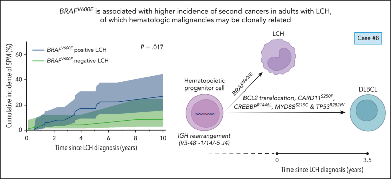
In this retrospective study, BRAF mutation status did not correlate with disease extent or (event-free) survival in 156 adults with Langerhans cell histiocytosis. BRAFV600E was associated with an increased incidence of second malignancies, often comprising hematological cancers, which may be clonally related.
TO THE EDITOR:
Langerhans cell histiocytosis (LCH) is a rare hematologic disorder with variable clinical presentations.1 The discovery of somatic mutations in the mitogen-activated protein kinase (MAPK) signaling pathway established LCH as a clonal neoplasm.2 In pediatric LCH, BRAFV600E is detected in 50% to 60% of patients and associated with severe disease forms.3,4 In adults, the prevalence and clinical associations of BRAFV600E remain incompletely characterized. We present data from a large BRAFV600E genotyped cohort of adults with LCH. In addition, we systematically reviewed published literature pertaining to frequency and clinical associations of BRAFV600E in this population and provide a meta-analysis of relevant data.
We performed an observational cohort study of adults with LCH diagnosed and/or treated at Mayo Clinic or 1 of 3 Dutch academic medical centers. Ethical approval, or waiver of consent, was obtained from all Institutional Review Boards. Mutational analysis of LCH tissue samples was performed as part of routine diagnostics or in the context of this study using published methods.4 Techniques included BRAFV600E allele-specific polymerase chain reaction (PCR), next-generation-sequencing (NGS), whole-exome sequencing, and/or BRAF-VE1 immunohistochemistry.4,5 The latter was internally validated for the evaluation of histiocytoses by cross-testing with NGS.6 Disease extent was categorized into single-system (SS) and multisystem (MS) disease and subcategorized per consensus recommendations.1 Events were defined as LCH progression, relapse, or death from any cause. Associations of BRAFV600E with clinical characteristics were evaluated with Fisher exact or Mann-Whitney tests. Event-free survival (EFS) and overall survival were estimated by the Kaplan-Meier method and compared with log-rank tests. Cumulative incidences were calculated and compared by competing risk analysis, with death as a competing event using the Fine-Gray method. Standardized incidence ratios (SIRs) were calculated as the ratio of observed-to-expected numbers of second cancers, as previously described.7 Age-, sex-, and calendar year–specific cancer rates from The Netherlands Cancer Registry from 1989 to 2020 were used to calculate expected numbers; rates of 1989 were used for the period before 1989, and rates of 2020 were used for the period after 2020. Methods for systematic review and meta-analysis are detailed in the supplemental Methods (available on the Blood website).
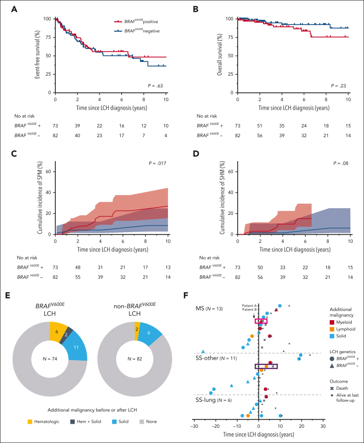
We included 156 adults with LCH and known lesional BRAFV600E status, diagnosed between 1984 and 2021 (85 from Mayo Clinic and 71 from The Netherlands). Patients from Mayo Clinic had more severe disease presentations and received chemotherapy more frequently (supplemental Table 1), likely representing referral bias. In the combined cohort, median age at diagnosis was 41 years (range, 18-88 years). Disease extent included 58 patients (37%) with MS-LCH, 22 patients (14%) with SS-pulmonary LCH, and 76 patients (49%) with nonpulmonary SS-LCH. BRAFV600E was detected in 74 of 156 (47%) patients (Table 1 and supplemental Figure 1). In the BRAFV600E-negative group, MAP2K1 mutations were detected in 20 of 41 cases (49%), BRAF exon 12 insertions/deletions were detected in 2 of 24 cases (8%), and a BRAFR662K or a LMTK2:BRAF fusion was detected in single cases8 (supplemental Figure 1 and supplemental Table 2). BRAFV600E did not correlate with age, sex, disease extent, sites of disease, or first-line therapy subtype (Table 1 and supplemental Figure 1). BRAFV600E also did not correlate with reduced EFS (P = .63; Figure 1A) or overall survival (P = .23; Figure 1B), even when excluding patients who received targeted therapy (supplemental Figure 2). However, BRAFV600E was associated with an increased incidence of second primary malignancies (SPMs) after LCH diagnosis, with a 5-year cumulative incidence of 17.3% (95% confidence interval [CI], 6.7%-27.9%) for BRAFV600E-positive vs 4.1% (95% CI, 0.0%-8.7%) for BRAFV600E-negative patients (Gray test P = .017; Figure 1C). This association was confirmed in multivariable analysis (supplemental Table 3). In addition, BRAFV600E-positive but not BRAFV600E-negative patients had a significantly elevated risk of SPMs when compared with the age-, sex-, and calendar year–matched general population of The Netherlands (SIR BRAFV600E: 5.72; 95% CI, 3.13-9.60; SIR non-BRAFV600E: 1.71; 95% CI, 0.47-4.37; supplemental Table 4). Of 18 patients with SPMs, 9 presented with second hematologic malignancies (SHMs), including 4 with acute myeloid leukemia (AML). Additional solid malignancies comprised many subtypes (supplemental Table 5). Both BRAFV600E-positive and BRAFV600E-negative patients had a significantly elevated risk of SHMs when compared with the age-, sex-, and calendar year–matched general population of The Netherlands (SIR BRAFV600E: 32.71; 95% CI, 13.15-67.39; SIR non-BRAFV600E: 9.54; 95% CI, 1.16-34.48; supplemental Table 4). Comparison of cumulative incidence of SHMs between BRAFV600E-positive and BRAFV600E-negative patients was not statistically significant (Gray test P = .08; Figure 1D), possibly due to lack of power. Notably, SHMs were not confined to patients with MS-LCH. Moreover, none of the patients with SHMs had prior exposure to alkylating agents or topoisomerase inhibitors, but all 4 with secondary AML had received cladribine (supplemental Table 6). When including malignancies occurring before LCH diagnosis, a higher frequency of additional malignancies in BRAFV600E patients remained apparent (Figure 1E); particularly, the 2 patients with hematologic malignancies before LCH harbored BRAFV600E (Figure 1F).
Table 1.
Clinical characteristics according to lesional BRAFV600E status
| Characteristic | BRAFV600E positive | BRAFV600E negative | P value |
|---|---|---|---|
| Patients, no. | 74 | 82 | |
| Age at diagnosis, median (range), y | 42.9 (19-88) | 39.0 (18-73) | .19 |
| Sex | |||
| Male | 38 (51.4) | 48 (58.5) | .42 |
| Female | 36 (48.6) | 34 (41.5) | |
| Disease extent at diagnosis | |||
| MS | 27 (36.5) | 31 (37.8) | .87 |
| SS | 47 (63.5) | 51 (62.2) | |
| Detailed subtype∗ | |||
| MS, RO+† | 9 (12.2) | 6 (7.3) | .42 |
| MS, RO- | 18 (24.3) | 25 (30.5) | .47 |
| SS, bone | 29 (39.2) | 29 (35.4) | .74 |
| SS, UFB | 24 (32.4) | 24 (29.3) | .73 |
| SS, MFB | 5 (6.8) | 5 (6.1) | 1 |
| SS, skin | 6 (8.1) | 5 (6.1) | .76 |
| SS, lung | 10 (13.5) | 12 (14.6) | 1 |
| SS, other | 2 (2.7) | 5 (6.1) | .45 |
| Disease site(s) at diagnosis | |||
| Bone | 51 (68.9) | 51 (62.2) | .40 |
| Lung | 24 (32.4) | 32 (39.0) | .41 |
| Skin | 14 (18.9) | 11 (13.4) | .39 |
| Central nervous system‡ | 9 (12.2) | 9 (11.0) | 1 |
| Lymph node | 6 (8.1) | 12 (14.6) | .22 |
| RO† | 9 (12.2) | 7 (8.5) | .60 |
| Gastrointestinal tract | 2 (2.7) | 1 (1.2) | .60 |
| First-line therapy | |||
| Chemotherapy§ | 20 (27.0) | 22 (26.8) | 1 |
| Radiotherapy | 8 (10.8) | 7 (8.5) | .79 |
| Targeted therapy|| | 3 (4.1) | 1 (1.2) | .35 |
| None/other therapy | 43 (58.1) | 51 (62.2) | .63 |
| Unknown | 0 (0.0) | 1 (1.2) | 1 |
| Follow-up, median (range), y¶ | 4.4 (0-36) | 4.2 (0-31) | .40 |
Data are given as number (percentage) of each group, unless otherwise indicated. A graphical summary of these data is provided in supplemental Figure 1.
MFB, multifocal bone; RO, risk organ; UFB, unifocal bone.
Fisher exact tests comparing patients with vs without a disease extent subtype are shown.
The hematopoietic system, liver, and spleen were considered ROs, extrapolating from pediatric data.3,4
Given that the posterior pituitary and pituitary stalk are direct extensions of the hypothalamus, pituitary tumors are classified as central nervous system involvement.
With or without additional local therapy. Among BRAFV600E-positive patients, 1 of 20 also received radiotherapy. Among BRAFV600E-negative patients, 3 of 22 also received radiotherapy.
BRAF and/or MEK inhibitors.
Estimated with the reverse Kaplan-Meier method.
Figure 1.
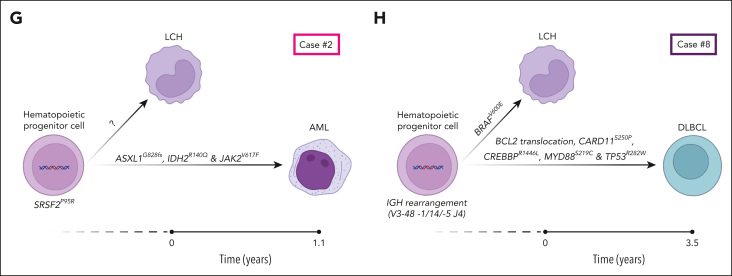
Clinical outcomes according to lesional BRAFV600E status. (A) EFS according to BRAFV600E status. Event was defined as LCH progression, relapse, or death from any cause. (B) Overall survival according to BRAFV600E status. (C) Cumulative incidence of SPMs after LCH diagnosis according to BRAFV600E status, with death as a competing event. Basal cell carcinoma was excluded from analyses of SPMs, given lack of clinical relevance. Colored ribbons indicate 95% confidence intervals. (D) Cumulative incidence of SHMs after LCH diagnosis according to BRAFV600E status, with death as a competing event. Using the etmCIF R function, the curve depicting BRAFV600E-positive patients terminates at 6.57 years, because there are no events (SHMs or death) in this group after this time point. (E) Proportion of patients with ≥1 additional malignancies, irrespective of the occurrence before or after LCH diagnosis, according to LCH lesional BRAFV600E status. (F) Time between diagnosis of LCH and the 35 additional malignancies diagnosed in 30 patients from our cohort. Each virtual horizontal line represents 1 patient. Case 2 is indicated by the pink box; case 8 is indicated by the dark purple box. Patients are grouped by LCH disease extent at diagnosis. (G) Proposed clonal relationship of the LCH and AML in case 2. In separate LCH (time [t] = 0) and AML (t = 1 year) samples, the same SRSF2P95R mutation was identified. The LCH sample was BRAFV600E negative by sequencing and BRAF-VE1 immunohistochemistry; no other MAPK pathway gene alteration was identified. In the AML specimen, additional mutations were detected in ASXL1, IDH2, and JAK2, which were absent in the LCH sample. (H) Proposed clonal relationship of the LCH and DLBCL in case 8. In separate LCH (t = 0) and DLBCL (t = 3.5 years) samples, an identical IGH gene rearrangement was identified by EuroClonality-NGS panels. In addition, BRAFV600E was detected in the LCH sample, but not in the DLBCL sample by BRAF-VE1 immunohistochemistry, allele-specific droplet digital PCR, and NGS. In the DLBCL sample, a BCL2 translocation was observed by fluorescence in situ hybridization, and CARD11S250P (NM_032415: c.T748C), CREBBPR1446L (NM_004380: c.G4337T), MYD88S219C (NM_002468: c.C656G), and TP53R282W (NM_000546: c.C844T) mutations were detected by NGS, which were not present in the LCH sample. Hem, hematologic.
In case 2 with AML diagnosed 1 year after BRAFV600E-negative MS-LCH, SRSF2P95R was identified in both the LCH sample (variant allele frequency, 26%) and AML specimen (variant allele frequency, 49%). In addition, ASXL1, IDH2, and JAK2 alterations were detected in the AML specimen, but not in the LCH sample (Figure 1G). In case 8 with diffuse large B-cell lymphoma (DLBCL) diagnosed 3.5 years after SS-bone LCH (treated surgically), PCR–based IdentiClone analysis suggested identical immunoglobulin heavy chain (IGH) and immunoglobulin kappa light chain gene rearrangements in separate LCH and DLBCL samples (supplemental Figures 3 and 4). Additional NGS-based clonality assessment confirmed the presence of an identical IGH rearrangement (V3-48-1/14/-5 J4) in the DLBCL sample and microdissected LCH tissue devoid of B cells (supplemental Figure 5). In addition, distinct oncogenic genetic alterations unique to the LCH (BRAFV600E) or DLBCL (BCL2 translocation; CARD11S250P; CREBBPR1446L; MYD88S219C; and TP53R282W) were detected (Figure 1H).
Regarding the meta-analysis (supplemental Figures 6 and 7), BRAFV600E frequency in 712 evaluated adults with LCH was 38.9% (95% CI, 35.3%-42.5%). There was no correlation of BRAFV600E with MS disease (P = .07; odds ratio, 0.75; 95% CI, 0.53-1.05) or with events (P = .14; odds ratio, 0.80; 95% CI, 0.54-1.18). No meta-analysis could be performed for SPMs, due to lack of reported data.
Our study represents the largest study to date focusing on frequency and clinical associations of BRAFV600E in adults with LCH. BRAFV600E frequencies in our cohort (47%) and meta-analysis (39%) were lower when compared with large studies of children with LCH.3,4 Contrary to findings in pediatric LCH,3,4 BRAFV600E was not associated with clinical features or with inferior EFS. These findings are supported by prior reports9, 10, 11 included in our meta-analysis. A limitation of our and other studies is that organ involvement and response to treatment were not uniformly assessed. Nevertheless, these data underscore that caution is warranted when adopting insights from pediatric LCH to the adult population.
Interestingly, BRAFV600E correlated with a higher incidence of SPMs (Figure 1C and supplemental Table 4). A previous cohort study reported a high prevalence (32%) of additional malignancies in 132 adult patients with LCH.12 Moreover, an increased risk of SPMs among patients with LCH was found in a recent population-based study,13 which also revealed that SPMs were the most common cause of death in adults with LCH. However, no genomic data for these patients were reported. Strikingly, 9 of 18 patients (50%) with SPMs in our cohort had SHMs (Figure 1F). This is in accordance with a recent study of LCH-associated malignancies.14 Furthermore, a high prevalence of myeloid malignancies has been reported in patients with Erdheim-Chester disease,15 a closely related histiocytic neoplasm also frequently characterized by BRAFV600E. Although both BRAFV600E-positive and BRAFV600E-negative patients from our cohort had a significantly elevated risk of SHMs compared with the general Dutch population, patients with BRAFV600E-positive LCH seem to be at highest risk of developing SHMs (Figure 1D). Given that LCH may arise from mutated bone marrow–derived progenitors with oligo- or multipotent differentiation capacity,5 SHMs may have shared clonal origins with LCH in some patients. Several cases with LCH and diverse hematologic malignancies harboring the same mutations, supporting this notion, have been reported.16 Our finding of identical genomic alterations in separate LCH and SHM samples (cases 2 and 8) suggests a shared clonal origin of both hematologic diseases and demonstrates the importance of analyzing such cases beyond BRAFV600E. Notably, clonal immunoglobulin and T-cell receptor gene rearrangements have previously been detected in LCH cases, including patients without (known) SHMs,17 supporting the concept that histiocytoses may occasionally be derived from lymphoid-committed cells. Analysis of clonal relations in other cases from our cohort was not feasible due to unavailability of specimens. Prospective collection and genomic analysis of tissue, blood, and/or bone marrow samples of adult patients with LCH is warranted, as this will shed more light on associations between LCH and additional neoplasms.14 Finally, further studies are needed to identify prognostic factors beyond lesional BRAFV600E status.
Conflict-of-interest disclosure: G.G. served on an advisory board for Opna Bio LLC, received royalties from UpToDate, and is a consultant to 2nd.MD. W.O.T. has received research funding from Mallinckrodt Inc, the Center for Multiple Sclerosis and Autoimmune Neurology at Mayo Clinic, and the National Institutes of Health; speaking fees from DKBMed, NeurologyLive, and the American Association for Clinical Chemistry; and publishing royalties from the publication of Mayo Clinic Cases in Neuroimmunology. The remaining authors declare no competing financial interests.
Acknowledgments
The authors thank the molecular diagnostics staff of the Department of Pathology of Leiden University Medical Center, as well as Michelle van der Klift, Fleur de Groot, Patty Jansen, and Karoly Szuhai for their help with genetic analyses. Sofie Breuking is acknowledged for prior work on the systematic review. The authors also thank Wichor Bramer from the Erasmus MC Medical Library for developing and updating the search strategies for the systematic review. Figure 1G-H was made with BioRender.com software.
This study was supported by grants from Stichting 1000 Kaarsjes voor Juultje to A.G.S.v.H., the University of Iowa/Mayo Clinic Lymphoma SPORE P50 CA97274 to G.G. and J.P.A., and the Walter B. Frommeyer, Jr, Fellowship Award in Investigative Medicine, University of Alabama at Birmingham, to G.G. P.G.K. received an MD/PhD grant from Leiden University Medical Center.
Authorship
Contribution: C.v.d.B., J.A.M.v.L., R.S.G., G.G., and A.G.S.v.H. conceived the study; A.A.A.-M., P.G.K., T.C.E.Z., and J.P.A. collected clinical data; P.G.K., J.F.-B., E.C.S., S.D., R.A.L.d.G., A.W.L., T.v.W., and A.G.S.v.H. performed additional molecular genetic analyses outside routine diagnostics; P.G.K. analyzed the combined clinicogenomic data set and drafted the (supplemental) figures and tables; T.C.E.Z. performed the systematic review and meta-analysis, with P.G.K. as second assessor; E.A.M.F. supervised the systematic review and statistical analyses; J.C.T. helped with calculating standardized incidence ratios for second cancers; A.A.A.-M., P.G.K., T.C.E.Z., R.S.G., G.G., and A.G.S.v.H. drafted the manuscript; A.A.A.-M., J.P.A., N.N.B., S.M.S., M.V.S., C.D.-P., M.J.K., J.H.R., R.V., W.O.T., J.R.Y., K.R., A.R., A.H.G.C., R.M.V., C.J.M.v.N., B.V.B., G.J.B., P.S., J.A.M.B., J.S.P.V., M.A.J.v.d.S., E.F.S., S.H.T., C.v.d.B., J.A.M.v.L., R.S.G., and G.G. were involved in clinical care for included patients; and all authors critically reviewed and approved the manuscript.
Footnotes
∗A.A.A.-M., P.G.K., and T.C.E.Z. are joint first authors and contributed equally.
†G.G. and A.G.S.v.H. are joint last authors and contributed equally.
Presented at the 39th Annual Meeting of the Histiocyte Society, 21 to 24 October 2023, Athens, Greece.
Data are available on reasonable request from the corresponding authors.
The online version of this article contains a data supplement.
Contributor Information
Gaurav Goyal, Email: ggoyal@uabmc.edu.
Astrid G. S. van Halteren, Email: a.vanhalteren@erasmusmc.nl.
Supplementary Material
References
- 1.Goyal G, Tazi A, Go RS, et al. International expert consensus recommendations for the diagnosis and treatment of Langerhans cell histiocytosis in adults. Blood. 2022;139(17):2601–2621. doi: 10.1182/blood.2021014343. [DOI] [PubMed] [Google Scholar]
- 2.Allen CE, Beverley PCL, Collin M, et al. The coming of age of Langerhans cell histiocytosis. Nat Immunol. 2020;21(1):1–7. doi: 10.1038/s41590-019-0558-z. [DOI] [PubMed] [Google Scholar]
- 3.Héritier S, Emile J-F, Barkaoui M-A, et al. BRAF mutation correlates with high-risk Langerhans cell histiocytosis and increased resistance to first-line therapy. J Clin Oncol. 2016;34(25):3023–3030. doi: 10.1200/JCO.2015.65.9508. [DOI] [PMC free article] [PubMed] [Google Scholar]
- 4.Kemps PG, Zondag TCE, Arnardóttir HB, et al. Clinicogenomic associations in childhood Langerhans cell histiocytosis: an international cohort study. Blood Adv. 2023;7(4):664–679. doi: 10.1182/bloodadvances.2022007947. [DOI] [PMC free article] [PubMed] [Google Scholar]
- 5.Xiao Y, van Halteren AGS, Lei X, et al. Bone marrow–derived myeloid progenitors as driver mutation carriers in high- and low-risk Langerhans cell histiocytosis. Blood. 2020;136(19):2188–2199. doi: 10.1182/blood.2020005209. [DOI] [PubMed] [Google Scholar]
- 6.Acosta-Medina AA, Abeykoon JP, Go RS, et al. BRAF testing modalities in histiocytic disorders: comparative analysis and proposed testing algorithm. Am J Clin Pathol. Published online 17 July 2023. [DOI] [PubMed]
- 7.Teepen JC, van Leeuwen FE, Tissing WJ, et al. Long-term risk of subsequent malignant neoplasms after treatment of childhood cancer in the DCOG LATER study cohort: role of chemotherapy. J Clin Oncol. 2017;35(20):2288–2298. doi: 10.1200/JCO.2016.71.6902. [DOI] [PubMed] [Google Scholar]
- 8.Zanwar S, Abeykoon JP, Dasari S, et al. Clinical and therapeutic implications of BRAF fusions in histiocytic disorders. Blood Cancer J. 2022;12(6):97. doi: 10.1038/s41408-022-00693-7. [DOI] [PMC free article] [PubMed] [Google Scholar]
- 9.Jouenne F, Chevret S, Bugnet E, et al. Genetic landscape of adult Langerhans cell histiocytosis with lung involvement. Eur Respir J. 2020;55(2) doi: 10.1183/13993003.01190-2019. [DOI] [PubMed] [Google Scholar]
- 10.Cao X, Duan M, Zhao A, et al. Treatment outcomes and prognostic factors of patients with adult Langerhans cell histiocytosis. Am J Hematol. 2022;97(2):203–208. doi: 10.1002/ajh.26412. [DOI] [PubMed] [Google Scholar]
- 11.Stathi D, Yavropoulou MP, Allen CE, et al. Prevalence of the BRAF V600E mutation in Greek adults with Langerhans cell histiocytosis. Pediatr Hematol Oncol. 2022;39(6):540–548. doi: 10.1080/08880018.2022.2029988. [DOI] [PubMed] [Google Scholar]
- 12.Ma J, Laird JH, Chau KW, Chelius MR, Lok BH, Yahalom J. Langerhans cell histiocytosis in adults is associated with a high prevalence of hematologic and solid malignancies. Cancer Med. 2019;8(1):58–66. doi: 10.1002/cam4.1844. [DOI] [PMC free article] [PubMed] [Google Scholar]
- 13.Goyal G, Parikh R, Richman J, et al. Spectrum of second primary malignancies and cause-specific mortality in pediatric and adult Langerhans cell histiocytosis. Leuk Res. 2023;126 doi: 10.1016/j.leukres.2023.107032. [DOI] [PubMed] [Google Scholar]
- 14.Bagnasco F, Zimmermann SY, Egeler RM, et al. Langerhans cell histiocytosis and associated malignancies: a retrospective analysis of 270 patients. Eur J Cancer. 2022;172:138–145. doi: 10.1016/j.ejca.2022.03.036. [DOI] [PubMed] [Google Scholar]
- 15.Papo M, Diamond EL, Cohen-Aubart F, et al. High prevalence of myeloid neoplasms in adults with non–Langerhans cell histiocytosis. Blood. 2017;130(8):1007–1013. doi: 10.1182/blood-2017-01-761718. [DOI] [PMC free article] [PubMed] [Google Scholar]
- 16.Kemps PG, Hebeda KM, Pals ST, et al. Spectrum of histiocytic neoplasms associated with diverse haematological malignancies bearing the same oncogenic mutation. J Pathol Clin Res. 2021;7(1):10–26. doi: 10.1002/cjp2.177. [DOI] [PMC free article] [PubMed] [Google Scholar]
- 17.Chen W, Wang J, Wang E, et al. Detection of clonal lymphoid receptor gene rearrangements in Langerhans cell histiocytosis. Am J Surg Pathol. 2010;34(7):1049–1057. doi: 10.1097/PAS.0b013e3181e5341a. [DOI] [PubMed] [Google Scholar]
Associated Data
This section collects any data citations, data availability statements, or supplementary materials included in this article.


