Abstract
Studies of the proteins synthesized by Salmonella typhimurium during growth within tissue culture cells have previously focused on a single cell type. In the present study we examine the different protein patterns exhibited by S. typhimurium during growth within three different cell types relevant to those it would encounter throughout the course of a natural infection, including intestinal epithelial cells (Intestine-407), macrophages (J774.A, rat bone marrow-derived macrophages, and mouse bone marrow-derived macrophages), and liver cells (NMuLi). Side-by-side comparisons reveal that S. typhimurium responds to these different cellular environments with specific patterns of protein synthesis unique to each cell type. The numbers of proteins detected in each cell line are as follows: 142 proteins in Intestine-407, of which 58 appear to be unique to growth within this cell line; 413 proteins in J774.A, of which 157 appear to be unique; 260 proteins in rat bone marrow-derived macrophages, of which 40 appear to be unique; 336 proteins in mouse bone marrow-derived macrophages, of which 113 appear to be unique; and 183 proteins in NMuLi, of which 91 appear to be unique.
Facultative anaerobes such as Salmonella typhimurium respond dynamically to the particular constraints of their diverse environments through the expression and/or repression of groups of genes, each designed to confer a selective advantage under specific environmental conditions (17, 22–24, 27, 28). This phenomenon is termed global regulation (9). Studies have been done which delineate the groups of proteins salmonellae synthesize while growing in culture under various environmental conditions, as well as within cultured mammalian cells, and not surprisingly, many of the genes involved in global regulation also affect virulence of the organism (15, 16). Salmonellae are able to attach to, invade (8), and survive within eukaryotic cells (7). During the course of an infection, the organism invades a number of different cell types, including the intestinal epithelium (25, 26), various macrophages (2), and deep tissues such as the liver and spleen (5, 6). The mechanisms which facilitate tropism toward such a broad spectrum of host cells have not been specifically addressed. Although numerous studies have been done on the intracellular protein synthesis patterns of S. typhimurium in cultured cell lines, most laboratories have limited their investigations to a single cell type (1, 12) or occasionally to various cell lines representing a single cell type (2). In an effort to address the extent to which S. typhimurium is capable of responding to each unique intracellular environment, protein synthesis was analyzed with a single virulent SL1344 strain, χ3339, during growth within three distinct cell types: intestinal epithelium, macrophages, and normal murine liver. These cell types are representative of those which would be encountered by S. typhimurium during the natural course of infection. The different cell lines were all infected with S. typhimurium, and the intracellular protein synthesis patterns were examined by two-dimensional (2-D) gel electrophoresis. The organism was observed to synthesize proteins specific to each cell type examined, presenting evidence that S. typhimurium dynamically responds to each unique environment encountered during the establishment of systemic infection.
Maintenance and growth conditions of the mammalian and bacterial cells.
The three commercially available established cell lines used for this study were all purchased from the American Type Culture Collection (ATCC), Rockville, Md. They include the human embryonic intestinal epithelial cell line Intestine-407 (Int-407; purchased as ATCC CCL6 [11]), the murine macrophage-like cell line J774.A (ATCC TIB67 [21]), and the murine normal liver epithelial cell line NMuLi (ATCC CRL1638 [19]). These cell lines were maintained in Eagle’s minimal essential medium (Gibco BRL, Gaithersburg, Md.) (EMEM) containing 10% fetal calf serum (FCS; Sigma, St. Louis, Mo.) and 2 mM glutamine at 37°C in a 5% CO2 environment. Working stocks were passaged every 3 to 5 days for up to 15 passages. Cells were seeded at 5 × 10⁵/ml/well into six-well plates (Costar, Cambridge, Mass.) and used the following day at near 95% confluency for purposes of the labeling procedure.
Bone marrow-derived macrophages were prepared by culturing bone marrow cells from femurs and tibias of either Sprague-Dawley rats (Harlan Sprague Dawley, Indianapolis, Ind.) or BALB/c mice (Harlan Sprague Dawley) following previously described procedures (2). Cells were maintained in 75-cm2 flasks in Dulbecco’s modified Eagle medium (Gibco BRL) plus 10% FCS, 5% horse serum (Sigma, St. Louis, Mo.), 10% conditioned medium from L929 cells (L-cells), 1 mM glutamine, and 1% penicillin for 24 h in a 5% CO2 environment. Nonadherent bone marrow cells were removed after 24 h, spent medium was replaced, and the cells were incubated for an additional 5 days. Prior to their use in infection experiments, macrophages were scraped from the surface of the tissue culture flask; resuspended in fresh Dulbecco’s modified Eagle medium supplemented with 10% FCS, 5% horse serum, and 10% L-cell-conditioned medium (without antibiotics); and used to seed six-well tissue culture plates at a concentration of 5 × 10⁵/ml and used the following day at 95% confluency.
The bacterial strain used in this study was S. typhimurium χ3339, an animal-passaged isolate of the virulent SL1344 wild type (10). χ3339 was first grown in Luria broth (13) as an aerated overnight culture at 37°C. It was then inoculated at a dilution of 1:100 into 30 ml of prewarmed EMEM and incubated statically overnight in a 5% CO2 atmosphere at 37°C. Titers in EMEM were comparable to those obtained in Luria broth (1.9 × 10⁸). The culture was centrifuged at 8,800 × g for 10 min and resuspended in 10 ml of fresh EMEM, and 1 ml per well was added to the monolayers at a multiplicity of infection of approximately 100:1.
Labeling the proteins synthesized intracellularly by χ3339.
Int-407, J774.A, and NMuLi cells were seeded at 5 × 10⁵/ml and allowed to grow to near 95% confluency in six-well plates. Primary macrophage cultures were plated as described above. Monolayers were washed twice with prewarmed Hanks’ balanced salt solution, and each well received 1 ml of the χ3339 suspension described above at a multiplicity of infection of approximately 100:1. All incubations were done in a 5% CO2 atmosphere at 37°C. Attachment and invasion were allowed to proceed for 2 h. Supernatant fluid was then removed, the monolayers were washed three times with Hanks’ balanced salt solution, and fresh EMEM containing gentamicin (100 μg/ml), cycloheximide (200 μg/ml), and Tran35S Label (150 μCi/ml; specific activity, 1,092 Ci/mmol) (ICN Radiochemicals, Irvine, Calif.) was added. This labeling solution contains a mixture of [35S]methionine and [35S]cysteine. Control monolayers containing no bacteria were also treated with cycloheximide and were maintained under the same conditions for each cell line. One milliliter of the remaining bacterial suspension was placed in a microcentrifuge tube and allowed to incubate under the same conditions as the infected monolayers. Labeling continued for 2 h, and the monolayers were then washed three times with phosphate-buffered saline and lysed with 1 ml of 0.1% sodium deoxycholate. Ten microliters was removed to calculate the titer. The numbers of bacteria recovered from each cell type were 5.1 × 10⁶ (J774.A), 4.2 × 10⁶ (Int-407), 2.5 × 10⁶ (rat bone marrow-derived macrophages), 1.9 × 10⁶ (mouse bone marrow-derived macrophages), and 1.1 × 10⁶ (NMuLi). The remaining bacteria were collected by centrifugation in a microcentrifuge tube for 10 min at 8,800 × g. The 1-ml sample of control bacteria was prepared in the same manner. Bacterial pellets were resuspended in 40 μl of sample buffer I (SB-I; 0.3% [wt/vol] sodium dodecyl sulfate, 200 mM dithiothreitol, 28 mM Tris-HCl, 22 mM Tris base) and boiled for 5 min. The suspensions were treated with 4 μl of sample buffer II (SB-II; 24 mM Tris base, 476 mM Tris-HCl, 50 mM MgCl2, DNase [1 mg/ml], RNase A [0.25 mg/ml]), chilled on ice for 10 min, and then treated with 160 μl of sample buffer III (SB-III; 9.9 M urea, 4% Nonidet P-40, 2.2% preblended ampholytes [pH 3 to 10 and 5 to 7], 100 mM dithiothreitol). Ten microliters was removed from each sample to determine the counts per minute. Samples were stored at −70°C until analyzed by 2-D electrophoresis. Noninfected control monolayers were washed three times with phosphate-buffered saline, lysed with 0.5 ml of boiling-hot SB-I, boiled further for 5 min, and then placed on ice for 5 min. Twenty-four microliters of SB-II was added, and the sample was further chilled for 10 min. Samples were centrifuged at 8,800 × g for 10 min, the supernatant was removed, and the pellets were allowed to air dry for 5 min. The pellets were resuspended in 240 μl of a 1:4 mixture of SB-I and SB-III and analyzed by 2-D electrophoresis. A 10-μl sample was removed to determine the counts per minute, and these samples were loaded on the basis of the amount of protein (approximately 100 μg). All of the bacterial samples were normalized to 500,000 cpm, except those from NMuLi (50,000 cpm), for which the total counts obtained were much lower. These experiments were repeated three times for the Int-407, NMuLi, and J774.A cell lines and twice for the mouse bone marrow-derived macrophages, each time with a single culture of χ3339 and simultaneous infection of the cell lines. The rat bone marrow-derived macrophages were examined separately, and the experiment was repeated twice. Samples from each trial were analyzed as described below.
2-D electrophoresis of the intracellular proteins.
2-D electrophoresis was performed essentially by the method of O’Farrell (18), with some modifications. (All reagents were obtained from Oxford Glycosystems, Bedford, Mass.; the equipment used was the Millipore 2-D Investigator, which is now available from ESA, Chelmsford, Mass.). Protein samples were loaded onto 4.1% acrylamide tube gels for isoelectric focusing in the first dimension. The ampholytes used were in a preblended mixture with a pH range of 3 to 10, reinforced in the pH 5 to 7 range. Isoelectric focusing was carried out for 18,000 V · h. The second dimension consisted of large-format (22- by 22-cm) 11.5% polyacrylamide slab gels which were run for 6 h at a maximum voltage of 500. The 2-D parameters described in this study allow detection of proteins having pIs primarily within the 3.5 to 6.5 range and molecular masses of approximately 14 to 100 kDa. Gels were fixed in a solution of isopropanol-water-acetic acid (65:25:10 [vol/vol/vol]) for 30 min, soaked in Amplify (Amersham International, Arlington Heights, Ill.) for 30 min, dried, and exposed to Kodak X-Omat AR X-ray film (Eastman Kodak Co., Rochester, N.Y.) at −70°C. The number of bacteria obtained from NMuLi cells was lower than that obtained from the others, as were the counts obtained. We were able to load only 50,000 cpm for preparations obtained from NMuLi cells, which resulted in the need for a longer exposure time, 4 days. The gels from the rest of the samples were exposed for 2 days. Samples of the uninfected, cycloheximide-treated monolayers of each cell type were also run on 2-D gels. These gels were exposed for the same length of time as the gels containing the bacterial samples from that cell type. The autoradiograms revealed no proteins, indicating that host cell protein synthesis was inhibited throughout the time course of the assay (data not shown).
Image acquisition and analysis of protein patterns.
A description of the equipment and software used for image acquisition and analysis has been given previously (4). We have used the technique of 2-D polyacrylamide gel electrophoresis extensively in this laboratory to analyze proteins synthesized by virulent strains of S. typhimurium in response to diverse environmental conditions in vitro. These analyses have led to the development of a database of proteins synthesized under an extensive range of growth conditions (unpublished data), including intracellular environments with a variety of cell lines and various stresses, including in vitro conditions which represent some aspects of the macrophage intracellular environment (heat shock, acid shock, detergent shock, nutrient deprivation, and oxidative stress). This database was used as a source of comparison for analysis of the present study.
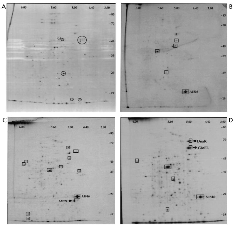
Our analyses of the bacterial samples revealed that the fewest proteins were synthesized within the epithelial cell lines Int-407 and NMuLi (Table 1). There were 142 total detectable proteins synthesized by χ3339 during growth within the Int-407 cell line (Fig. 1B). The most prominent of these proteins was designated A1816, which represented 36% of the total integrated intensity of all proteins combined. A1816 exhibited a pI of 4.77 and a molecular mass of 20.5 kDa. The intracellular pattern was compared to the pattern exhibited by χ3339 grown in EMEM (Fig. 1A), and 66 proteins were found to be present under both conditions. Three of the common proteins were induced at threefold or higher levels during intracellular growth. The remaining 76 proteins were synthesized only during intracellular growth, and of this group, 58 appeared to be synthesized only in response to the Int-407 intracellular environment (Table 1).
TABLE 1.
Cell-specific patterns of protein synthesis by χ3339
| No. of proteinsa | Presence of protein(s) after growth in:
|
|||||
|---|---|---|---|---|---|---|
| EMEM | Int-407 | NMuLi | J774.A | RBMb | MBMc | |
| 85 | × | |||||
| 58 | × | |||||
| 91 | × | |||||
| 157 | × | |||||
| 40 | × | |||||
| 113 | × | |||||
| 9d | × | × | × | × | × | × |
| 0 | × | × | × | × | × | |
| 5 | × | × | × | × | ||
| 27e | × | × | × | |||
| 1f | × | × | × | |||
Indicates the number of proteins found to be unique to each of these specified groups.
Rat bone marrow-derived macrophages.
Mouse bone marrow-derived macrophages.
Proteins present under all conditions, both intra- and extracellular.
Macrophage-specific proteins.
Proteins found only within cultured cell lines.
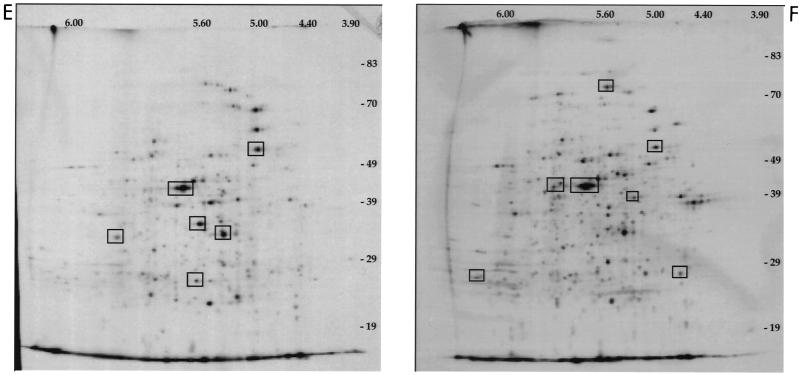
FIG. 1.
2-D sodium dodecyl sulfate-polyacrylamide gel electrophoresis and autofluorography of χ3339 whole-cell proteins synthesized in the presence of Tran35S Label. Circles show representative proteins that are missing or reduced during intracellular growth. Squares show representative proteins that are enhanced or unique to intracellular growth. (A) Whole-cell proteins synthesized by χ3339 during growth within EMEM. (B) Whole-cell proteins synthesized by χ3339 during growth within Int-407 cells. Protein A1816 is labeled. (C) Whole-cell proteins synthesized by χ3339 during growth within NMuLi cells. Protein A1816 is labeled. The arrow indicates a predominant protein, AX324, which is found only during growth within NMuLi. (D) Whole-cell proteins synthesized by χ3339 during growth within J774.A cells. Protein A1816 is labeled. The heat shock proteins DnaK and GroEL are also labeled. (E) Whole-cell proteins synthesized by χ3339 during growth within rat bone marrow-derived macrophages. Protein A1816 is not detected during growth within these cells. (F) Whole-cell proteins synthesized by χ3339 during growth within mouse bone marrow-derived macrophages.
There were 183 detectable proteins synthesized by χ3339 when this organism was grown within the NMuLi cell line (Fig. 1C); 87 of them matched the pattern seen during growth within EMEM. The remaining 96 proteins were synthesized only during intracellular growth, and 91 of them were found to be unique to growth within NMuLi cells (Table 1). The most prominent protein synthesized solely within NMuLi cells was designated AX324, which represented 12% of the total integrated intensity. AX324 exhibited a pI of 4.83 and a molecular mass of 24.2 kDa. A1816, seen during growth within Int-407 cells, was also synthesized within NMuLi cells. It represented 23.8% of the total integrated intensity of all proteins combined.
The greatest number of proteins was synthesized within the macrophage cells (Table 1). One possible explanation for this result is that de novo synthesis of a larger set of proteins may be required for survival and multiplication within these cells than within the epithelial cell lines, which do not mount an actively antimicrobial response upon invasion by bacteria. There were 413 proteins detected in the J774.A cell line (Fig. 1D) (including A1816; 10% of total integrated intensity), of which 163 matched the in vitro pattern (Fig. 1A). Of the 163 that matched the pattern in EMEM, 123 were induced at threefold or higher levels during intracellular growth. There were 250 proteins which were synthesized only during intracellular growth, 157 of which appeared to be unique to the J774 environment (Table 1).
There were 260 proteins detected during growth within rat bone marrow-derived macrophages, 161 of which matched the pattern seen in EMEM. Of the remaining 99 proteins, 40 were unique to the rat bone marrow-derived-macrophage environment (Fig. 1E; Table 1).
There were 336 proteins detected during growth within mouse bone marrow-derived macrophages, 144 of which matched the pattern seen for growth in EMEM (Fig. 1F). Of the remaining 192, 113 were observed to be unique to this cell line. Interestingly, the A1816 protein, previously seen only in the established cell lines, was detected by computer-assisted analysis during growth within this primary cell line, although at levels significantly lower than those in the established lines. A1816 represented only 0.6% of the total integrated intensity (Table 2). The difference in protein pattern between the primary macrophages and the immortalized cell line was not surprising, since it has been shown that J774.A cells allow higher rates of intracellular survival than do primary macrophage cultures (2, 29) and, subsequently, a greater recovery of bacteria from this cell line as compared with the primary macrophages, which could affect the number of detectable proteins. An alternative explanation for this difference in protein number may be that the timing involved in synthesis of proteins varies among cell types, even among the different macrophages. It is possible that the primary macrophages respond more rapidly than J774.A cells. In order to see the entire repertoire of proteins made in each cell type it would be necessary to perform studies of bacteria harvested at a number of different time points in each cell line. However, even within the time constraints of the present study, we were able to identify proteins unique to each cell type and line.
TABLE 2.
Percent integrated intensity of two prominent proteins synthesized within each cell line
| Protein | pI | Molecular massa | % Integrated intensity in:
|
|||||
|---|---|---|---|---|---|---|---|---|
| EMEM | Int-407 | NMuLi | J774.A | RBMb | MBMc | |||
| A1816 | 4.77 | 26.4 | NDd | 35.8 | 23.8 | 10.4 | ND | 0.6 |
| C1807 | 5.59 | 45.5 | 0.4 | 8.4 | 4.5 | 10.4 | 10.5 | 8.4 |
In kilodaltons.
Rat bone marrow-derived macrophages.
Mouse bone marrow-derived macrophages.
ND, not detectable.
Abshire and Neidhardt (1) observed the repression of 12 proteins and the induction of 22 proteins within four hours of phagocytosis in U937 cells. However, computer imaging and detection analysis revealed that 40 proteins were induced and 100 were repressed over the course of intracellular growth (1). The regulation of this larger number of proteins corresponds well with our own observations.
In addition to the identification of groups of proteins which were uniquely associated with each cell line or primary cell culture, several interesting patterns were observed. Nine proteins were identified that were present under all conditions, both intra- and extracellular (Table 1). A group of 27 proteins was synthesized only within macrophages and the macrophage-like cell line J774.A. A comparison to our database of proteins induced by in vitro stresses revealed that 2 of these 27 proteins appear to be completely unique to the macrophage environment, since the synthesis of either of these proteins was not observed to be induced by any of the stresses previously tested in our laboratory (unpublished data). An indication of the differences among the intracellular environments tested is apparent in the fact that no proteins were identified that were synthesized under all intracellular conditions, although a group of five proteins was observed which were synthesized in all of the cell lines except the liver cell line, NMuLi. This observation could be the result of the lower number of bacteria and counts obtained from the NMuLi cell line. It should also be kept in mind that there may be many other proteins synthesized in such small amounts that we are unable to detect them within the limits of this particular technique. Therefore, the groups discussed here should be considered only as starting points for further study and not as definitive lists of all cell-specific proteins synthesized by S. typhimurium.
Table 2 provides a comparison among cell types of two prominent proteins, A1816 and C1807. A1816 was greatly induced almost exclusively within the immortalized cell lines, with the exception of the mouse bone marrow-derived macrophages. Although A1816 was synthesized within these cells, its synthesis occurred at a significantly reduced level. C1807 was seen to be enhanced under all intracellular conditions, regardless of cell type.
It is evident from this study that the S. typhimurium SL1344 strain χ3339 responded differently to each cell line which it invaded or into which it was phagocytosed. A systematic study of cell-specific responses has not been previously undertaken, although several instances of differential response of salmonellae to specific cell lines have been previously noted (1, 7). It has been reported that S. typhimurium (ATCC 14028) heat shock proteins were prominently upregulated following phagocytosis by cells of the murine macrophage-like cell line J774 (3), but these same proteins were not upregulated by S. typhimurium SR-11 following phagocytosis by the human macrophage-like cell line U937 (1). In the present study the levels of the heat shock proteins DnaK and GroEL were upregulated within the J774.A cell line (Fig. 1D) but were not seen to be significantly enhanced in either of the primary cell cultures of mouse or rat bone marrow-derived macrophages.
The extent of variation within the SL1344 intracellular protein patterns in this study indicates that the systems used for sensing environmental conditions and regulating gene expression provide Salmonella a reasonably high degree of complexity in controlling its response to each specific intracellular environment. S. typhimurium uses a series of two-component regulatory systems to coordinately regulate genes in response to a diverse set of environmental signals (14). It is reasonable to assume that host cells possess unidentified factors which serve as signals to induce the synthesis of proteins necessary for survival or multiplication within those cells. These hypothetical signals may vary considerably between different cell types, providing Salmonella with the opportunity to specifically adapt to each new environment, thereby increasing the likelihood for survival. This view is certainly supported by the present comparative analysis of the SL1344 protein synthesis patterns within a variety of different cell types.
The obvious and significant differences in protein patterns presented here provide a starting point for the elucidation of cell tropism in Salmonella. Since many of these proteins are synthesized only during intracellular growth, they cannot be identified through comparisons with the SL1344 2-D reference map generated from in vitro-cultured bacteria (20). Therefore, the cell-specific proteins A1816 (enhanced primarily in established cell lines) and AX324 (unique to growth within NMuLi) are currently being purified for microsequence analysis. It is hoped that the identification of these proteins will lead to a better understanding of the responses of S. typhimurium to the various intracellular environments encountered during a systemic infection as well as to the identification of proteins that may be necessary to establish that infection.
Acknowledgments
This work was supported by grant AI24533 from the National Institutes of Health and an unrestricted grant from Bristol Myers Squibb.
We thank our colleagues for helpful discussions.
REFERENCES
- 1.Abshire K Z, Neidhardt F C. Analysis of proteins synthesized by Salmonella typhimurium during growth within a host macrophage. J Bacteriol. 1993;175:3734–3743. doi: 10.1128/jb.175.12.3734-3743.1993. [DOI] [PMC free article] [PubMed] [Google Scholar]
- 2.Buchmeier N A, Heffron F. Intracellular survival of wild-type Salmonella typhimurium and macrophage-sensitive mutants in diverse populations of macrophages. Infect Immun. 1989;57:1–7. doi: 10.1128/iai.57.1.1-7.1989. [DOI] [PMC free article] [PubMed] [Google Scholar]
- 3.Buchmeier N A, Heffron F. Induction of Salmonella stress proteins upon infection of macrophages. Science. 1990;248:730–732. doi: 10.1126/science.1970672. [DOI] [PubMed] [Google Scholar]
- 4.Burns-Keliher L L, Portteus A, Curtiss R., III Specific detection of Salmonella typhimurium proteins synthesized intracellularly. J Bacteriol. 1997;179:3604–3612. doi: 10.1128/jb.179.11.3604-3612.1997. [DOI] [PMC free article] [PubMed] [Google Scholar]
- 5.Conlan J W, North R J. Early pathogenesis of infection in the liver with the facultative intracellular bacteria Listeria monocytogenes, Francisella tularensis, and Salmonella typhimurium involves lysis of infected hepatocytes by leukocytes. Infect Immun. 1992;60:5164–5171. doi: 10.1128/iai.60.12.5164-5171.1992. [DOI] [PMC free article] [PubMed] [Google Scholar]
- 6.Dunlap N E, Benjamin W B, Berry A K, Eldridge J H, Briles D E. A ‘safe-site’ for Salmonella typhimurium is within splenic polymorphonuclear cells. Microb Pathog. 1992;13:181–190. doi: 10.1016/0882-4010(92)90019-k. [DOI] [PubMed] [Google Scholar]
- 7.Finlay B B, Falkow S. Salmonella as an intracellular parasite. Mol Microbiol. 1989;3:1833–1841. doi: 10.1111/j.1365-2958.1989.tb00170.x. [DOI] [PubMed] [Google Scholar]
- 8.Galan J E, Curtiss R., III Expression of Salmonella typhimurium genes required for invasion is regulated by changes in DNA supercoiling. Infect Immun. 1990;58:1879–1885. doi: 10.1128/iai.58.6.1879-1885.1990. [DOI] [PMC free article] [PubMed] [Google Scholar]
- 9.Gottesman S. Bacterial regulation: global regulatory networks. Annu Rev Genet. 1984;18:415–441. doi: 10.1146/annurev.ge.18.120184.002215. [DOI] [PubMed] [Google Scholar]
- 10.Gulig P A, Curtiss R., III Plasmid-associated virulence of Salmonella typhimurium. Infect Immun. 1987;55:2891–2901. doi: 10.1128/iai.55.12.2891-2901.1987. [DOI] [PMC free article] [PubMed] [Google Scholar]
- 11.Henle G, Deinhardt F. The establishment of strains of human cells in tissue culture. J Immunol. 1957;79:54–59. [PubMed] [Google Scholar]
- 12.Kusters J G, Mulders-Kremers G A W M, van Doornik C E M, van der Zeijst B A M. Effects of multiplicity of infection, bacterial protein synthesis, and growth phase on adhesion to and invasion of human cell lines by Salmonella typhimurium. Infect Immun. 1993;61:5013–5020. doi: 10.1128/iai.61.12.5013-5020.1993. [DOI] [PMC free article] [PubMed] [Google Scholar]
- 13.Luria S E, Burrous J W. Hybridization between Escherichia coli and shigella. J Bacteriol. 1957;74:461–476. doi: 10.1128/jb.74.4.461-476.1957. [DOI] [PMC free article] [PubMed] [Google Scholar]
- 14.Mekalanos J J. Environmental signals controlling expression of virulence determinants in bacteria. J Bacteriol. 1992;174:1–7. doi: 10.1128/jb.174.1.1-7.1992. [DOI] [PMC free article] [PubMed] [Google Scholar]
- 15.Miller J F, Mekalanos J J, Falkow S. Coordinate regulation and sensory transduction in the control of bacterial virulence. Science. 1989;243:916–922. doi: 10.1126/science.2537530. [DOI] [PubMed] [Google Scholar]
- 16.Miller S I, Kukral A M, Mekalanos J J. A two-component regulatory system (phoP-phoQ) controls Salmonella typhimurium virulence. Proc Natl Acad Sci USA. 1989;86:5054–5058. doi: 10.1073/pnas.86.13.5054. [DOI] [PMC free article] [PubMed] [Google Scholar]
- 17.Neidhardt F C, VanBogelen R A, Vaughn V. The genetics and regulation of heat shock proteins. Annu Rev Genet. 1984;18:295–329. doi: 10.1146/annurev.ge.18.120184.001455. [DOI] [PubMed] [Google Scholar]
- 18.O’Farrell P H. High resolution two-dimensional electrophoresis of proteins. J Biol Chem. 1975;250:4007–4021. [PMC free article] [PubMed] [Google Scholar]
- 19.Owens R B, Smith H S, Hackett A J. Epithelial cell cultures from normal glandular tissue of mice. J Natl Cancer Inst (Bethesda) 1974;52:1375–1378. doi: 10.1093/jnci/53.1.261. [DOI] [PubMed] [Google Scholar]
- 20.Qi S-Y, Moir A, O’Connor C D. Proteome of Salmonella typhimurium SL1344: identification of novel abundant cell envelope proteins and assignment to a two-dimensional reference map. J Bacteriol. 1996;178:5032–5038. doi: 10.1128/jb.178.16.5032-5038.1996. [DOI] [PMC free article] [PubMed] [Google Scholar]
- 21.Ralph P, Nakoinz I. Phagocytosis and cytocytosis by a macrophage tumour and its cloned cell line. Nature. 1975;257:393–394. doi: 10.1038/257393a0. [DOI] [PubMed] [Google Scholar]
- 22.Smith M W, Neidhardt F C. Proteins induced by anaerobiosis in Escherichia coli. J Bacteriol. 1983;154:336–343. doi: 10.1128/jb.154.1.336-343.1983. [DOI] [PMC free article] [PubMed] [Google Scholar]
- 23.Smith M W, Neidhardt F C. Proteins induced by aerobiosis in Escherichia coli. J Bacteriol. 1983;154:344–350. doi: 10.1128/jb.154.1.344-350.1983. [DOI] [PMC free article] [PubMed] [Google Scholar]
- 24.Spector M P, Aliabadi A, Gonzalez T, Foster J W. Global control in Salmonella typhimurium: two-dimensional electrophoretic analysis of starvation-, anaerobiosis-, and heat shock-inducible proteins. J Bacteriol. 1986;168:420–424. doi: 10.1128/jb.168.1.420-424.1986. [DOI] [PMC free article] [PubMed] [Google Scholar]
- 25.Takeuchi A. Electron microscopic studies of experimental Salmonella infection. I. Penetration into the intestinal epithelium by Salmonella typhimurium. Am J Pathol. 1967;50:109–136. [PMC free article] [PubMed] [Google Scholar]
- 26.Takeuchi A, Sprinz H. Electron-microscope studies of experimental Salmonella infection in the preconditioned guinea pig. II. Response of the intestinal mucosa to the invasion by Salmonella typhimurium. Am J Pathol. 1967;51:137–161. [PMC free article] [PubMed] [Google Scholar]
- 27.Walker G C. Mutagenesis and inducible responses to deoxyribonucleic acid damage in Escherichia coli. Microbiol Rev. 1984;48:60–93. doi: 10.1128/mr.48.1.60-93.1984. [DOI] [PMC free article] [PubMed] [Google Scholar]
- 28.Wanner B L, McSharry R. Phosphate-controlled gene expression in Escherichia coli K-12 using Mud1-directed lacZ fusions. J Mol Biol. 1982;158:347–363. doi: 10.1016/0022-2836(82)90202-9. [DOI] [PubMed] [Google Scholar]
- 29.Wilmes-Riesenberg M R, Bearson B, Foster J W, Curtiss R., III The role of the acid tolerance response in the virulence of Salmonella typhimurium. Infect Immun. 1995;64:1085–1092. doi: 10.1128/iai.64.4.1085-1092.1996. [DOI] [PMC free article] [PubMed] [Google Scholar]


