Abstract
Background
Actinomyces turicensis is rarely responsible of clinically relevant infections in human. Infection is often misdiagnosed as malignancy, tuberculosis, or nocardiosis, therefore delaying the correct identification and treatment. Here we report a case of a 55-year-old immunocompetent adult with brain abscess caused by A. turicensis. A systematic review of A. turicensis infections was performed.
Methods
A systematic review of the literature was performed according to the Preferred Reporting Items for Systematic Reviews and Meta-Analyses (PRISMA) guidelines. The databases MEDLINE, Embase, Web of Science, CINAHL, Clinicaltrials.gov and Canadian Agency for Drugs and Technology in Health (CADTH) were searched for all relevant literature.
Results
Search identified 47 eligible records, for a total of 67 patients. A. turicensis infection was most frequently reported in the anogenital area (n = 21), causing acute bacterial skin and skin structure infections (ABSSSI) including Fournier’s gangrene (n = 12), pulmonary infections (n = 8), gynecological infections (n = 6), cervicofacial district infections (n = 5), intrabdominal or breast infections (n = 8), urinary tract infections (n = 3), vertebral column infections (n = 2) central nervous system infections (n = 2), endocarditis (n = 1). Infections were mostly presenting as abscesses (n = 36), with or without concomitant bacteremia (n = 7). Fever and local signs of inflammation were present in over 60% of the cases. Treatment usually involved surgical drainage followed by antibiotic therapy (n = 51). Antimicrobial treatments most frequently included amoxicillin (+clavulanate), ampicillin/sulbactam, metronidazole or cephalosporins. Eighty-nine percent of the patients underwent a full recovery. Two fatal cases were reported.
Conclusions
To the best of our knowledge, we hereby present the first case of a brain abscess caused by A. turicensis and P. mirabilis. Brain involvement by A. turicensis is rare and may result from hematogenous spread or by dissemination of a contiguous infection. The infection might be difficult to diagnose and therefore treatment may be delayed. Nevertheless, the pathogen is often readily treatable. Diagnosis of actinomycosis is challenging and requires prompt microbiological identification. Surgical excision and drainage and antibiotic treatment usually allow for full recovery.
Supplementary Information
The online version contains supplementary material available at 10.1186/s12879-024-08995-w.
Keywords: Actinomyces turicensis, Schaalia, Actinomycosis, Systematic review, Case report
Background
Actinomyces are filamentous Gram-positive anaerobic bacteria [1], generally found as commensals of the oropharynx and gastrointestinal or urogenital tracts [2]. Actinomycosis is a non-opportunistic and generally polymicrobial progressive granulomatous disease, characterized by subacute or chronic abscess formation, frequently misdiagnosed as malignancy, tuberculosis, or nocardiosis [1, 3]. It is characterized by tiny yellow clumps called sulfur granules, constituted by a biofilm of bacteria. These, together with necrosis and filamentous Gram-positive fungal-like bacteria, are the typical microscopic findings [3].
Actinomycosis generally involves the cervicofacial region (50%), the thoraco-pulmonary (30%) or the abdominopelvic tract (20%) [1]. The infection is acquired by minor trauma or aspiration rather than via hematogenous spread [4]. Actinomyces israelii is the most common species in human infections and in most clinical forms of actinomycosis, while A. turicensis is rarely responsible for clinically relevant infections in humans [3, 4].
The disease is generally readily treatable but often misdiagnosed [2]. The microbiological identification of the pathogen is mandatory, especially since the infection is often polymicrobial. In addition to culture, which takes at least 5 days and up to 15–20 days and could frequently result sterile, genotypic methods, such as comparative 16S ribosomal RNA (rRNA) gene sequencing and matrix-assisted laser desorption ionization time-of-flight (MALDI-TOF), are quicker and more accurate tools for Actinomyces identification. Actinomyces generally retain sensitivity to a wide spectrum of antimicrobials, including β-lactams, clarithromycin, erythromycin, doxycycline, and clindamycin. Long-term treatments are generally required, in addition to surgical debridement.
We report the case of a 55-year-old man with polymicrobial brain abscesses involving Actinomyces turicensis; to the best of our knowledge this is the first case in an adult patient with a history of previous alcohol abuse but no other reasons for immunosuppression. We also performed a systematic review of the literature, to summarize cases of infections due to A. turicensis. Because of the paucity of reports, we believe this work might be of interest to Infectious Diseases and Internal Medicine practitioners, to better understand the clinical presentations, diagnostic approach, and current treatment strategies of actinomycosis due to A. turicensis.
Case report
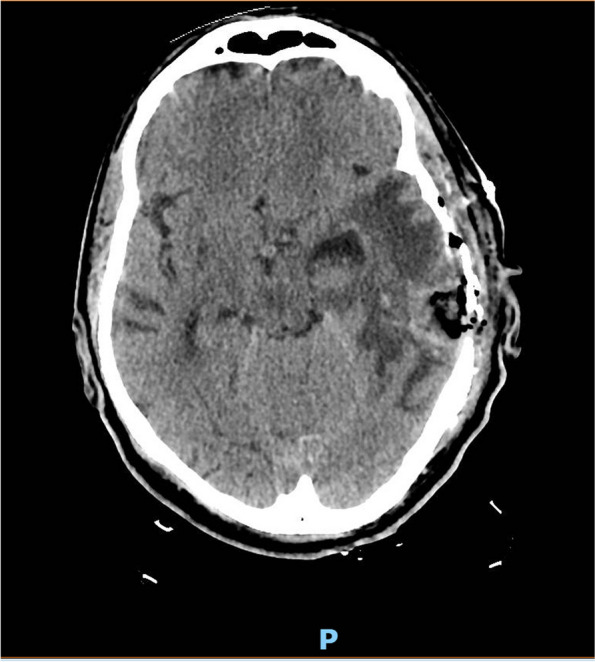
A 55-year-old man with a history of alcohol abuse and recurrent otitis was found on the ground and brought to the emergency room with confusion and seizures. On physical examination, he presented with hypotension and severe hypothermia. He had a Glasgow Coma Scale (GCS) of 8 and was intubated for airway protection. The initial laboratory analysis revealed an increase in inflammatory markers (white blood cell [WBC] count 22.570 /μL, C-reactive protein [CRP] 218 mg/L [reference range 0–5], procalcitonin [PCT] 8.16 ng/mL) and blood tests were compatible with signs of rhabdomyolysis (creatin kinase [CK] 1602 UI/L, creatinine 2.35 mg/dl, lactate dehydrogenase [LDH] 376 U/L, myoglobin 3075 ng/ml). Brain computed tomography (CT) was performed, which showed two brain lesions in the left temporal-occipital site, measuring 3.9 × 1.8 cm and 2.4 × 1.5 cm respectively, with vasogenic edema and 0.9 cm left-to-right midline shift. Signs of inflammation of the paranasal sinuses were also reported (Fig. 1).
Fig. 1.

Brain CT-scan, showing left temporomandibular abscesses of 3.9 × 1.8 cm (lateral) and 2.4 × 1.5 cm (medial) respectively with hyperdense margins on baseline scans and post-contrast enhancement
Chest and abdominal CT scan were also performed in order to rule out local pathologies and possible septic embolisms. Blood cultures resulted negative and transthoracic echocardiogram showed no vegetations or signs of endocarditis. Serology for HIV and Toxoplasma gondii resulted negative. Antiedema (mannitol) and anticonvulsant (valproate) therapy was initiated along with empiric antibiotic treatment with ceftriaxone, 2 g every 12 hours, metronidazole, 500 mg every 6 hours, and linezolid 600 mg every 12 hours. The culture of the brain abscess aspirate, collected during neurosurgery, identified Actinomyces turicensis and Proteus mirabilis on two different samples. Specifically, an intraoperative sample was collected in Amies elution medium and cultivated on three agar plates (Sabouraud dextrose agar, Columbia CNA agar and MacConkey agar), while another sample was collected in the absence of medium and cultivated on the same plates plus two additional ones (Chocolate agar and microaerophilic Columbia CNA agar). The plates were incubated at 37° degrees and first bacterial growth was observed at 36 hours. Microbiological identification was performed by MALDI-TOF (Bruker Biotyper®), showing high log (score) value (2.17 and 1.97 for each sample respectively). The antimicrobial susceptibility testing was performed by microdilution and Vitek-2 (bioMerieux®) automated system respectively for the anaerobic and the aerobic bacteria (Table 1).
Table 1.
Antimicrobial susceptibility testing for A. turicensis and P. mirabilis isolated on patient
| A. turicensis | Antibiotic | MIC | Susceptibility |
| ampicillin | < 0.25 μg/mL | S | |
| ceftaroline | < 0.25 μg/mL | S | |
| linezolid | 2 μg/mL | S | |
| moxifloxacin | < 0.125 μg/mL | S | |
| gentamicin | > 4 μg/mL | R | |
| P. mirabilis | |||
| amoxicillin/clavulanic acid | 8 μg/mL | S | |
| ceftazidime | < 0.12 μg/mL | S | |
| piperacillin/tazobactam | < 4 μg/mL | S | |
| meropenem | < 0.25 μg/mL | S | |
| gentamicin | < 1 μg/mL | S | |
| colistin | > 16 μg/mL | R | |
| ciprofloxacin | 2 μg/mL | R |
MIC Minimum inhibitory concentration S sensitive, R resistant
After obtaining the antimicrobial susceptibility test results, antibiotic therapy was simplified to ceftriaxone 2 g every 12 hours. Metronidazole and linezolid were discontinued.
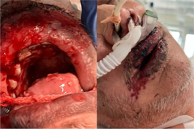
After treatment optimization, the patient developed a fever and an initially vesiculopapular, then necrotizing, lesion of the upper lip and oral cavity (Fig. 2).
Fig. 2.

Vesiculopapular and necrotic lesions of the oral cavity and perioral area
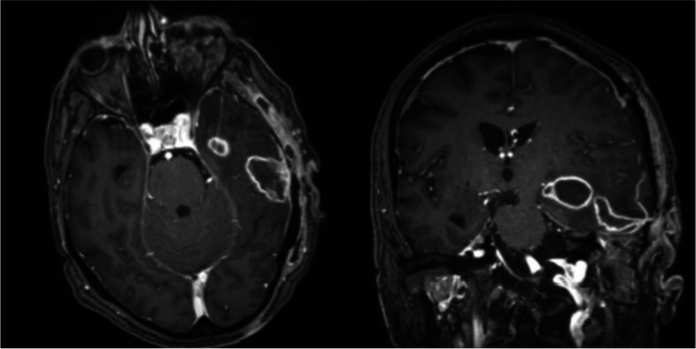
In the suspicion of a herpetic lesion, patient was started on acyclovir for 5 days, with progressive resolution of the lesion. To rule out a possible cutaneous involvement by A. turicensis, a wound swab was performed, resulting positive for Herpes simplex virus-1 (HSV-1) and a carbapenem-resistant Acinetobacter baumannii. Therefore, antimicrobial therapy was enhanced with the addition of ampicillin/sulbactam 3 g every 6 hours for improved coverage of both the brain abscess (A. turicensis) and the mucosal lesion isolate (A. baumannii). Five weeks after surgery, a brain magnetic resonance (MR) showed a reduction of the abscesses and resolution of edema and midline shift (Fig. 3).
Fig. 3.

T1-weighted MR scans of brain, 5 weeks following neurosurgery
The patient was then discharged to a neurorehabilitation facility with indication to continue the antimicrobial treatment with oral amoxicillin-clavulanate for a total of 8 weeks of therapy.
Systematic review
Materials and methods
The present study was conducted and reported according to the Preferred Reporting Items for Systematic Reviews and Meta-Analyses (PRISMA) statement [5].
Search strategy and database selection
The search was conducted on the databases MEDLINE, EMBASE, Web of Science, CINAHL, Clinicaltrials.gov and Canadian Agency for Drugs and Technology in Health (CADTH), including all available records from inception to August 30th, 2023. Each included database was searched with the search term “Actinomyces turicensis” as an all-terms strategy. No filter was applied to the search engines. The search strategy as elaborated by the search engine, together with the corresponding records found divided by database is available in additional files (see Additional file 1).
Obtained records were merged on the online tool Rayyan, where duplicates were identified and removed from the included list. The first round of selection for relevance and eligibility was performed on the same platform [6]. Search and selection were performed in blind. Discrepancies in selection were resolved by discussion. A list of records obtained after the primary screening by title and abstract was then downloaded and entered into a computerized database for further analysis by reading the full text of the study. A final list of included records was then generated, and each study was examined for relevant data. Extracted information included author and journal information, year, study design, demographic information about included patient/s, site of infection, clinical presentation, diagnostic procedures, treatment, and outcome. Additional anamnestic information about possible predisposing conditions was also gathered. All extracted information was then summarized in figures and tables and added to the present study.
Inclusion and exclusion criteria
Records were identified as eligible if they reported clinical data about infections by A. turicensis. No restrictions were made in terms of study design, peer-review, year of publication, country, language, patient age, or type of patient. In vitro or animal studies were excluded. Records reporting aggregated data only were excluded as well.
Quality appraisal of included studies
Included studies were evaluated for their risk of bias by means of the most appropriate eligible reference scale when their design was either interventional or observational. For observational and randomized studies, the Newcastle-Ottawa scale (NOS) and the Cochrane Risk of Bias Tool 2 (ROB2) were used, respectively [7, 8]. The risk of bias analysis was performed, in blind, by AI, LVR and ADL. Discrepancies were solved by discussion.
Results
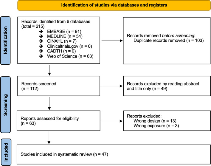
Our search on the six databases has identified 215 records, of which 103 were duplicates and were removed. Therefore, 112 records were screened for relevance and eligibility from the analysis of abstract and title only, resulting in 63 records. A subsequent examination of the relevant data in the full text was conducted, resulting in the exclusion of 16 records. At the end of the study selection process, 47 records were included in the systematic review. A flowchart describing the selection process is reported below (Fig. 4).
Fig. 4.
PRISMA flowchart of included studies
Included records were published between 2002 and 2023, with a prevalence in the last 5 years (26/47, 55%). Most of the studies were conducted in the USA (19/47, 40%), Europe (15/47, 32%) and China (3/47, 6%). Among the included records, we observed 43 Case reports, [1, 9–50] and 4 Case series [51–54], resulting in an overall population of 67 patients.
Clinical, demographics and microbiological records of the overall population are reported below (Tables 2 and 3).
Table 2.
Demographic features and underlying conditions of the patients
| References | Age | Sex | Predisposing risk factors | Immune system impairment |
|---|---|---|---|---|
| Panwar K et al., 2019 [9] | 45 | M | None | Diabetes, obesity |
| Saca J et al., 2023 [10] | 49 | M | Diabetic foot ulcer | Diabetes |
| Unigarro et al., 2023 [11] | 58 | F | Cervical cancer | Chemotherapy |
| Baher H et al., 2022 [12] | 36 | M | Endovenous drug use | None |
| Gandhi K et al., 2022 [13] | 10 | F | Surgical infection | None |
| Böttger S et al., 2022 [54] | 74 | M | Impacted decayed tooth with periodontitis | None |
| Fisher M et al., 2022 [14] | 74 | F | Pulmonary sequestration and COPD | None |
| Lin J et al., 2021 [15] | 36 | M | None | None |
| Sarumathi D et al., 2020 [55] | 42 | M | Nephrotic syndrome | Corticosteroids |
| Herrmann, AA et al., 2019 [17] | 71 | M | Stage IV esophageal cancer | Chemotherapy, immunotherapy |
| Lowry D et al., 2019 [18] | 56 | M | None | Diabetes, psoriatic arthritis on adalimumab treatment |
| Denham, J.D. et al., 2018 [19] | 71 | F | None | None |
| Snead, J.A. et al. 2018 [20] | 79 | M | Infected sacral decubitus ulcer | Prostate cancer |
| Gibson AL et al., 2018 [21] | 3 | F | Neurosurgical and spinal interventions | None |
| Elborno, D. et al., 2016 [22] | 13 | F | Microperforate hymen | None |
| Matela, A.et al., 2015 [23] | 52 | M | Dental procedure | None |
| Nickoloff, S et al., 2014 [56] | 62 | M | Poor dentition | Smoking |
| Shkolnik, I.et al., 2014 [25] | 37 | F | Poor dentition | Smoking, alcohol abuse |
| Palacios D et al., 2023 [26] | 42 | F | None | None |
| Doldán L et al., 2023 [27] | 59 | F | Cervical cancer | None |
| Cronin JT et al., 2023 [28] | 70 | M | Mini-open rotator cuff repair | Corticosteroid local injection |
| Tan CY et al., 2022 [51] | 15 (IQR 8–52) | M (7), F (8) | N.A. | N.A. |
| Khan A et al., 2022 [29] | 61 | M | Benign prostatic hyperplasia | None |
| Mao TC et al., 2022 [30] | 67 | M | None | None |
| Tabaksert A et al., 2021 [1] | 56 | M | None | None |
| Nia A et al., 2021 [31] | 42 | M | None | None |
| Agrafiotis AC et al., 2021 [32] | 51 | M | None | Smoking, alcohol abuse, corticosteroids |
| Johnson SW et al., 2021 [33] | 33 | M | None | Obesity, diabetes |
| Barnes A et al., 2020 [34] | 53 | M | None | None |
| Jin W et al., 2020 [35] | 50 | F | None | None |
| Kansara T et al., 2020 [36] | 52 | F | None | None |
| Le Bihan A et al., 2019 [37] | 43 | F | Chronic lactation from breast | Smoking |
| Vassa N et al., 2019 [38] | 61 | M | None | Chemotherapy and radiation |
| Kocsis B et al., 2018 [39] | 43 | M | Mastoiditis | Alcohol abuse, smoking |
| Cobo F., 2018 [40] | 44 | F | Mastitis | None |
| Gatti M et al., 2017 [41] | 64 | F | None | Obesity |
| Eenhuis LL et al., 2016 [42] | 42 | F | Intra-uterine contraceptive device | None |
| Oh HB et al., 2015 [43] | 25 | F | None | None |
| Hagiya H et al., 2015 [44] | 80 | F | None | None |
| Kottam A et al., 2015 [45] | 30 | F | Intra-uterine contraceptive device | None |
| Miller S et al., 2014 [46] | 5 | M | Recurrent otitis media | None |
| Abdulrahman GO Jr. et al., 2015 [47] | 22 | F | Nipple piercing | Smoking |
| Ong C et al., 2012 [48] | 73 | F | None | Smoking |
| Chudácková E et al., 2010 [52] | 28 (IQR 20–30) | M (4), F (3) | None | Diabetes (2), none (5) |
| Zautner AE et al., 2019 [49] | 23 | M | Femur hypoplasia | None |
| Attar KH et al., 2007 [53] | 33 | F | Bilateral nipple piercing | Steroid, smoke, obesity |
| Riegert-Johnson DL et al., 2002 [50] | 59 | M | Dental care | None |
COPD chronic obstructive pulmonary disease, IQR interquartile range, N. A not available
Table 3.
Clinical presentation and microbiological findings
| References | Infection | Microbiological findings | Coinfections | Symptoms |
|---|---|---|---|---|
| Panwar K et al., 2019 [9] | Necrotizing fasciitis | Monomicrobial | Nil | Nausea, vomit, fever |
| Saca J et al., 2023 [10] | Osteomyelitis and necrotizing fasciitis | Polymicrobial | S. agalactiae, P. denticola, S. moorei | Foot pain, fever, tachycardia |
| Unigarro et al., 2023 [11] | Septic shock after uterine perforation | Monomicrobial | Nil | Dysuria, abdominal pain, nausea, vomit, drowsyness, hypotension |
| Baher H et al., 2022 [12] | Pleural empyema | Monomicrobial | Nil | Fever, tachycardia, tachypnea, hypotension |
| Gandhi K et al., 2022 [13] | Abscess of the cartilagineous helix | Monomicrobial | Nil | Pain, erythema at previous surgical site |
| Böttger S et al., 2022 [54] | Odontogenic craniofacial necrotizing fasciitis | Polymicrobial | B. thetaiotaomicron, S. epidermidis | Black blisters, anesthesia of the skin, livid erythema |
| Fisher M et al., 2022 [14] | Pulmonary abscess | Monomicrobial | Nil | Dyspnea and cough |
| Lin J et al., 2021 [15] | Abscess of the buttocks | Monomicrobial | Nil | Pain, erythema, purulent cutaneous discharge |
| Sarumathi D et al., 2020 [55] | UTI | Monomicrobial | Nil | Fever, dysuria, and loose stools |
| Herrmann, AA et al., 2019 [17] | Spinal epidural abscess | Polymicrobial | E. cloacae, S. milleri | Back pain, fever |
| Lowry D et al., 2019 [18] | Pulmonary abscess | Monomicrobial | Nil | Dyspnea |
| Denham, J.D. et al., 2018 [19] | Pyometra | Monomicrobial | Nil | Purulent vaginal discharge |
| Snead, J.A. et al. 2018 [20] | Bacteremia | Monomicrobial | Nil | Fever, chills, tachycardia, hypotension, altered mental status |
| Gibson AL et al., 2018 [21] | Epidural abscess | Polymicrobial | A. europaeus | Fever, lethargy |
| Elborno, D. et al., 2016 [22] | Tubo-ovarian abscess | Monomicrobial | Nil | N.A. |
| Matela, A.et al., 2015 [23] | Pulmonary abscess | Polymicrobial | S. viridans | Chest pain, fever |
| Nickoloff, S et al., 2014 [56] | Empyema | Monomicrobial | Nil | Chest pain, fever, weight loss |
| Shkolnik, I.et al., 2014 [25] | Pulmonary abscess | Monomicrobial | Nil | Weight loss, cough, chest pain |
| Palacios D et al., 2023 [26] | Recurrent peri-clitoral abscess | Monomicrobial | Nil | Recurrent peri-clitoral mass |
| Doldán L et al., 2023 [27] | Para-uterine abscess | Monomicrobial | Nil | Purulent vaginal discharge, fever |
| Cronin JT et al., 2023 [28] | Surgical site infection | Monomicrobial | Nil | Purulent surgical wound dehiscence |
| Tan CY et al., 2022 [51] | Pilonidal (11), Perianal (4) | Monomicrobial (1), polimicrobial (14) | Mixed anaerobes, S. milleri, S. aureus, Citrobacter spp., Coliform | N.A. |
| Khan A et al., 2022 [29] | Fournier’s gangrene | Polymicrobial | H. haemolyticus, S. anginosus, P harei | Diarrhea, fever, penile swelling, dysuria, hematuria, hypotension |
| Mao TC et al., 2022 [30] | Fournier’s gangrene | Monomicrobial | Nil | Scrotum swelling |
| Tabaksert A et al., 2021 [1] | Parapharingeal and mediastinal abscess | Polymicrobial | E. faecalis, S. anginosus, S. constellatus | Fever, dysfagia |
| Nia A et al., 2021 [31] | Hip abscess | Polymicrobial | F. nucleatum | Pain, fever |
| Agrafiotis AC et al., 2021 [32] | Pleural empyema | Polymicrobial | F. necrogenes, M. micros | N.A. |
| Johnson SW et al., 2021 [33] | Pleural empyema | Polymicrobial | F. nucleatum | Chest pain, cough, fever |
| Barnes A et al., 2020 [34] | Prostatic abscess and Mandibular abscess | Polymicrobial | Peptostreptococcus spp. | Shock, inguinal pain, fever, vomit, dysuria |
| Jin W et al., 2020 [35] | Adrenal gland abscess | Polymicrobial | E. coli, P. mirabilis, plus others in mNGs | Back pain, fever |
| Kansara T et al., 2020 [36] | Pyelonephritis and abscess | Monomicrobial | Nil | Abdominal pain, vomit, fever |
| Le Bihan A et al., 2019 [37] | Breast abscess | Polymicrobial | P. harei | Breast swelling |
| Vassa N et al., 2019 [38] | Ludwig angina | Monomicrobial | Nil | Oral bleeding |
| Kocsis B et al., 2018 [39] | Meningitis | Monomicrobial | Nil | Unconsciousness, fever |
| Cobo F., 2018 [40] | Breast abscess | Monomicrobial | Nil | Pain, fever |
| Gatti M et al., 2017 [41] | Abdominal wall | Monomicrobial | Nil | Hypotension, necrotic abdominal wall |
| Eenhuis LL et al., 2016 [42] | Peritonitis | Monomicrobial | Nil | Hypotension, fever, abdominal pain |
| Oh HB et al., 2015 [43] | Pilonidal abscess | Polymicrobial | P. bivia, Peptostreptococcus spp. | Swelling of sacral region, fever |
| Hagiya H et al., 2015 [44] | Pyometra | Polymicrobial | C. clodtridioforme | Fever |
| Kottam A et al., 2015 [45] | Endocarditidis and pelvis and liver microabscesses | Monomicrobial | Nil | N.A. |
| Miller S et al., 2014 [46] | Cerebellar abscess | Polymicrobial | P. mirabilis, P. harei, B. thetaiotaomicron, A. hydrogenalis | Otorrhoea, anorexia, vomit, lethargy |
| Abdulrahman GO Jr. et al., 2015 [47] | Breast abscess | Polymicrobial | P.harei | Breast pain |
| Ong C et al., 2012 [48] | Left iliac fossa and liver abscesses | Monomicrobial | Nil | Abdominal pain, fever |
| Chudácková E et al., 2010 [52] | Pilonidal (2), cutaneous (2), anal (1), perianal (1), gas gangrene (1) | Monomicrobial (2), polimicrobial (5) | B. ureolyticus, F. nucleatum, S. milleri, P. anaerobius, S. aureus, P. acnes, Prevotella spp. | N.A. |
| Zautner AE et al., 2019 [49] | Fistula of the knee | Polymicrobial | A. europaeus | Swelling of the knee |
| Attar KH et al., 2007 [53] | Breast abscess | Monomicrobial | Nil | Pain, sweeling, fever |
| Riegert-Johnson DL et al., 2002 [50] | Hepatic abscess | Polymicrobial | B. fragilis | Fever, vomit |
Key: mNGs metagenomic next-generation sequencing, N.A. not available, UTI urinary tract infection
Some of the included cases did not provide enough information about immunosuppression conditions, symptoms, or treatments; therefore, the lack of data was considered when calculating the incidences, to minimize underestimation of the data.
Demographic features and underlying conditions
Published cases showed an almost equal distribution of males and females (35 vs. 32) with a median age of 42 (IQR 23–57). From the analysis of the patient anamnestic data, 21 patients (21 out of the 52 patients for which data was available, i.e. 40%) resulted to have had some cause of comorbidity or immunosuppression, particularly smoking (9), diabetes (6), obesity (5), chemotherapy or immunotherapy (4), high dose steroids (3), alcohol abuse (3). Moreover, in relation to the site of infection, a supposed predisposing condition was reported in 27 patients (27/52, 52%). No information about predisposing condition or immunosuppression were reported for 15 patients.
Site of infection and associated symptoms
Among the overall population, we observed 21 infections of the anogenital district, 12 Acute Bacterial Skin and Skin Structure Infections (ABSSSI) of which 2 were defined as Fournier’s gangrene, 8 lung infections (4 empyema and 4 abscesses), 6 gynecological infections, 5 infections of the cervicofacial district, 4 infections of the breast, 4 abdominal infections (1 peritonitis, 2 liver abscesses, 1 infection of the adrenal gland), 3 urinary tract infections, 2 infections of the vertebral column, 2 central nervous system infections, 1 endocarditis. One patient had both the cervicofacial region and urinary tract infections. Interestingly, 36 (36/67, 54%) infections presented as abscesses and 7 infections (7/67, 19.4%) presented with a concomitant bacteremia. Among the symptoms described at admission, fever (25 out of the 42 patients for which such data were available, i.e. 60%), local pain (18/42, 43%), local swelling and erythema (8/42, 19%), vomiting (6/42, 1%), dysuria (4/42, 10%), were the most frequently reported. Furthermore, 7 patients (7/42, 17%) presented with hypotension or shock and 5 patients (5/42, 12%) presented with altered state of consciousness. In the case of 25 patients, no information about symptoms was reported.
Microbiology
In all cases where the data were available, the microbiological identification of A. turicensis was allowed by culture examination. This was conducted on tissue samples (31/62, 50%), purulent drainage fluid (14/62, 22.5%), intraoperative samples (6/62, 9,6%), blood samples (7/62, 11.2%), Broncho-Alveolar Lavage (BAL) fluid (2/62, 3.2%), cerebrospinal fluid (1/62, 1.6%), urine sample (1/62, 1.6%). Fifty-seven percent of the infections were polymicrobial (n = 38). Reported co-infections were identified by tissue/pus culture or molecular assays and are reported in Table 3. Co-infecting agents were almost invariably part of the anaerobic flora.
Treatment
Out of the 67 cases described in the literature, abscess drainage was performed in 10 patients (15%), surgical debridement was performed in 41 cases (61%), an antibiotic approach without surgery was chosen for 15 patients (22%), while no information about surgical procedures was reported for one patient. Surgery was considered curative, i.e. without any antibiotic therapy, in 8 out of 67 patients, though insufficient data was reported for the antibiotic treatment for 11 patients. Specifically, 4 received an unspecified broad-spectrum antibiotic regimen, while for 7 patients no data was reported.
In the other 48 cases, a wide range of antibiotic use was reported, as summarized in Table 4.
Table 4.
Treatment strategies and clinical outcome
| References | Source control | Administered antibiotics | Duration of therapy (days) | Outcome |
|---|---|---|---|---|
| Panwar K et al., 2019 [9] | Surgical debridement | VAN, TZP | N.A. | Full recovery |
| Saca J et al., 2023 [10] | Surgical debridement, | AMC, SAM | N.A. | Recurrence and superinfection |
| Unigarro et al., 2023 [11] | None | CARBA, LZD, CLI | 9 | Full recovery |
| Baher H et al., 2022 [12] | None | AMC, MTZ | N.A. | Full recovery |
| Gandhi K et al., 2022 [13] | None | AMC | 180 | Full recovery |
| Böttger S et al., 2022 [54] | Surgical debridement | CARBA | N.A. | Full recovery |
| Fisher M et al., 2022 [14] | None | N.A. | N.A. | Full recovery |
| Lin J et al., 2021 [15] | None | STX | 90 | Full recovery |
| Sarumathi D et al., 2020 [55] | None | MTZ, AMP | N.A. | Full recovery |
| Herrmann, AA et al., 2019 [17] | None | N.A. | N.A. | Death |
| Lowry D et al., 2019 [18] | None | N.A. | N.A. | Full recovery |
| Denham, J.D. et al., 2018 [19] | None | AMC | 180 | Full recovery |
| Snead, J.A. et al. 2018 [20] | None | TZP | 42 | Full recovery |
| Gibson AL et al., 2018 [21] | N.A. | N.A. | N.A. | N.A. |
| Elborno, D. et al., 2016 [22] | Drainage | AMX, MTZ | 365 | Full recovery |
| Matela, A.et al., 2015 [23] | Surgical debridement | TZP, AMC | N.A. | Full recovery |
| Nickoloff, S et al., 2014 [56] | Drainage | AMC | N.A. | Full recovery |
| Shkolnik, I.et al., 2014 [25] | Drainage | CRO, MTZ | 42 | N.A. |
| Palacios D et al., 2023 [26] | Drainage | AMX | 14 | Recurrence |
| Doldán L et al., 2023 [27] | Drainage | AMX | 90 | Full recovery |
| Cronin JT et al., 2023 [28] | Surgical debridement | AMX | 420 | Full recovery |
| Tan CY et al., 2022 [51] | Surgical debridement | N.A. | 0 (0–6.5) | N.A. |
| Khan A et al., 2022 [29] | Surgical debridement | TZP, VAN, CLI, SAM, AMC | 21 | Full recovery |
| Mao TC et al., 2022 [30] | Surgical debridement | CFP, TZP, CLI | N.A. | Full recovery |
| Tabaksert A et al., 2021 [1] | Surgical debridement | CARBA, MTZ, AMX | 180 | Full recovery |
| Nia A et al., 2021 [31] | Surgical debridement | AMC, MTZ | 42 | Full recovery |
| Agrafiotis AC et al., 2021 [32] | Surgical debridement | AMC | 180 | Full recovery |
| Johnson SW et al., 2021 [33] | Dreinage | SAM, AMC | 180 | Full recovery |
| Barnes A et al., 2020 [34] | Surgical debridement | VAN, TZP, SAM, CRO, AMC | 210 | Full recovery |
| Jin W et al., 2020 [35] | Drainage | CARBA | 91 | Full recovery |
| Kansara T et al., 2020 [36] | None | MTZ, CARBA, VAN, CRO | 15 | Full recovery |
| Le Bihan A et al., 2019 [37] | None | AMX, MTZ | 70 | Full recovery |
| Vassa N et al., 2019 [38] | None | VAN, TZP, PEN, LVX, MTZ, SAM | 42 | Full recovery |
| Kocsis B et al., 2018 [39] | Surgical debridement | CRO, VAN, AMP | N.A. | Death |
| Cobo F., 2018 [40] | None | AMX | 10 | Full recovery |
| Gatti M et al., 2017 [41] | Surgical debridement | DAP, RIF, TZP, AMP | 35 | Full recovery |
| Eenhuis LL et al., 2016 [42] | Surgical debridement | CRO, GEN, and MTZ, PEN, | 210 | Full recovery |
| Oh HB et al., 2015 [43] | Surgical debridement | AMC | 7 | Full recovery |
| Hagiya H et al., 2015 [44] | Drainage | SAM | 30 | Full recovery |
| Kottam A et al., 2015 [45] | Surgical debridement | PEN, CRO, MTZ, CARBA | 60 | Full recovery |
| Miller S et al., 2014 [46] | Surgical debridement | CTX, MTZ, PEN,CIP, AMX | 210 | Full recovery |
| Abdulrahman GO Jr. et al., 2015 [47] | Drainage | AMC, PEN, AMX | 194 | Full recovery |
| Ong C et al., 2012 [48] | None | PEN, AMX | 180 | Full recovery |
| Chudácková E et al., 2010 [52] | Surgical debridement | N.A. | N.A. | N.A. |
| Zautner AE et al., 2019 [49] | Surgical debridement | PEN, GEN | 14 | Recurrence and superinfection |
| Attar KH et al., 2007 [53] | Surgical debridement | VAN, CXM | 21 | Full recovery |
| Riegert-Johnson DL et al., 2002 [50] | Drainage | CRO, MTZ | 150 | Full recovery |
VAN vancomycin, TZP piperacilline/tazobactam, SAM ampicillin/sulbactam, AMC amoxicillin/clavulanic acid, CARBA carbapenem, LZD linezolid, CLI clindamycin, MTZ metronidazole, STX trimethoprim/sulfamethoxazole, AMP ampicillin, AMX amoxicillin, CRO ceftriaxone, CFP cefoperazone, PEN penicillin, LVX levofloxacin, DAP daptomycin, RIF rifampin, GEN gentamicin, CTX cefotaxime, CIP ciprofloxacin, CXM cefuroxime
Broad-spectrum antibiotics, active on both Gram-positive and Gram-negative bacteria, were the most frequent first choice treatment, favoring intravenous administration in severe infections. Particularly, piperacillin/tazobactam was used in 7 patients, vancomycin was prescribed in 6 cases, carbapenems where the treatment of choice in 5 patients, while metronidazole or cephalosporin were used in 3 cases each. Regarding targeted therapy, the most frequently administered antibiotics were amoxicillin/clavulanate (n.17 cases), amoxicillin (n.13 cases), ampicillin/sulbactam (n.6 cases), penicillin (n. 6 cases) and ampicillin (n. 4 cases). Metronidazole (n.15 cases) or cephalosporin (n.6 cases) were added in case of suspected or documented polymicrobial infections.
Regarding the overall duration of therapy, data were available for 46 out of 67 patients. Mean treatment duration was 80 days, while median duration was 38.5 days (IQR 7.5–172.5). Shorter treatment, i.e. less than 1 month, was the most frequently observed (14/46, 30%), followed by a duration of 1–3 months (10/46, 22%), 3–6 months (8/46, 17%) and more than 6 months (6/46, 13%). The remaining cases underwent no antimicrobial therapy as surgery was considered curative (8/46, 17%). As expected, longer treatments were reported in cases of abscesses.
Outcome
Among the included studies, clinical outcome data were available for 44 out of 67 cases (65.6%). Thirty-nine patients (89%) showed a full recovery, while 3 patients (7%) experienced recurrence or superinfection and 2 patients (5%) died.
Discussion and conclusions
To the best of our knowledge, this is the first case in the literature of a brain abscess caused by A. turicensis and P. mirabilis in an adult patient. Brain involvement in actinomycosis is uncommon [57, 58], generally resulting from hematogenous spread or contiguous infection of the ear, sinus, and cervicofacial region [46, 58, 59]. In our case, the brain CT showed inflammation of the paranal sinuses but excluded ear involvement, even if a history of frequent otitis was reported.
Brain abscesses caused by opportunistic pathogens are frequently in patient with Human Immunodeficiency virus (HIV) infection or other causes of immunosuppression, whereas bacteria are the most common cause in immunocompetent patients [60]. While actinomycosis is a non-opportunistic disease, central nervous system involvement is very rare. Therefore, possible causes of immunosuppression must always be excluded. Our patient had a history of alcohol abuse [61, 62], which is considered a pro-inflammatory and nutritionally impaired condition often associated with immune deficiency.
The diagnosis of actinomycosis is challenging and requires an invasive approach for diagnosis. Literature suggests a surgical intervention for any brain abscess measuring at least 2.5 cm in diameter [63, 64]. Our patient underwent surgical excision of abscesses with consequent microbiological identification. Brain abscesses are frequently polymicrobial [46, 65, 66]; indeed P. mirabilis was also identified in our case [66].
Furthermore, growth of Actinomyces is generally slow and the bacteriological identification is difficult. Culture could frequently result sterile due to previous antibiotic therapy, concomitant microorganisms and inadequate sampling or incubation conditions. Surgical sampling of biopsy or pus seems to be the most appropriate clinical specimen [3].
Although often difficult to diagnose, actinomycosis is generally readily treatable, showing susceptibility to many antimicrobials including β-lactams, clarithromycin, erythromycin, doxycycline, and clindamycin. Therefore, thanks to the wide susceptibility and availability of treatment, several are the drugs of choice and there is no univocal indication. However, penicillin G or amoxicillin are the most used [3].
In our case, ceftriaxone was considered as target therapy with addiction to ampicillin/sulbactam for a week, as strengthening of the brain abscesses treatment. The prompt clinical and laboratory response in our patient allowed the switch to oral therapy with amoxicillin-clavulanic acid, which has proven to be non-inferior to standard intravenous treatment [67].
Our systematic review of the literature identified 47 articles reporting infections caused by A. turicensis. All included records are case reports (43) and case series (4), with an increased number of published papers in the last 20 years, probably due to the improvement of microbiological techniques, spectrometry, and molecular assay, that allow to better identification of Actinomyces species. Since the diagnosis of actinomycosis requires bacteriological identification, a lack of correct microbiological data, in the past, may have led to a misinterpretation of the risk and an underestimate of the incidence.
Although A. israelii is the main cause of disease within the species [4], we identified 67 cases of infections due to A. turicensis. From the present literature revision, most A. turicensis cases were anogenital, gynecological and urinary tract infections (30), lung infections (8) or cervicofacial infections (5).
As reported in the literature, actinomycosis is generally due to local dissemination of the pathogen rather than hematogenous spread [4]. Among the analyzed articles, a concomitant bacteremia was indeed found in 10% (7/67) of cases only, while a predisposing condition of local dissemination was supposed in at least 40% (27/52) of cases. Notably, while actinomycosis is a non-opportunistic disease, a reason for immune system impairment has been found in at least 52% (21/52%) of the cases.
Interestingly, only two central nervous system infections were reported among the included records, both presenting a history of ear infections (i.e. mastoiditis and otitis). In our cases, although a previous history of recurrent otitis was reported, no acute ear infection was present at patient admission. Concerning treatment options and outcome, a wide range of therapies is reported and a relatively low mortality (5%), confirming to be a readily treatable infection when promptly diagnosed [2].
In 76% of cases drainage or surgical debridement was performed, representing not only a therapeutical approach but also as a diagnostic procedure.
In conclusion, diagnosis of actinomycosis is challenging and requires prompt microbiological identification. Surgical excision or drainage together with long-term antibiotics is essential to achieve clinical recovery. Further investigations are needed to assess the optimal antibiotic regimen and its duration.
Supplementary Information
Acknowledgements
Not applicable.
Abbreviations
- ABSSSI
Acute bacterial skin and skin structure infection
- AMC
Amoxicillin/clavulanic acid
- AMP
Ampicillin
- AMX
Amoxicillin
- BAL
Broncho alveolar lavage
- CADTH
Canadian agency for drugs and technology in health
- CARBA
Carbapenem
- CFP
Cefoperazone
- CIP
Ciprofloxacin
- CK
Creatine Kinase
- CLI
Clindamycin
- CRO
Ceftriaxone
- CRP
C Reactive Protein
- CT
Computed tomography
- CTX
Cefotaxime
- CXM
Cefuroxime
- DAP
Daptomycin
- GEN
Gentamicin
- HIV
Human immunodeficiency virus
- HSV
Herpes Simplex Virus
- IQR
InterQuartile Range
- LDH
Lactate dehydrogenase
- LVX
Levofloxacin
- LZD
Linezolid
- MALDI-TOF
Matrix assisted laser desorption ionization – time of flight
- MIC
Minimum inhibitory concentration
- MR
Magnetic resonance
- MTZ
Metronidazole
- N.A.
Not Available
- NOS
Newcastle-ottawa scale
- GCS
Glasgow coma scale
- PCT
Procalcitonin
- PEN
Penicillin
- PRISMA
Preferred reporting items for systematic reviews and meta-analyses
- RIF
Rifampin
- S
Sentitive
- SAM
Ampicillin/sulbactam
- STX
Trimethoprim/sulfamethoxazole
- R
Resistant
- TZP
piperacilline/tazobactam
- VAN
Vancomycin
Authors’ contributions
Study was designed by AI and LVR. LVR, AI, and ADL performed all phases of the systematic review. Data extraction was performed by LVR and AI. Extracted data was checked by ADL, MI, LS and VM. Microbiological data were provided and controlled by AA, CDA, SM and MCB. DGB, MD and IG and all other authors were involved in patient care, and substantially contributed to the production of the final manuscript. All authors read and approved the final manuscript.
Funding
This research received no external funding.
Availability of data and materials
All data generated or analyzed during this study are included in this article and its supplementary materials.
Declarations
Ethics approval and consent to participate
Not applicable.
Consent for publication
A written informed consent was obtained from the patient described in the case report for publication of both clinical information, pictures, and radiological scans.
Competing interests
LS received a research grant from Gilead and fee for lectures and expertise from Merck, Gilead, Pfizer. MA reports honoraria for lectures and research grants from Merk, Gilead, Abbvie, Angelini SpA. V.M. received honoraria for lectures from Janssen-Cilag. M.I. received honoraria for lectures from Biogen Italia, AIM Educational, MICOM srl and research grants from Gilead.
Footnotes
Publisher’s Note
Springer Nature remains neutral with regard to jurisdictional claims in published maps and institutional affiliations.
References
- 1.Tabaksert A, Kumar R, Raviprakash V, Srinivasan R. Actinomyces turicensis parapharyngeal space infection in an immunocompetent host: first case report and review of literature. Access Microbiol. 2021;3:000241. doi: 10.1099/acmi.0.000241. [DOI] [PMC free article] [PubMed] [Google Scholar]
- 2.Wong VK, Turmezei TD, Weston VC. Actinomycosis. BMJ. 2011;343:d6099. doi: 10.1136/bmj.d6099. [DOI] [PubMed] [Google Scholar]
- 3.Valour F, Sénéchal A, Dupieux C, Karsenty J, Lustig S, Breton P, et al. Actinomycosis: etiology, clinical features, diagnosis, treatment, and management. Infect Drug Resist. 2014;7:183–197. doi: 10.2147/IDR.S39601. [DOI] [PMC free article] [PubMed] [Google Scholar]
- 4.Olson TS, Seid AB, Pransky SM. Actinomycosis of the middle ear. Int J Pediatr Otorhinolaryngol. 1989;17:51–55. doi: 10.1016/0165-5876(89)90293-0. [DOI] [PubMed] [Google Scholar]
- 5.Page MJ, McKenzie JE, Bossuyt PM, Boutron I, Hoffmann TC, Mulrow CD, et al. The PRISMA 2020 Statement: an updated guideline for reporting systematic reviews. BMJ. 2021;n71. [DOI] [PMC free article] [PubMed]
- 6.Ouzzani M, Hammady H, Fedorowicz Z, Elmagarmid A. Rayyan—a web and mobile app for systematic reviews. Syst Rev. 2016;5:210. doi: 10.1186/s13643-016-0384-4. [DOI] [PMC free article] [PubMed] [Google Scholar]
- 7.Sterne JAC, Savović J, Page MJ, Elbers RG, Blencowe NS, Boutron I, et al. RoB 2: a revised tool for assessing risk of bias in randomised trials. BMJ. 2019:l4898. [DOI] [PubMed]
- 8.Lo CK-L, Mertz D, Loeb M. Newcastle-Ottawa scale: comparing reviewers’ to authors’ assessments. BMC Med Res Methodol. 2014;14:45. doi: 10.1186/1471-2288-14-45. [DOI] [PMC free article] [PubMed] [Google Scholar]
- 9.Panwar K, Duane TM, Tessier JM, Patel K, Sanders JM. Actinomyces turicensis necrotizing soft-tissue infection of the thigh in a diabetic male. Surg Infect. 2019;20:431–433. doi: 10.1089/sur.2018.149. [DOI] [PubMed] [Google Scholar]
- 10.Alaq Al-Abayechi, MD1, Divya Chandramohan, MD2, Hasan Baher, MD1, James Saca, MD1. RARE PATHOGENS ENCOUNTERED IN MAGGOT-INFESTED FOOT WOUNDS. Abstract published at SHM Converge 2023. Abstract 458 Journal of Hospital Medicine.
- 11.Unigarro L, Marín K, Alvear C, Salgado J, Basantes E. Choque séptico por Actinomyces turicensis, asociado a cáncer de cérvix. Reporte de caso. Mexican J Oncol. 2023;22:128–132. [Google Scholar]
- 12.Baher H, Jones L, Al-Abayechi A, Peters JI. A rare case of actinomyces turicensis empyema in an iv drug user. Chest. 2022;162:A1378. doi: 10.1016/j.chest.2022.08.1164. [DOI] [Google Scholar]
- 13.Gandhi K, van der Woerd BD, Graham ME, Barton M, Strychowsky JE. Cervicofacial Actinomycosis in the pediatric population: presentation and management. Ann Otol Rhinol Laryngol. 2022;131:312–321. doi: 10.1177/00034894211021273. [DOI] [PMC free article] [PubMed] [Google Scholar]
- 14.Fisher M, Soller D, Khaskia Y, Schellenberg J. Double rarity: a CASE of ACTINOMYCES in pulmonary sequestration. CHEST. 2021;160:A1713. doi: 10.1016/j.chest.2021.07.1559. [DOI] [Google Scholar]
- 15.Lin Jinming XB, Zhu Q, Xu ZY, Chen Q. A case of multiple sinuses in the buttocks caused by Actinomyces turicensis. Chin J Dermatol. 2021;54:155–7. 10.35541/cjd.20190779.
- 16.JCDR - Actinomycetaceae, Bacteremia, Kidney disorder https://www.jcdr.net/article_fulltext.asp?issn=0973-709x&year=2020&month=June&volume=14&issue=6&page=DD01&id=13734. Accessed 8 Sep 2023.
- 17.Herrmann AA, Othman SI, DeFoe KM, Carolan EJ, Rosenbloom MH. Spinal epidural abscess: esophageal fistula as a potential infection source. Interdiscip Neurosurg. 2019;16:42–43. doi: 10.1016/j.inat.2018.12.007. [DOI] [Google Scholar]
- 18.Lowry D, Grossman C, Boakye-Wenzel HN, Warren M, Dy RV. D57. Atypical pneumonias and other infections. American Thoracic Society; 2019. An Atypical case of waxing and waning lung lesions due to Actinomyces; pp. A6827–A6827. [Google Scholar]
- 19.Pyometra. Consultant360. 2018. https://www.consultant360.com/article/consultant360/womens-health/pyometra. Accessed 8 Sep 2023.
- 20.Snead JA, Ruggiero N, Sangha R, Joseph L, Mukherji R. Actinomyces turicensis bacteremia secondary to a decubitus ulcer: a case report and review of the literature. Infect Dis Clin Pract. 2018;26:e16. doi: 10.1097/IPC.0000000000000574. [DOI] [Google Scholar]
- 21.Gibson AL, Liu S, Naifeh M. Actinomyces epidural abscess: a virtually unheard of process in the virtual age. United States, New Orleans, LA: J. Invest. Med; 2018. pp. 487–488. [Google Scholar]
- 22.Elborno D, Pandya L, Chor J. Case report: pelvic Actinomyces in an adolescent with microperforate hymen. J Pediatr Adolesc Gynecol. 2016;29:191. doi: 10.1016/j.jpag.2016.01.079. [DOI] [Google Scholar]
- 23.Matela A, Ali Z, Changawala N, Desai A, Musta A, Nair GB. A48. Pulmonary infections: CASE studies (bacterial) American Thoracic Society; 2015. An unusual case of Actinomyces Turicensis pulmonary infection presenting as a lung mass; pp. A1840–A1840. [Google Scholar]
- 24.Abstracts from the 37th annual meeting of the Society of General Internal Medicine. J Gen Intern Med. 2014;29:1–545. [DOI] [PMC free article] [PubMed]
- 25.Shkolnik I, Hassani A, Miceli M, Big C, Bagdasarian N. A unique case of Actinomyces turicensis pulmonary abscess. Infect Dis Clin Pract. 2014;22:e37. doi: 10.1097/IPC.0b013e318291c873. [DOI] [Google Scholar]
- 26.Palacios D, Wallace HC. Recurrent Peri-clitoral abscess with positive Actinomyces turicensis culture. Case Rep Obstet Gynecol. 2023;2023:9912910. doi: 10.1155/2023/9912910. [DOI] [PMC free article] [PubMed] [Google Scholar]
- 27.Doldán L, Huarachi-Chirilla Y, Vargas C, Domínguez C, Chediack V, Cunto E. Cervical carcinoma abscessed by Schaalia turicensis. Medicina (B Aires) 2023;83:341. [PubMed] [Google Scholar]
- 28.Cronin JT, Richards BW, Skedros JG. Schaalia (formerly Actinomyces) turicensis infection following open rotator cuff repair. Cureus. 2023;15:e34242. doi: 10.7759/cureus.34242. [DOI] [PMC free article] [PubMed] [Google Scholar]
- 29.Khan A, Gidda H, Murphy N, Alshanqeeti S, Singh I, Wasay A, et al. An Unusual Bacterial Etiology of Fournier’s Gangrene in an Immunocompetent Patient. Cureus. 2022;14:e26616. doi: 10.7759/cureus.26616. [DOI] [PMC free article] [PubMed] [Google Scholar]
- 30.Mao T, Zhou X, Tian M, Zhang Y, Wang S. A rare case of male Fournier’s gangrene with mixed Actinomyces turicensis infection. BMC Urol. 2022;22:25. doi: 10.1186/s12894-022-00975-z. [DOI] [PMC free article] [PubMed] [Google Scholar]
- 31.Nia A, Ungersboeck A, Uffmann M, Leaper D, Assadian O. Septic hip abscess due to Fusobacterium nucleatum and Actinomyces turicensis in an immunocompetent SARS-CoV-2 positive patient. Anaerobe. 2021;71:102420. doi: 10.1016/j.anaerobe.2021.102420. [DOI] [PMC free article] [PubMed] [Google Scholar]
- 32.Agrafiotis AC, Lardinois I. Pleural empyema caused by Actinomyces turicensis. New Microbes New Infect. 2021;41:100892. doi: 10.1016/j.nmni.2021.100892. [DOI] [PMC free article] [PubMed] [Google Scholar]
- 33.Johnson SW, Billatos E. Polymicrobial empyema; a novel case of Actinomyces turicensis. Respir Med Case Rep. 2021;32:101365. doi: 10.1016/j.rmcr.2021.101365. [DOI] [PMC free article] [PubMed] [Google Scholar]
- 34.Barnes A, Kaur A, Augenbraun M. An unusual presentation of prostatic abscess due to Actinomyces turicensis and Peptostreptococcus. Cureus. 2020;12:e8665. doi: 10.7759/cureus.8665. [DOI] [PMC free article] [PubMed] [Google Scholar]
- 35.Jin W, Miao Q, Wang M, Zhang Y, Ma Y, Huang Y, et al. A rare case of adrenal gland abscess due to anaerobes detected by metagenomic next-generation sequencing. Ann Transl Med. 2020;8:247. doi: 10.21037/atm.2020.01.123. [DOI] [PMC free article] [PubMed] [Google Scholar]
- 36.Kansara T, Majmundar M, Doshi R, Ghosh K, Saeed M. A case of life-threatening Actinomyces turicensis bacteremia. Cureus. 2020;12:e6761. doi: 10.7759/cureus.6761. [DOI] [PMC free article] [PubMed] [Google Scholar]
- 37.Le Bihan A, Ahmed F, O’Driscoll J. An uncommon cause for a breast abscess: Actinomyces turicensis with Peptoniphilus harei. BMJ Case Rep. 2019;12:e231194. doi: 10.1136/bcr-2019-231194. [DOI] [PMC free article] [PubMed] [Google Scholar]
- 38.Vassa N, Mubarik A, Patel D, Muddassir S. Actinomyces turicensis: an unusual cause of cervicofacial actinomycosis presenting as ludwig angina in an immunocompromised host - case report and literature review. IDCases. 2019;18:e00636. doi: 10.1016/j.idcr.2019.e00636. [DOI] [PMC free article] [PubMed] [Google Scholar]
- 39.Kocsis B, Tiszlavicz Z, Jakab G, Brassay R, Orbán M, Sárkány Á, et al. Case report of Actinomyces turicensis meningitis as a complication of purulent mastoiditis. BMC Infect Dis. 2018;18:686. doi: 10.1186/s12879-018-3610-y. [DOI] [PMC free article] [PubMed] [Google Scholar]
- 40.Cobo F. Breast abscess due to Actinomyces turicensis in a non-puerperal woman. Enferm Infecc Microbiol Clin (Engl Ed) 2018;36:388–389. doi: 10.1016/j.eimc.2017.09.014. [DOI] [PubMed] [Google Scholar]
- 41.Gatti M, Gasparini LE, Grimaldi CM, Abbati D, Clemente S, Brioschi PR, et al. Septic shock due to NSTI caused by Actinomyces Turicensis: the role of clinical pharmacology. Case report and review of the literature. J Chemother. 2017;29:372–375. doi: 10.1080/1120009X.2017.1306154. [DOI] [PubMed] [Google Scholar]
- 42.Eenhuis LL, de Lange ME, Samson AD, Busch ORC. Spontaneous bacterial peritonitis due to Actinomyces mimicking a perforation of the proximal jejunum. Am J Case Rep. 2016;17:616–620. doi: 10.12659/AJCR.897956. [DOI] [PMC free article] [PubMed] [Google Scholar]
- 43.Oh HB, Abdul Malik MH, Keh CHL. Pilonidal abscess associated with primary Actinomycosis. Ann Coloproctol. 2015;31:243–245. doi: 10.3393/ac.2015.31.6.243. [DOI] [PMC free article] [PubMed] [Google Scholar]
- 44.Hagiya H, Ogawa H, Takahashi Y, Kimura K, Hasegawa K, Otsuka F. Actinomyces turicensis bacteremia secondary to Pyometra. Intern Med. 2015;54:2775–2777. doi: 10.2169/internalmedicine.54.4637. [DOI] [PubMed] [Google Scholar]
- 45.Kottam A, Kaur R, Bhandare D, Zmily H, Bheemreddy S, Brar H, et al. Actinomycotic endocarditis of the eustachian valve: a rare case and a review of the literature. Tex Heart Inst J. 2015;42:44–49. doi: 10.14503/THIJ-13-3517. [DOI] [PMC free article] [PubMed] [Google Scholar]
- 46.Miller S, Walls T, Atkinson N, Zaleta S. A case of otitis media complicated by intracranial infection with Actinomyces turicensis. JMM Case Rep. 2014;1:e004408. doi: 10.1099/jmmcr.0.004408. [DOI] [PMC free article] [PubMed] [Google Scholar]
- 47.Abdulrahman GO, Gateley CA. Primary actinomycosis of the breast caused by Actinomyces turicensis with associated Peptoniphilus harei. Breast Dis. 2015;35:45–47. doi: 10.3233/BD-140381. [DOI] [PubMed] [Google Scholar]
- 48.Ong C, Barnes S, Senanayake S. Actinomyces turicensis infection mimicking ovarian tumour. Singap Med J. 2012;53:e9–11. [PubMed] [Google Scholar]
- 49.Zautner AE, Schmitz S, Aepinus C, Schmialek A, Podbielski A. Subcutaneous fistulae in a patient with femoral hypoplasia due to Actinomyces europaeus and Actinomyces turicensis. Infection. 2009;37:289–291. doi: 10.1007/s15010-008-7392-9. [DOI] [PubMed] [Google Scholar]
- 50.Riegert-Johnson DL, Sandhu N, Rajkumar SV, Patel R. Thrombotic thrombocytopenic purpura associated with a hepatic abscess due to Actinomyces turicensis. Clin Infect Dis. 2002;35:636–637. doi: 10.1086/342327. [DOI] [PubMed] [Google Scholar]
- 51.Tan CY, Javed MA, Artioukh DY. Emerging presence of Actinomyces in perianal and pilonidal infection. J Surg Case Rep. 2022;2022:rjac367. doi: 10.1093/jscr/rjac367. [DOI] [PMC free article] [PubMed] [Google Scholar]
- 52.Chudácková E, Geigerová L, Hrabák J, Bergerová T, Liska V, Scharfen J. Seven isolates of Actinomyces turicensis from patients with surgical infections of the anogenital area in a Czech hospital. J Clin Microbiol. 2010;48:2660–2661. doi: 10.1128/JCM.00548-10. [DOI] [PMC free article] [PubMed] [Google Scholar]
- 53.Attar KH, Waghorn D, Lyons M, Cunnick G. Rare species of actinomyces as causative pathogens in breast abscess. Breast J. 2007;13:501–505. doi: 10.1111/j.1524-4741.2007.00472.x. [DOI] [PubMed] [Google Scholar]
- 54.Böttger S, Zechel-Gran S, Schmermund D, Streckbein P, Wilbrand J-F, Knitschke M, et al. Odontogenic Cervicofacial necrotizing fasciitis: microbiological characterization and Management of Four Clinical Cases. Pathogens. 2022;11:78. doi: 10.3390/pathogens11010078. [DOI] [PMC free article] [PubMed] [Google Scholar]
- 55.Sarumathi D, Gopichand P, Sejpal K, Priyamvada P, Mandal J. Actinomyces turicensis causing urinary tract infection in nephrotic syndrome patient- a case report. JCDR. 2020; 10.7860/JCDR/2020/44091.13734.
- 56.Nickoloff S, Sharma A, Martin J. An uncommon presentation of a rare pathogen: considering the cause of prolonged hiccups. San Diego, CA, United States: Journal of General Internal Medicine; 2014. pp. S309–S310. [Google Scholar]
- 57.Rahiminejad M, Hasegawa H, Papadopoulos M, MacKinnon A. Actinomycotic brain abscess. BJR Case Rep. 2016;2:20150370. doi: 10.1259/bjrcr.20150370. [DOI] [PMC free article] [PubMed] [Google Scholar]
- 58.Perez A, Syngal G, Fathima S, Sandkovsky U. Actinomyces causing a brain abscess. Proc (Bayl Univ Med Cent) 2021;34:698–700. doi: 10.1080/08998280.2021.1945354. [DOI] [PMC free article] [PubMed] [Google Scholar]
- 59.Smego RA., Jr Actinomycosis of the central nervous system. Rev Infect Dis. 1987;9:855–865. doi: 10.1093/clinids/9.5.855. [DOI] [PubMed] [Google Scholar]
- 60.Sonneville R, Ruimy R, Benzonana N, Riffaud L, Carsin A, Tadié J-M, et al. An update on bacterial brain abscess in immunocompetent patients. Clin Microbiol Infect. 2017;23:614–620. doi: 10.1016/j.cmi.2017.05.004. [DOI] [PubMed] [Google Scholar]
- 61.Watzl B, Watson RR. Role of alcohol abuse in nutritional immunosuppression. J Nutr. 1992;122(3 Suppl):733–737. doi: 10.1093/jn/122.suppl_3.733. [DOI] [PubMed] [Google Scholar]
- 62.Zhang P, Bagby GJ, Happel KI, Raasch CE, Nelson S. Alcohol abuse, immunosuppression, and pulmonary infection. Curr Drug Abuse Rev. 2008;1:56–67. doi: 10.2174/1874473710801010056. [DOI] [PubMed] [Google Scholar]
- 63.Kameda-Smith MM, Ragulojan M, Hart S, Duda TR, MacLean MA, Chainey J, et al. A Canadian National Survey of the neurosurgical management of intracranial abscesses. Can J Neurol Sci. 2022;1–8. [DOI] [PubMed]
- 64.Mamelak AN, Mampalam TJ, Obana WG, Rosenblum ML. Improved management of multiple brain abscesses: a combined surgical and medical approach. Neurosurgery. 1995;36:76–85. doi: 10.1227/00006123-199501000-00010. [DOI] [PubMed] [Google Scholar]
- 65.Abo-Zed A, Yassin M, Phan T. A rare case of polymicrobial brain abscess involving Actinomyces. Radiol Case Rep. 2021;16:1123–1126. doi: 10.1016/j.radcr.2021.02.042. [DOI] [PMC free article] [PubMed] [Google Scholar]
- 66.Muddassir R, Khalil A, Singh R, Taj S, Khalid Z. Intracranial Abscess and Proteus mirabilis: A Case Report and Literature Review. Cureus. 2020;12:e12326. doi: 10.7759/cureus.12326. [DOI] [PMC free article] [PubMed] [Google Scholar]
- 67.Bodilsen J, Brouwer MC, van de Beek D, Tattevin P, Tong S, Naucler P, et al. Partial Oral antibiotic treatment for bacterial brain abscess: an open-label randomized non-inferiority trial (ORAL) Trials. 2021;22:796. doi: 10.1186/s13063-021-05783-8. [DOI] [PMC free article] [PubMed] [Google Scholar]
Associated Data
This section collects any data citations, data availability statements, or supplementary materials included in this article.
Supplementary Materials
Data Availability Statement
All data generated or analyzed during this study are included in this article and its supplementary materials.


