Abstract
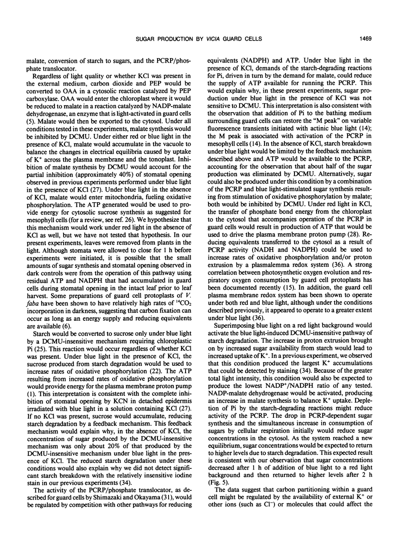
Concentrations of soluble sugars in guard cells in detached, sonicated epidermis from Vicia faba leaves were analyzed quantitatively by high performance liquid chromatography to determine the extent to which sugars could contribute to changes in the osmotic potentials of guard cells during stomatal opening. Stomata were illuminated over a period of 4 hours with saturating levels of red or blue light, or a combination of red and blue light. When stomata were irradiated for 3 hours with red light (50 micromoles per square meter per second) in a solution of 5 millimolar KCl and 0.1 millimolar CaCl2, stomatal apertures increased a net maximum of 6.7 micrometers and the concentration of total soluble sugar was 289 femtomoles per guard cell (70% sucrose, 30% fructose). In an identical solution, 2.5 hours of irradiation with 25 micromoles per square meter per second of blue light caused a maximum net increase of 7.1 micrometers in stomatal aperture and the total soluble sugar concentration was 550 femtomoles per guard cell (91% sucrose, 9% fructose). Illumination with blue light at 25 micromoles per square meter per second in a solution lacking KCl caused a maximum net increase in stomatal aperture of 3.5 micrometers and the sugar concentration was 382 femtomoles per guard cell (82% sucrose, 18% fructose). In dual beam experiments, stomata irradiated with 50 micromoles per square meter per second of red light opened steadily with a concomitant increase in sugar production. Addition of 25 micromoles per square meter per second of blue light caused a further net gain of 3.7 micrometers in stomatal aperture and, after 2 hours, sugar concentrations had increased by an additional 138 femtomoles per guard cell. Experiments with 3-(3,4-dichlorophenyl)-1,1-dimethylurea (DCMU) were performed with epidermis illuminated with 50 micromoles per square meter per second of red light or with 25 micromoles per square meter per second of blue light in solutions containing or lacking KCl. DCMU completely inhibited sugar production under red light, had no effect on guard cell sugar production under blue light when KCl was present, and inhibited sugar production by about 50% when guard cells were illuminated with blue light in solutions lacking KCl. We conclude that soluble sugars can contribute significantly to the osmoregulation of guard cells in detached leaf epidermis of V. faba. These results are consistent with the operation of two different sugar-producing pathways in guard cells: a photosynthetic carbon reduction pathway and a pathway of blue light-induced starch degradation.
Full text
PDF


Images in this article
Selected References
These references are in PubMed. This may not be the complete list of references from this article.
- Assmann S. M. Enhancement of the Stomatal Response to Blue Light by Red Light, Reduced Intercellular Concentrations of CO(2), and Low Vapor Pressure Differences. Plant Physiol. 1988 May;87(1):226–231. doi: 10.1104/pp.87.1.226. [DOI] [PMC free article] [PubMed] [Google Scholar]
- Fischer R. A. Role of Potassium in Stomatal Opening in the Leaf of Vicia faba. Plant Physiol. 1971 Apr;47(4):555–558. doi: 10.1104/pp.47.4.555. [DOI] [PMC free article] [PubMed] [Google Scholar]
- Gotow K., Tanaka K., Kondo N., Kobayashi K., Syōno K. Light Activation of NADP-Malate Dehydrogenase in Guard Cell Protoplasts from Vicia faba L. Plant Physiol. 1985 Nov;79(3):829–832. doi: 10.1104/pp.79.3.829. [DOI] [PMC free article] [PubMed] [Google Scholar]
- Gotow K., Taylor S., Zeiger E. Photosynthetic Carbon Fixation in Guard Cell Protoplasts of Vicia faba L. : Evidence from Radiolabel Experiments. Plant Physiol. 1988 Mar;86(3):700–705. doi: 10.1104/pp.86.3.700. [DOI] [PMC free article] [PubMed] [Google Scholar]
- Green D. B., Dodge S. M., Lee J. R., Tallman G. Effect of sodium hexanitrocobaltate (III) decomposition on its staining of intracellular potassium ions. Stain Technol. 1990;65(1):15–24. doi: 10.3109/10520299009105603. [DOI] [PubMed] [Google Scholar]
- Humble G. D., Raschke K. Stomatal opening quantitatively related to potassium transport: evidence from electron probe analysis. Plant Physiol. 1971 Oct;48(4):447–453. doi: 10.1104/pp.48.4.447. [DOI] [PMC free article] [PubMed] [Google Scholar]
- Macallum A. B. On the distribution of potassium in animal and vegetable cells. J Physiol. 1905 Feb 28;32(2):95–198.3. doi: 10.1113/jphysiol.1905.sp001068. [DOI] [PMC free article] [PubMed] [Google Scholar]
- Mawson B. T., Zeiger E. Blue light-modulation of chlorophyll a fluorescence transients in guard cell chloroplasts. Plant Physiol. 1991 Jul;96(3):753–760. doi: 10.1104/pp.96.3.753. [DOI] [PMC free article] [PubMed] [Google Scholar]
- Outlaw W. H., Manchester J. Guard cell starch concentration quantitatively related to stomatal aperture. Plant Physiol. 1979 Jul;64(1):79–82. doi: 10.1104/pp.64.1.79. [DOI] [PMC free article] [PubMed] [Google Scholar]
- Outlaw W. H., Mayne B. C., Zenger V. E., Manchester J. Presence of Both Photosystems in Guard Cells of Vicia faba L: IMPLICATIONS FOR ENVIRONMENTAL SIGNAL PROCESSING. Plant Physiol. 1981 Jan;67(1):12–16. doi: 10.1104/pp.67.1.12. [DOI] [PMC free article] [PubMed] [Google Scholar]
- Reckmann U., Scheibe R., Raschke K. Rubisco activity in guard cells compared with the solute requirement for stomatal opening. Plant Physiol. 1990 Jan;92(1):246–253. doi: 10.1104/pp.92.1.246. [DOI] [PMC free article] [PubMed] [Google Scholar]
- Robinson N. L., Preiss J. Localization of Carbohydrate Metabolizing Enzymes in Guard Cells of Commelina communis. Plant Physiol. 1987 Oct;85(2):360–364. doi: 10.1104/pp.85.2.360. [DOI] [PMC free article] [PubMed] [Google Scholar]
- Serrano E. E., Zeiger E., Hagiwara S. Red light stimulates an electrogenic proton pump in Vicia guard cell protoplasts. Proc Natl Acad Sci U S A. 1988 Jan;85(2):436–440. doi: 10.1073/pnas.85.2.436. [DOI] [PMC free article] [PubMed] [Google Scholar]
- Shimazaki K., Terada J., Tanaka K., Kondo N. Calvin-Benson Cycle Enzymes in Guard-Cell Protoplasts from Vicia faba L: Implications for the Greater Utilization of Phosphoglycerate/Dihydroxyacetone Phosphate Shuttle between Chloroplasts and the Cytosol. Plant Physiol. 1989 Jul;90(3):1057–1064. doi: 10.1104/pp.90.3.1057. [DOI] [PMC free article] [PubMed] [Google Scholar]
- Tallman G., Zeiger E. Light quality and osmoregulation in vicia guard cells : evidence for involvement of three metabolic pathways. Plant Physiol. 1988 Nov;88(3):887–895. doi: 10.1104/pp.88.3.887. [DOI] [PMC free article] [PubMed] [Google Scholar]
- Vani T., Raghavendra A. S. Tetrazolium Reduction by Guard Cells in Abaxial Epidermis of Vicia faba: Blue Light Stimulation of a Plasmalemma Redox System. Plant Physiol. 1989 May;90(1):59–62. doi: 10.1104/pp.90.1.59. [DOI] [PMC free article] [PubMed] [Google Scholar]
- Widholm J. M. The use of fluorescein diacetate and phenosafranine for determining viability of cultured plant cells. Stain Technol. 1972 Jul;47(4):189–194. doi: 10.3109/10520297209116483. [DOI] [PubMed] [Google Scholar]


