Abstract
Background
Cystic fibrosis (CF) lung disease is characterised by progressive airway wall thickening and widening. We aimed to validate an artificial intelligence-based algorithm to assess dimensions of all visible bronchus-artery (BA) pairs on chest CT scans from patients with CF.
Methods
The algorithm fully automatically segments the bronchial tree; identifies bronchial generations; matches bronchi with the adjacent arteries; measures for each BA-pair bronchial outer diameter (Bout), bronchial lumen diameter (Bin), bronchial wall thickness (Bwt) and adjacent artery diameter (A); and computes Bout/A, Bin/A and Bwt/A for each BA pair from the segmental bronchi to the last visible generation. Three datasets were used to validate the automatic BA analysis. First BA analysis was executed on 23 manually annotated CT scans (11 CF, 12 control subjects) to compare automatic with manual BA-analysis outcomes. Furthermore, the BA analysis was executed on two longitudinal datasets (Copenhagen 111 CTs, ataluren 347 CTs) to assess longitudinal BA changes and compare them with manual scoring results.
Results
The automatic and manual BA analysis showed no significant differences in quantifying bronchi. For the longitudinal datasets the automatic BA analysis detected 247 and 347 BA pairs/CT in the Copenhagen and ataluren dataset, respectively. A significant increase of 0.02 of Bout/A and Bin/A was detected for Copenhagen dataset over an interval of 2 years, and 0.03 of Bout/A and 0.02 of Bin/A for ataluren dataset over an interval of 48 weeks (all p<0.001). The progression of 0.01 of Bwt/A was detected only in the ataluren dataset (p<0.001). BA-analysis outcomes showed weak to strong correlations (correlation coefficient from 0.29 to 0.84) with manual scoring results for airway disease.
Conclusion
The BA analysis can fully automatically analyse a large number of BA pairs on chest CTs to detect and monitor progression of bronchial wall thickening and bronchial widening in patients with CF.
Keywords: Bronchiectasis, Cystic Fibrosis, Imaging/CT MRI etc, Paediatric Lung Disaese
WHAT IS ALREADY KNOWN ON THIS TOPIC
Cystic fibrosis (CF) lung disease is characterised by progressive airway disease consisting of thickening and widening of the bronchial wall and widening. To diagnose this, the radiologist is comparing by eyeballing the dimensions of a limited number of larger airways to that of the adjacent arteries. The manual objective measurement of all visible bronchial-artery pairs has been shown to be very sensitive to detecting and monitoring bronchial wall thickening and widening even of small airways. However, this manual method is extremely time-consuming, and therefore, needs to be automated before it can be used for clinical care.
WHAT THIS STUDY ADDS
The automatic and manual bronchial-artery (BA) analysis shows equal ability to detect bronchial widening and wall thickening on chest CT scans of CF patients.
The automatic BA analysis was shown to be sensitive to detect and monitor airways disease in two longitudinal cohorts.
BA results match those of the validated manual Perth-Rotterdam annotated grid morphometric analysis-CF scoring system (PRAGMA-CF).
HOW THIS STUDY MIGHT AFFECT RESEARCH, PRACTICE OR POLICY
This validated sensitive fully automatic BA analysis can be used in clinical trials to assess the effect of interventions on bronchial dimensions.
The automatic BA analysis will be used to analyse 5–10 000 chest CTs to add BA outcomes to the European CF Society Patient Registry.
In the near future, the automatic BA analysis can be integrated into the hospital picture archiving and communication system (PACS) systems to be used for clinical care for the sensitive detection and monitoring of airway disease not only in CF but also for other chronic lung diseases.
Introduction
Cystic fibrosis (CF) lung disease is characterised by progressive structural lung changes.1 Chest CT is the most sensitive imaging modality to detect and monitor structural lung changes in patients with CF.2 The most important structural lung changes related to CF lung disease are airway wall thickening, mucus plugging and bronchiectasis. These abnormalities start in infancy and progress throughout life.3–6 In clinical practice, these abnormalities on CT are described by radiologists in a non-quantitative manner. Several (semi) quantitative scoring systems have been developed to detect and monitor disease progression in clinical practice,7–9 such as the Perth-Rotterdam annotated grid morphometric analysis for CF (PRAGMA-CF).10 PRAGMA-CF was shown to be sensitive to detect disease progression in multiple cohorts.3 10 11 To date, PRAGMA-CF is mostly used for clinical research but not for clinical care, due to the need of trained observers and the relatively time-consuming manual analysis. An alternative quantitative image analysis method is to manually measure the dimensions of all visible bronchus-artery (BA) pairs on a chest CT scan.12 13 This manual BA analysis, previously known as AA analysis, has been shown to be sensitive to detect and monitor CF-related airway disease (airway wall thickening and widening) even in young children with CF. Potentially the BA analysis is more sensitive than PRAGMA-CF as it measures with precision the dimensions of a large number of BA pairs on a chest CT, in contrast to eyeballing as is the case for PRAGMA-CF. However, a major disadvantage of the manual BA analysis is that it is extremely time-consuming, taking up 1–2 days per CT scan in a preschool child and up to 1 week per CT in an adult, to measure all BA pairs. Because of this limitation, it is not feasible to implement the BA analysis in clinical practice or to use it for clinical trials. Recently, a fully automated algorithm for the detection and quantification of BA pairs was developed using state-of-the-art artificial intelligence (AI) techniques. The availability of this fully automated BA analysis could be of great value as an outcome measure for clinical trials for CF and to support clinical decision-making.
For this study, we analysed three datasets from different centres as clinical validation for the automatic BA analysis. The first BA analysis was executed on manually annotated CT scans to compare the automatic BA-analysis outcomes with the manual outcomes. Furthermore, the BA analysis was run on two longitudinal datasets which allowed to assess changes over time of BA results and to compare them with PRAGMA-CF scoring results.
Materials and methods
The BA analysis of this paper is a pre-released version of the artificial intelligence based CE-certified LungQ V.2.0.1 (Thirona, Nijmegen, The Netherlands). The main components of LungQ-BA analysis are bronchi segmentation, BA matching and quantification of BA dimensions. Since the quantification part of the analysis is not based on a supervised deep learning approach, no training data were needed for this. The bronchi segmentation and BA matching algorithms, on the other hand, are based on supervised classification and required training data. Most of the training was already performed in prior versions of the product, but to ensure a robust performance in patients with CF, additional training was performed on around 1.5 million training samples (including data augmentation techniques) from BA matches in CT scans of CF patients. During and at the conclusion of the development, trained data analysts visually verified the results of the algorithms (eg, if bronchi are properly found or if a correct BA match was provided), to ensure the high performance of each individual component. More technical details are detailed in online supplemental appendix S1.
thorax-2023-220021supp001.pdf (6.2MB, pdf)
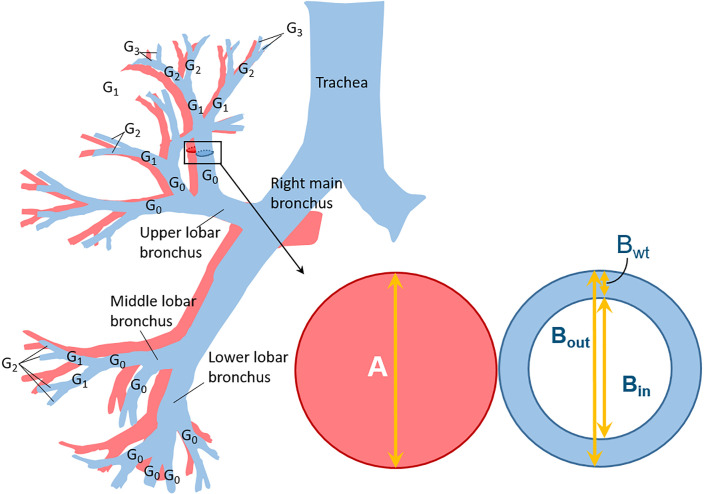
In short, the automatic BA analysis comprises five main steps: (1) segmentation of the bronchial tree; (2) matching of the adjacent artery for each detected bronchial branch (BA pair); (3) identification of the generation number (G) for each BA pair starting from the segmental bronchi (G0) down to the last detectable generation (figure 1); (4) computation of the following cross-sectional dimensions for each BA pair: bronchial outer diameter (Bout), bronchial lumen diameter (Bin), bronchial wall thickness (Bwt) which is computed as (Bout−Bin)/2 and diameter of the adjacent artery (A) (figure 1) and (5) computation of the following ratios for each BA pair: Bout/A, Bin/A and Bwt/A. The BA dimensions of each BA pair are the mean value of a large number of measurements from each bronchial branch. A schematic overview of generation definition and the BA ratio computation is shown in figure 1. In addition, the BA dimensions of bronchi distally from a bronchus obstructed by a mucus plug are excluded by the algorithm.
Figure 1.
The schematic view of the bronchial tree and of an bronchus-artery (BA) pair in cross-section showing the measurements taken for each bronchus. The bronchial tree (blue) with its accompanying artery system (pink) is shown on the left. The segmental bronchi are defined as G0 and the subsegmental bronchi as G1. When a bronchus splits into two or more, the generation number increases by one. On the right a BA pair is shown, the bronchus in blue and the adjacent artery in pink. The arrows depict the bronchus and artery dimensions that can be measured by the automatic BA analysis: bronchial outer diameter (Bout); bronchial lumen diameter (Bin), bronchial wall thickness (Bwt) and adjacent artery diameter (A). From these dimensions, BA ratios Bout/A and Bin/A are computed to detect bronchial widening and Bwt/A to detect bronchial wall thickening.
Clinical validation datasets
The clinical validation of the automatic BA analysis was done on three datasets collected from different centres. Chest CT scans were included for this study if they fulfilled the following requirements: inspiratory chest CT series; continuous helical CT acquisition; slice thickness equal or less than 1.5 mm; imaging of the entire lung parenchyma and no major artefacts.
The first cross-sectional dataset (CF control) has been previously reported and consisted of 11 randomly selected children with CF from the Erasmus MC Sophia Children's Hospital CF-CT cohort and 12 age-matched control subjects without CT abnormalities as evaluated by two independent radiologists.12 On these 23 inspiratory CT scans, dimensions of all visible BA pairs were measured manually. Measurements were made perpendicular to the longitudinal bronchial axis with an elliptic tool (Myrian, Montpellier, France; V.1.16.2) outlining the contours of bronchi and arteries to define the inner bronchial areas and outer bronchial areas and adjacent artery area. Subsequently, for each generation average diameters of bronchial inner wall, bronchial outer wall, and adjacent artery were computed from annotated surface areas. In total 4853 BA pairs were measured manually which served as a ground truth for comparison with the automatic BA-analysis outcomes.
The second dataset (Copenhagen cohort) consisted of 111 spirometry-controlled inspiratory chest CT scans from 57 children obtained from the longitudinal Copenhagen CF cohort.14 This dataset consisted of 56 baseline and 55 follow-up CT scans made at a 2-year interval, which were made as part of a prospective study comparing chest CT outcomes to multiple breath washout outcomes.
The third dataset (ataluren cohort) consisted of 347 inspiratory chest CT scans from 197 patients with CF from 36 CF sites in 11 countries in North America and Europe that participated in the ataluren study.15 This dataset consisted of 167 baseline CT scans and 180 follow-up CT scans over a 48-week interval.
For the Copenhagen and ataluren cohorts, all CT scans were previously analysed using the manual PRAGMA-CF analysis.6 14 PRAGMA-CF10 is a quantitative hierarchical scoring method for the quantification of bronchiectasis, mucus plugging, airway wall thickening, atelectasis and trapped air. Each component is expressed as a percentage (%) of the total lung volume. Furthermore, a composite score, %Disease, is computed representing the percentage of total lung volume occupied by airway abnormalities by summing the following components: %Bronchiectasis, %Mucus plugging and %Airway Wall Thickening. The Copenhagen cohort did not show significant progression of PRAGMA-CF outcomes over 2 years.14 For the ataluren cohort significant progression of PRAGMA-CF %Disease and %Airway Wall Thickening and a trend for %Bronchiectasis over 48 weeks were shown.6
Statistical analysis
Validating with manual measurements
To validate the measurement of BA ratios (Bout/A, Bin/A and Bwt/A) assessed by automatic BA analysis, mixed-effects models were used to investigate the difference between manual and automatic BA analysis on measurements in each segmental generation (from G1 to G5) on the CF-control dataset. We included: method (manual/automatic BA analysis), disease status (control/CF), age, gender and total lung volume as fixed effects, and subjects and segmental generations as random effects for analysis.
To evaluate the sensitivity of the manual and automatic BA analysis in discriminating abnormal airways and normal airways, the median of each BA ratio per generation per subject was used to obtain the area under the curve for Bout/A, Bin/A and Bwt/A for each segmental generation (G1–G5) in the CF control dataset. Then, we compared the area under the curve between the two methods using DeLong’s test.16
Defining cut-off values for bronchial widening and wall thickening
The BA analysis results in continuous scores for bronchial dimensions. However, in order to compare the BA output with the PRAGMA-CF score a dichotomous cut-off to define bronchial widening and wall thickening had to be chosen. Both the Bout/A and Bin/A have previously been used as markers of bronchiectasis.17 As there is no universally accepted cut-off value for Bout/A and Bin/A to define bronchiectasis18 cut-off values were computed from the CF-control dataset as follows:
for Bout/A and Bin/A, threshold values corresponding to receiver operating curve (Youden test) in CF-control dataset and 97.5 percentile from control subjects of the same dataset were compared to obtain the optimal cut-off values for G1–G5. In addition, a conservative cut-off value of 1.5 to define bronchiectasis for adults was used for Bout/A and Bin/A according to recent recommendations.19 To define the cut-off value for bronchial wall thickening (Bwt/A) we used the same approach as described above for bronchial widening using the CF-control dataset.
Correlation between the automatic BA analysis and PRAGMA-CF
For the comparison between PRAGMA-CF %Bronchiectasis and the automatic BA-analysis results (percentage of BA pairs above cut-off values) for bronchial widening in the two longitudinal datasets, we computed the correlation between these parameters both for baseline and for follow-up CT scans. This was not done for bronchial wall thickening as the reproducibility of PRAGMA-CF %Airway Wall Thickening is mostly poor. Spearman (or Pearson) correlation coefficients were used depending on whether the data distribution was skewed. In general, a correlation coefficient of 0–0.10 is regarded as negligible correlation, 0.10–0.39 as weak correlation, 0.40–0.69 as moderate correlation, 0.70–0.89 as strong correlation and 0.90–1.00 as very strong correlation.20
Monitoring disease progression
We computed the number of BA pairs above cut-off values for Bout/A, Bin/A and Bwt/A ratios for baseline and for follow-up of the Copenhagen and ataluren datasets. The number of abnormal airways (bronchial widening and wall thickening) is presented in median (range).
To investigate the progression in actual measurements of BA ratios in segmental generations in both longitudinal datasets, linear mixed-effects analysis were used. Higher generations are less visible, therefore, the numbers of measured BA pairs can become low which can introduce a bias. Since this conclusion is data driven, we performed sensitivity analyses assuming all or fewer generations. At first, a sensitivity analysis was performed on groups of generations (using G1–G5 for Copenhagen, G1–G6 for ataluren) or all generations for BA ratios to determine the optimal number of segmental generations to be included in the BA analysis to monitor disease progression for both longitudinal datasets. For both longitudinal dataset analyses, we included visiting time point, baseline age, gender, baseline height and total lung volume as fixed effects and subjects, lobes and segmental generations as random effects. For the BA-ratio outcomes (Bout/A, Bin/A and Bwt/A ratios), the logarithmic scale was used for Bin/A of the ataluren dataset and the square root scale was used for the rest since the normality assumptions of the residuals were not met.
All statistical analyses were done using R, V.4.0.5 (R Foundation for statistical Computing. Vienna, 2005). A p<0.05 was considered as statistically significant.
Results
Study population
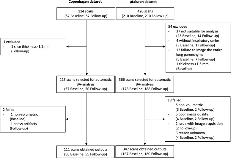
All 23 scans in the CF-control dataset were analysed successfully by the automatic BA analysis. In total, 113/114 (99.1%) CT scans in the Copenhagen dataset and 366/420 (87.1%) CT scans in the ataluren dataset met the inclusion criteria for the BA analysis. Of these, CTs 111/113 (98.2%) were analysed successfully for the Copenhagen dataset and 347/366 (96.8%) for the ataluren dataset. The reasons why CTs were excluded from analysis or why analysis failed are shown in figure 2. Thirty-seven out of 420 chest CT scans we received were not suitable for BA analysis due to movement artefacts or truncated images of the lung. In total 481 CT scans from 277 subjects aged 6–53 years were analysed successfully. The patients’ characteristics, number of CT scans and their BA pairs of the validation datasets are displayed in table 1.
Figure 2.
Flow chart of two longitudinal datasets. BA, bronchus artery.
Table 1.
Demographics of the three clinical validation datasets and their number of BA pairs assessed by the automatic BA analysis
| Status | CF-control dataset | Longitudinal datasets | ||||||
| Manual BA (n=23) |
Automatic BA (n=23) |
Copenhagen (n=57) |
Ataluren (n=197) |
|||||
| CF | Control | CF | Control | CF | CF | |||
| Time points | Baseline | Follow-up | Baseline | Follow-up | ||||
| Age (years)—median (IQR) | 11 (9.5–12.3) |
13.9 (8.7–15) |
11 (9.5–12.3) |
13.9 (8.7–15) |
12.6 (9.1–15.1) |
13.9 (11.1–17) |
21 (15–26) |
22 (16–26) |
| Male—n (%) | 6 (55) | 7 (58) | 6 (55) | 7 (58) | 28 (50) | 28 (51) | 80 (48) | 89 (49) |
| CT scans analysed (n) | 11 | 12 | 11 | 12 | 56 | 55 | 167 | 180 |
| Total BA pairs measured (n) | 3290 | 1563 | 3259 | 1443 | 12 798 | 14 648 | 54 840 | 65 673 |
| BA (n) pairs per CT scan—mean (SD) | 299 (104) | 130 (37) | 296 (167) | 120 (34) | 229 (106) | 266 (122) | 328 (161) | 365 (162) |
%, percentage; BA pairs, bronchus-artery pairs; CF, cystic fibrosis; (n), number.
Validating with manual measurements
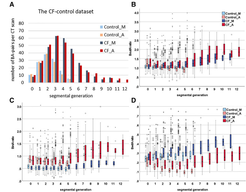
In the CF-control dataset, the automatic BA analysis was able to detect 4702 BA pairs against 4853 BA pairs12 by the manual method. In the control subjects, no BA pairs were found beyond G5 (figure 3, online supplemental figure S1) neither by the automatic BA analysis nor by the manual method. In children with CF, BA pairs were detected up to G11 for the manual method and G12 for the automatic BA analysis method. A number of BA pairs per CT were 130% and 147% higher in children with CF compared with the control subjects using the manual method and the automatic BA analysis, respectively (table 1).
Figure 3.
Number of BA pairs per CT scan and the distribution of BA ratios for the CF-control dataset assessed by the manual and automatic BA analysis for each segmental generation.Control_M: control subjects assessed by the manual method (light blue); Control_A: control subjects assessed by automatic BA analysis (pink); CF_M: subjects with cystic fibrosis assessed by the manual method (dark blue); CF_A: subjects with cystic fibrosis assessed by the automatic BA analysis (red); Bout/A ratio: ratio between bronchial outer diameter and adjacent artery diameter; Bin/A ratio: ratio between bronchial lumen diameter and adjacent artery diameter; Bwt/A ratio: ratio between bronchial wall thickness and adjacent artery diameter. Horizontal axis: 0 indicates the segmental bronchi and 1 and higher indicates consecutive subsegmental generations. (A) histogram shows the number of BA pairs per CT scan by segmental generations in the CF-control dataset. Boxplots (B–D) show boxplots for Bout/A ratio (B), Bin/A ratio (C), Bwt/A ratio (D) by segmental generation in the CF-control dataset. Each box shows median (horizontal line), IQR (solid box), 1.5×quartile range (whiskers) and outliers (circles and asterisks). BA, bronchus artery; CF, cystic fibrosis.
The difference between the manual method and automatic BA analysis in each segmental generation (from G1 to G5) is shown in the online supplement (online supplemental table S1). For Bout/A, no significant difference was detected (p=0.277). The Bin/A assessed by the automatic BA analysis was higher than for the manual method (mean difference 0.26; 95% CI 0.140 to 0.380, p<0.001). The Bwt/A assessed by the automatic BA analysis was lower than for the manual method (mean difference −0.13; 95% CI −0.176 to −0.084, p<0.001) (online supplemental figure S2). In addition, for Bout/A, Bin/A and Bwt/A, there was no significant difference in area under the curves between the automatic BA analysis and manual method from G1 to G5 (online supplemental tables S2–S4).
Cut-off values for bronchial widening and wall thickening
Based on measurements assessed by automatic BA analysis of the CF-control dataset, the optimal cut-off value for bronchial widening differentiating CF from control subjects was 1.1 for Bout/A and 0.8 for Bin/A. The best cut-off value for bronchial wall thickening was 0.14 for Bwt/A. (online supplemental table S5-1 and S5-2)
Correlation between the automatic BA analysis and PRAGMA-CF
The correlation coefficients between the automatic BA analysis outcomes and PRAGMA-CF outcome at baseline and follow-up were moderate or strong for the Copenhagen dataset and weak or moderate for ataluren datasets. The correlations results are shown in table 2 and online supplemental figure S3–S10.
Table 2.
Correlations between PRAGMA-CF and the automatic BA-analysis outcomes for the two longitudinal datasets
| |
%BE_PRAGMA-CF | |||
| Copenhagen | ataluren | |||
| Baseline | Follow-up | Baseline | Follow-up | |
| %BE_Automatic-BA | ||||
| Bout/A>1.1 | r=0.79 (0.64–0.88) | r=0.80 (0.65–0.88) | r=0.49 (0.34–0.61) | r=0.54 (0.41–0.65) |
| Bout/A>1.5 | r=0.75 (0.61–0.85) | r=0.84 (0.72–0.90) | r=0.59 (0.47–0.69) | r=0.66 (0.55–0.74) |
| Bin/A>0.8 | r=0.56 (0.34–0.72) | r=0.52 (0.26–0.72) | r=0.29 (0.13–0.43) | r=0.39 (0.26–0.51) |
| Bin/A>1.5 | r=0.47 (0.22–0.65) | r=0.71 (0.53–0.82) | r=0.47 (0.34–0.59) | r=0.57 (0.45–0.67) |
Results are shown as Spearman (or Pearson) correlation coefficient (r) and (95% CI). PRAGMA-CF: The Perth-Rotterdam Annotated Grid Morphometric Analysis for Cystic Fibrosis; BA: bronchial-artery; %BE_PRAGMA-CF: percentage of lung volume occupied by bronchiectasis on inspiratory chest CT scan assessed by PRAGMA-CF; %BE_Automatic-BA (Bout/A>1.1): percentage of BA pairs with Bout/A ratio greater than 1.1 quantified by the automatic BA analysis; %BE_Automatic-BA (Bout/A>1.5): percentage of BA pairs with Bout/A ratio greater than 1.5 quantified by the automatic BA analysis; %BE_Automatic-BA (Bin/A>0.8): percentage of BA pairs with Bin/A ratio greater than 0.8 quantified by the automatic BA analysis; %BE_Automatic-BA (Bin/A>1.5): percentage of BA pairs with Bin/A ratio greater than 1.5 quantified by the automatic BA analysis.
, ; BA, bronchus artery.
Monitoring disease progression
In the two longitudinal datasets, a number of BA pairs with Bout/A, Bin/A and Bwt/A ratios above cut-off values were higher at follow-up than at baseline. The number of BA pairs that were above each cut-off value in ataluren dataset was higher than that in the Copenhagen dataset (table 3).
Table 3.
The number of BA pairs with BA ratios above cut-off values per CT scan from paired subjects
| Copenhagen dataset (paired n=54) |
Ataluren dataset (paired n=150) |
|||
| Baseline | Follow-up after 2 years | Baseline | Follow-up after 48 weeks | |
| No of BA pairs per scan | 201 (88–527) | 254 (83–695) | 317 (21–807) | 332 (72–943) |
| No of BA pairs with Bout/A>1.1 | 90 (16–421) | 126 (21–568) | 259 (16–731) | 287 (16–871) |
| No of BA pairs with Bout/A>1.5 | 14 (1–189) | 20 (2–258) | 159 (5–536) | 178 (13–659) |
| No of BA pairs with Bin/A>0.8 | 85 (11–431) | 127 (8–546) | 234 (13–682) | 270 (12–834) |
| No of BA pairs with Bin/A>1.5 | 2 (1–36) | 3 (1–47) | 28 (1–319) | 39 (1–319) |
| No of BA pairs with Bwt/A>0.14 | 96 (30–487) | 115 (23–558) | 265 (18–744) | 281 (23–889) |
This table shows the median (range) number of BA pairs from G1 and higher per CT scan at baseline and follow-up for two longitudinal datasets classified as bronchial widening and bronchial wall thickness according to cut-off values. No.: number; no. of BA pairs with Bout/A>1.1: the number of BA pairs with the ratio of bronchial outer diameter and adjacent artery diameter greater than 1.1; No. of BA pairs with Bout/A>1.5: the number of BA pairs with Bout/A ratio greater than 1.5; no. of BA pairs with Bin/A>0.8: the number of BA pairs with the ratio of bronchial lumen diameter and adjacent artery diameter greater than 0.8; no. of BA pairs with Bin/A>1.5: the number of BA pairs with the Bin/A ratio greater than 1.5; no. of BA pairs with Bwt/A>0.14: the number of BA pairs with the ratio of bronchial wall thickness and adjacent artery diameter greater than 0.14.
BA, bronchus artery.
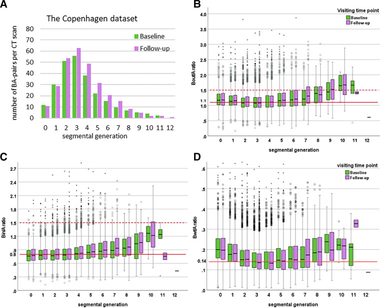
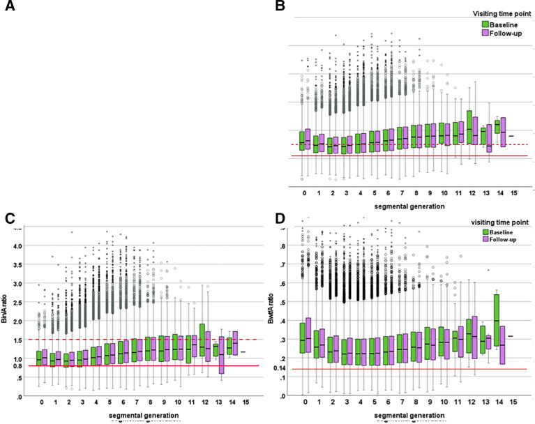
In the Copenhagen dataset, 27 446 BA pairs could be automatically analysed on 111 CT scans ranging from G0 to G12 (figure 4, online supplemental figure S11). The mean (SD) BA pairs per CT was 247 (116). At follow-up, the number of BA pairs per CT scan was 16.2% higher relative to baseline. The optimal number of segmental generations to investigate disease progression was G1 to G5. These generations included 89% of total BA pairs in all generations. Over 2 years, we detected that Bout/A and Bin/A increased significantly from G1 to G5 with a change of 0.02 for both Bout/A (95% CI −0.018 to 0.058; p<0.001) and Bin/A (95% CI −0.006 to 0.046; p<0.001) using mixed-effect models. However, there was just a trend for progression in Bwt/A (mean difference 0.002; 95% CI −0.009 to 0.013; p=0.03) from G1 to G5 (online supplemental table S6 and figure S12 for G1–G5, online supplemental figure 13 for sensitivity analysis).
Figure 4.
The number of bronchus-artery (BA) pairs for the Copenhagen dataset per CT scan and distribution of BA ratios at baseline and follow-up over 2 years for each segmental generation. Bout/A ratio: the ratio of bronchial outer diameter and adjacent artery diameter; Bin/A ratio: the ratio of bronchial lumen diameter and adjacent artery diameter; Bwt/A ratio: the ratio of bronchial wall thickness and adjacent artery diameter. Horizontal axis, 0 indicates the segmental bronchi and 1 and higher indicate consecutive subsegmental generations. (A) histogram shows the number of BA pairs per CT scan at baseline and follow-up by segmental generations. Boxplots (B–D) show the relation between Bout/A ratio (B), Bin/A ratio (C), Bwt/A ratio (D) at baseline and follow-up by segmental generation. Each box shows median (horizontal line), IQR (solid box), 1.5×quartile range (whiskers) and outliers (circles and asterisks). The red solid lines in B and D represent the computed cut-off values for bronchial widening and wall thickening. The red dotted line in B and C represents the conservative cut-off value for bronchiectasis of 1.5.19 BA, bronchus artery.
In the ataluren dataset, 120 513 BA pairs could be analysed on 347 CT scans ranging from G0 to G15 (figure 5, online supplemental figure S14). The mean (SD) number of BA pairs per CT was 347 (162). At follow-up, the number of BA pairs per CT scan was 11.3% higher relative to baseline. The optimal number of segmental generations to investigate disease progression was G1–G6. These generations included 90% of total BA pairs in all generations. Over 48 weeks, Bout/A, Bin/A and Bwt/A increased significantly from G1 to G6 with a change of 0.03 for Bout/A (95% CI −0.012 to 0.072; p<0.001), 0.02 for Bin/A (95% CI −0.003 to 0.043; p<0.001) and 0.01 for Bwt/A (95% CI 0.001 to 0.019; p<0.001) using mixed-effect models (online supplemental table S7 and figure S15 for G1–G6, online supplemental figure S16 for sensitivity analysis).
Figure 5.
The number of BA pairs for the ataluren dataset per CT scan and the distribution of BA ratios at baseline and follow-up over 48 weeks for each segmental generation. BA pair: BA pair; Bout/A ratio: the ratio of bronchial outer diameter and adjacent artery diameter; Bin/A ratio: the ratio of bronchial lumen diameter and adjacent artery diameter; Bwt/A ratio: the ratio of bronchial wall thickness and adjacent artery diameter. Horizontal axis, 0 indicates the segmental bronchi and 1 and higher indicate consecutive subsegmental generations. (A) histogram shows the number of BA pairs per CT scan at baseline and follow-up by segmental generations. Boxplots (B–D) show the relation between Bout/A ratio (B), Bin/A ratio (C), Bwt/A ratio (D) at baseline and follow-up by segmental generation. Each box shows median (horizontal line), IQR (solid box), 1.5×quartile range (whiskers) and outliers (circles and asterisks). The red solid lines in A, B and C represent the computed cut-off values for bronchial widening and wall thickening. The red dotted line represents the conservative cut-off value for bronchiectasis of 1.5.19 BA, bronchus artery.
Discussion
In this study, we validated a fully automatic BA analysis for measuring BA dimensions on chest CT scans of patients with CF aged from 6 to 54 years old for the detection and monitoring of bronchial widening and wall thickening. To the best of our knowledge, the automatic BA analysis is the first fully automated algorithm able to assess and measure large numbers of BA pairs on chest CT scans of patients with CF from three different datasets.
We showed that for the CF control dataset the automatic BA analysis was able to detect almost the same total number of BA pairs as the manual BA analysis. The agreement between the automatic and manual BA measurements was excellent for Bout/A. However, for Bin/A values were systematically higher, and for Bwt/A lower for the automated BA analysis than for the manual BA analysis. This can be explained by systematic differences between the two methods on how the inner and outer wall of a bronchus is detected. For the manual BA analysis, the observer draws the inner boundary of bronchial wall where a great intensity drop is perceived subjectively,21 while for the automatic analysis an objective cut-off is based on sub-resolution measurements which were validated on phantoms studies. Even though there were systematic differences in the BA measurements between the manual and automatic BA analysis, their sensitivity to detect bronchial widening and bronchial wall thickening was comparable. The automatic BA analysis allowed to detect and assess BA pairs up to G5 in control subjects, but up to G12 and further in the patients with CF lung disease caused by widening and thickening of the small airways making them visible also in the periphery of the lung. This difference between controls and CF was also observed by the manual measurements.
For the automated identification of bronchial widening, Bout/A ratio showed better discrimination than the Bin/A ratio in each segmental generation (from G1 to G5) (online supplemental tables S2 and S3). This was expected as the sensitivity of Bin/A to detect widening of the bronchi can be reduced due to mucus attached to the inner bronchial wall and/or folding of the mucosal layer especially at lung volumes below total lung capacity due to suboptimal inspiration.22 23 Our study, therefore, adds further evidence in support of the Bout/A ratio as a more precise parameter for the diagnosis of bronchial widening than the Bin/A ratio which is often used in clinical practice and clinical studies.24 25
Based on the automatic BA analysis of the CF-control dataset, we concluded that the optimal cut-off value for bronchial widening is 1.1 for Bout/A and 0.8 for Bin/A, instead of using the poorly validated cut-off of BA ratio greater than 1.26 The optimal cut-off for bronchial wall thickening is 0.14 for Bwt/A ratio. To compute this optimal cut-off between control subjects and patients with CF, we showed that including all BA measurements of G1-G5 resulted in the best discrimination between the two groups. This makes sense as G1–G5 includes a large fraction (81%–90%) of all the BA pairs that can be measured on a CT scan in most patients. In control subjects beyond generation G5 less BA pairs become visible due to the limited resolution of commonly used CT scanners which are not able to detect airways below 2 mm in diameter.27 In patients with CF a large number of BA pairs can still be observed for generations beyond G5 due to bronchial widening and wall thickening. The total number of BA pairs in G1–G5 was 1303 (90%) for controls vs 2841 (81%) for CF hence allowing for a fair comparison not biased by the higher number of BA pairs beyond G5 in CF.
For further validation of the BA analysis, we compared BA outcomes with those of the established PRAGMA-CF scoring method in a cross-sectional analysis. We observed a weak to strong correlation between the percentage of BA pairs showing bronchial widening for the automatic BA analysis and the fraction of total lung volume occupied by bronchiectasis for PRAGMA-CF. Comparing the manual PRAGMA-CF %Bronchiectasis with outcomes of the BA analysis for bronchial widening, it has become clear that trained observers are able to recognise the more severely widened bronchi with a BA-ratio of 1.5 or higher but have difficulties recognising bronchi with a BA-ratio between 1.1 and 1.5 as widened. This correlation was stronger for the Copenhagen dataset at baseline and follow-up relative to the ataluren dataset (table 2). The most likely explanation for this difference is that the ataluren study did not include a volume-controlled CT acquisition protocol resulting in a larger variability in lung volumes within and between subjects. Also, CT scanners and protocols varied from centre to centre (36 centres in total), in contrast to the single centre Copenhagen cohort where a strict volume control protocol was used both at baseline and at follow-up and all patients were scanned on the same CT scanner. Furthermore, the ataluren study consisted of older subjects with overall more advanced lung disease as illustrated by a forced vital capacity of 78% compared with a forced vital capacity of 98% for the Copenhagen cohort (online supplemental table S8).14 28 Overall, it can be concluded that the automatic BA analysis outcomes correlated well with comparable PRAGMA-CF outcomes in the cross-sectional analysis, however, volume control CT protocols are important for optimal BA analysis.
In monitoring of progression of CF lung disease, the BA analysis was sensitive to detect progression for bronchial widening (Bout/A and Bin/A) in the ataluren dataset as well as in the Copenhagen cohort. Using PRAGMA-CF progression of bronchiectasis was detected only in the ataluren cohort. This suggests higher sensitivity of the BA analysis to detect bronchial widening compared with PRAGMA-CF. Progression of bronchial wall thickening (Bwt/A) was only observed in the ataluren dataset but not in the Copenhagen dataset. These Bwt/A findings are in agreement with that of the longitudinal PRAGMA-CF analysis in the two cohorts.6 14
In addition, the BA ratios to define bronchial widening and wall thickening are computed based on adjacent arterial diameters. In control subjects, bronchi and adjacent arteries are expected to be of similar sizes.29 However, in patients with lung diseases such as CF, airway obstruction can result in hypoventilation of lung regions resulting in hypoxic pulmonary vasoconstriction.30 This could result in subtle changes of the arterial diameters visible on chest CT, and therefore, influence the sensitivity of the BA ratio to detect and monitor relevant structural airway disease. However, we do not think changes in arterial size have a major impact on our findings as we were unable to detect significant changes in arterial dimensions from baseline to follow-up in either Copenhagen or the ataluren dataset (online supplemental tables S9 and S10). This suggests that the progression in BA ratios found in these longitudinal cohorts is primarily caused by a change in bronchial dimensions. We speculate that hypoxic pulmonary vasoconstriction might be more relevant for the smaller airways as the accompanying arteries contain more smooth muscle relative to more central arteries,31 which might be unable to be detected by BA analysis on current CT resolution.
For longitudinal monitoring, we aimed to identify which generations needed to be included to have the highest sensitivity to detect change. For the Copenhagen dataset, we selected G1–G5 and for the ataluren dataset G1–G6 as the number of BA pairs from G1 to G6 accounted similar proportion (probably due to higher mean age and therefore also larger lung size) as the number of BA pairs from G1 to G5 of Copenhagen dataset. However, there were no major differences between the analysis results using the limited generations (G1–G5 or G1–G6) set compared with all detectable generations in our sensitivity analysis (online supplemental tables S6 and S7). In addition, it has been shown that geometrical changes in more central airways correlate well to those in the smaller airways.32 And the inflammation of the bronchial walls is more frequently present in the middle-size airways in patients with CF.33 34 Another reason for selecting fewer generations to investigate the change in Bout/A, Bin/A and Bwt/A is that geometrical changes in this compartment are very precise, as a large number of BA pairs can be measured. Overall, the automatic BA analysis of bronchus and artery dimensions in G1–G6 is an accurate and sensitive way to detect and quantify bronchial widening and bronchial wall thickening in CF. Therefore, we recommend to use G1–G5 in children and G1–G6 in adults to detect abnormal airways and to investigate disease progression in further studies to avoid large variability in detection of periphery.
Limitations
The limitations of our study are, first, the sample size of the CF-control dataset to compute thresholds for bronchial widening and wall thickening is relatively small. Given the extreme time-consuming process of manually annotating all visible BA pairs, it was not feasible to perform the BA analysis on a larger dataset. Despite the small number of patients in the dataset, the total number of assessed BA pairs is very large and observed cut-off BA-ratio values for bronchial widening and bronchial wall thickening are similar to those reported in the literature.35 Second, using chest CT, mucus cannot be differentiated from the bronchial wall when the bronchus is completely obstructed, however, the percentage of bronchi obstructed by mucus is relatively small. Despite this fact, the algorithm was sensitive enough to measure bronchial wall thickness and even detect a significant progression in ataluren dataset. The third limitation is the absence of a well-defined definition and cut-off value to diagnose bronchial widening being an important feature for the diagnosis of bronchiectasis.19 This leads to large variability between the observers. For evaluating BA dimensions in patient care, it is important to obtain robust BA-reference values of normal subjects in different age ranges, but such reference values are currently not available. We are in the process of analysing a large dataset of chest CT scans obtained from multiple centres without radiological evidence of lung disease. This dataset includes patients with normal chest CTs as evaluated by three independent radiologists that were acquired without strict volume control. Hence, additional reference values need to be collected including normal chest CTs acquired with volume control.
Future prospective
Potentially, the automated assessment of BA dimensions is highly relevant for a wide range of diseases such as asthma, chronic obstructive pulmonary disease, bronchiectasis and primary ciliary dyskinesia. For each of these diseases, further validation studies are needed. Compared with manual scoring methods this automatic BA analysis currently requires 20 minutes or less of computation time for the BA analysis of a CT scan to obtain objective, and sensitive clinically relevant outcomes related to airways disease. To make the BA analysis available for clinical use, it has to be implemented into the workflow of the radiologists. Finally, implementation of volume control during chest CT acquisition is needed to make optimal use of the BA analysis.
Conclusions
The automatic BA analysis is a newly developed AI-based algorithm to objectively assess a large number of BA dimensions of BA pairs on a chest CT. This BA analysis can be used for the quantitative diagnosis of bronchial widening, bronchial wall thickening and to assess the extent and progression of these structural changes. In this study, the BA analysis was more sensitive to detect progression of bronchial widening in mild CF lung disease compared with PRAGMA-CF. The BA analysis will contribute to the objective phenotyping and monitoring of the structural airway abnormalities in CF and potentially for other lung diseases in the future.
Acknowledgments
This publication was made possible in part thanks to a contribution from the European Regional Development Fund (EFRO) Oost-Nederland. The authors wish to thank Mariette P.C. Kemner-van de Corput for her data selection and Cristina Cretu for her reviewing and comments.
Footnotes
Contributors: QL has made substantial contributions to the conception, design and analysis of the work, drafted the manuscript, drew the figures, and gave final approval to the manuscript. LG-E contributed to developing LungQ, critically revised the manuscript, and gave final approval to the manuscript. E-RA contributed to the interpretation of data for this paper and critically revised the manuscript, and gave final approval to the manuscript. YC critically revised the manuscript and gave final approval to the manuscript. JPC contributed to the development of LungQ, revised the manuscript and gave final approval to the manuscript. RMS. contributed to the acquisition of data, revised the manuscript, and gave final approval to the manuscript. DC revised the manuscript and gave final approval to the manuscript. KGN contributed to the acquisition of data, revised the manuscript, and gave final approval to the manuscript. MdB critically revised the manuscript and gave final approval to the manuscript. PC contributed to the interpretation of image data, critically revised the manuscript, and gave final approval to the manuscript. HT contributed to the conception and design of the work, critically revised the manuscript, and gave final approval to the manuscript. HT is responsible for the overall content as the guarantor.
Funding: This study received funding from Nederlandse Cystic Fibrosis Stichting (NCFS)- Health Holland (PPS). (Project number: TKI-LSH-T2018).
Competing interests: HT has received in the last 5 years multiple grants from the following public and institutional grant institutions for lung structure and function research: NHMRC, NIH, CFF, ECFS, IMI, Sophia Foundation. He received unconditional grants for investigator-initiated research from Chiesi; Vectura, Novartis and Insmed. He has acted as consultant for Insmed, TBIO, Thirona, Neupharma and Boehringer. He has a part time position as chief medical officer for Thirona. He functions as vice chair and faculty for the Advance course sponsored by Vertex. He owns no shares. LG-E is a scientist working at Thirona. JPC is shareholder at Thirona. DC is director of the Erasmus MC-LungAnalysis laboratory. PC acted as consultant for Vertex and Chiesi Pharmaceuticals.
Provenance and peer review: Not commissioned; externally peer reviewed.
Supplemental material: This content has been supplied by the author(s). It has not been vetted by BMJ Publishing Group Limited (BMJ) and may not have been peer-reviewed. Any opinions or recommendations discussed are solely those of the author(s) and are not endorsed by BMJ. BMJ disclaims all liability and responsibility arising from any reliance placed on the content. Where the content includes any translated material, BMJ does not warrant the accuracy and reliability of the translations (including but not limited to local regulations, clinical guidelines, terminology, drug names and drug dosages), and is not responsible for any error and/or omissions arising from translation and adaptation or otherwise.
Data availability statement
Data are available on reasonable request. For data sharing, the permission is needed from Thirona and provider of CT scans.
Ethics statements
Patient consent for publication
Not applicable.
Ethics approval
This study involves human participants and was approved by Medical Ethical Review Committee Erasmus MCID: MEC-2013-338. Participants gave informed consent to participate in the study before taking part.
References
- 1. Sly PD, Gangell CL, Chen L, et al. Risk factors for bronchiectasis in children with cystic fibrosis. N Engl J Med 2013;368:1963–70. 10.1056/NEJMoa1301725 [DOI] [PubMed] [Google Scholar]
- 2. Murphy KP, Maher MM, O’Connor OJ. Imaging of cystic fibrosis and pediatric bronchiectasis. AJR Am J Roentgenol 2016;206:448–54. 10.2214/AJR.15.14437 [DOI] [PubMed] [Google Scholar]
- 3. Tiddens H, Chen Y, Andrinopoulou E-R, et al. The effect of inhaled hypertonic saline on lung structure in children aged 3-6 years with cystic fibrosis (SHIP-CT): a multicentre, randomised, double-blind, controlled trial. Lancet Respir Med 2022;10:669–78. 10.1016/S2213-2600(21)00546-4 [DOI] [PubMed] [Google Scholar]
- 4. Bouma NR, Janssens HM, Andrinopoulou E-R, et al. Airway disease on chest computed tomography of preschool children with cystic fibrosis is associated with school-age bronchiectasis. Pediatr Pulmonol 2020;55:141–8. 10.1002/ppul.24498 [DOI] [PMC free article] [PubMed] [Google Scholar]
- 5. Svedberg M, Gustafsson P, Tiddens H, et al. Risk factors for progression of structural lung disease in school-age children with cystic fibrosis. J Cyst Fibros 2020;19:910–6. 10.1016/j.jcf.2019.10.014 [DOI] [PubMed] [Google Scholar]
- 6. Tiddens HAWM, Andrinopoulou E-R, McIntosh J, et al. Chest computed tomography outcomes in a randomized clinical trial in cystic fibrosis: lessons learned from the first Ataluren phase 3 study. PLoS One 2020;15:e0240898. 10.1371/journal.pone.0240898 [DOI] [PMC free article] [PubMed] [Google Scholar]
- 7. Bhalla M, Turcios N, Aponte V, et al. Cystic fibrosis: scoring system with thin-section CT. Radiology 1991;179:783–8. 10.1148/radiology.179.3.2027992 [DOI] [PubMed] [Google Scholar]
- 8. Reiff DB, Wells AU, Carr DH, et al. CT findings in bronchiectasis: limited value in distinguishing between idiopathic and specific types. AJR Am J Roentgenol 1995;165:261–7. 10.2214/ajr.165.2.7618537 [DOI] [PubMed] [Google Scholar]
- 9. Brody AS, Kosorok MR, Li Z, et al. Reproducibility of a scoring system for computed tomography scanning in cystic fibrosis. J Thorac Imaging 2006;21:14–21. 10.1097/01.rti.0000203937.82276.ce [DOI] [PubMed] [Google Scholar]
- 10. Rosenow T, Oudraad MCJ, Murray CP, et al. A quantitative structural lung disease computed tomography outcome in young children with cystic fibrosis. Am J Respir Crit Care Med 2015;191:1158–65. 10.1164/rccm.201501-0061OC [DOI] [PubMed] [Google Scholar]
- 11. Wijker NE, Vidmar S, Grimwood K, et al. Early markers of cystic fibrosis structural lung disease: follow-up of the ACFBAL cohort. Eur Respir J 2020;55:1901694. 10.1183/13993003.01694-2019 [DOI] [PubMed] [Google Scholar]
- 12. Kuo W, Andrinopoulou E-R, Perez-Rovira A, et al. Objective airway artery dimensions compared to CT scoring methods assessing structural cystic fibrosis lung disease. J Cyst Fibros 2017;16:116–23. 10.1016/j.jcf.2016.05.015 [DOI] [PubMed] [Google Scholar]
- 13. Kuo W, Soffers T, Andrinopoulou E-R, et al. Quantitative assessment of airway dimensions in young children with cystic fibrosis lung disease using chest computed tomography. Pediatr Pulmonol 2017;52:1414–23. 10.1002/ppul.23787 [DOI] [PubMed] [Google Scholar]
- 14. Sandvik RM, Kongstad T, Green K, et al. Prospective longitudinal association between repeated multiple breath Washout measurements and computed tomography scores in children with cystic fibrosis. J Cyst Fibros 2021;20:632–40. 10.1016/j.jcf.2020.09.010 [DOI] [PubMed] [Google Scholar]
- 15. Kerem E, Konstan MW, De Boeck K, et al. Ataluren for the treatment of nonsense-mutation cystic fibrosis: a randomised, double-blind, placebo-controlled phase 3 trial. Lancet Respir Med 2014;2:539–47. 10.1016/S2213-2600(14)70100-6 [DOI] [PMC free article] [PubMed] [Google Scholar]
- 16. Anagnostopoulou P, Latzin P, Jensen R, et al. Normative data for multiple breath washout outcomes in school-aged Caucasian children. Eur Respir J 2020;55:1901302. 10.1183/13993003.01302-2019 [DOI] [PubMed] [Google Scholar]
- 17. Meerburg JJ, Veerman GDM, Aliberti S, et al. Diagnosis and quantification of bronchiectasis using computed tomography or magnetic resonance imaging: a systematic review. Respir Med 2020;170:105954. 10.1016/j.rmed.2020.105954 [DOI] [PubMed] [Google Scholar]
- 18. Tiddens H, Meerburg JJ, van der Eerden MM, et al. The radiological diagnosis of bronchiectasis: what's in a name? Eur Respir Rev 2020;29:190120. 10.1183/16000617.0120-2019 [DOI] [PMC free article] [PubMed] [Google Scholar]
- 19. Aliberti S, Goeminne PC, O’Donnell AE, et al. Criteria and definitions for the radiological and clinical diagnosis of bronchiectasis in adults for use in clinical trials: International consensus recommendations. Lancet Respir Med 2022;10:298–306. 10.1016/S2213-2600(21)00277-0 [DOI] [PubMed] [Google Scholar]
- 20. Schober P, Boer C, Schwarte LA. Correlation coefficients: appropriate use and interpretation. Anesth Analg 2018;126:1763–8. 10.1213/ANE.0000000000002864 [DOI] [PubMed] [Google Scholar]
- 21. Schmidt M, Karssemeijer N, Summers RM, et al. Reproducibility of airway wall thickness measurements. SPIE Medical Imaging; San Diego, California, USA.March 4, 2010. 10.1117/12.844453 [DOI] [Google Scholar]
- 22. Lambert RK. Role of bronchial basement membrane in airway collapse. J Appl Physiol 1991;71:666–73. 10.1152/jappl.1991.71.2.666 [DOI] [PubMed] [Google Scholar]
- 23. Mott LS, Park J, Murray CP, et al. Progression of early structural lung disease in young children with cystic fibrosis assessed using CT. Thorax 2012;67:509–16. 10.1136/thoraxjnl-2011-200912 [DOI] [PubMed] [Google Scholar]
- 24. Loeve M, Gerbrands K, Hop WC, et al. Bronchiectasis and pulmonary exacerbations in children and young adults with cystic fibrosis. Chest 2011;140:178–85. 10.1378/chest.10-1152 [DOI] [PubMed] [Google Scholar]
- 25. Chang AB, Bush A, Grimwood K. Bronchiectasis in children: diagnosis and treatment. Lancet 2018;392:866–79. 10.1016/S0140-6736(18)31554-X [DOI] [PubMed] [Google Scholar]
- 26. Díaz AA, Nardelli P, Wang W, et al. Artificial intelligence-based CT assessment of bronchiectasis: the Copdgene study. Radiology 2023;307:e221109. 10.1148/radiol.221109 [DOI] [PMC free article] [PubMed] [Google Scholar]
- 27. McGuinness G, Naidich DP, Leitman BS, et al. Bronchiectasis: CT evaluation. AJR Am J Roentgenol 1993;160:253–9. 10.2214/ajr.160.2.8424327 [DOI] [PubMed] [Google Scholar]
- 28. Wilschanski M, Miller LL, Shoseyov D, et al. Chronic Ataluren (PTC124) treatment of nonsense mutation cystic fibrosis. Eur Respir J 2011;38:59–69. 10.1183/09031936.00120910 [DOI] [PubMed] [Google Scholar]
- 29. O’Donnell AE. Bronchiectasis. Chest 2008;134:815–23. 10.1378/chest.08-0776 [DOI] [PubMed] [Google Scholar]
- 30. Diaz AA, Young TP, Maselli DJ, et al. Bronchoarterial ratio in never-smokers adults: implications for bronchial dilation definition. Respirology 2017;22:108–13. 10.1111/resp.12875 [DOI] [PMC free article] [PubMed] [Google Scholar]
- 31. Sylvester JT, Shimoda LA, Aaronson PI, et al. Hypoxic pulmonary vasoconstriction. Physiol Rev 2012;92:367–520. 10.1152/physrev.00041.2010 [DOI] [PMC free article] [PubMed] [Google Scholar]
- 32. Tiddens HA, Paré PD, Hogg JC, et al. Cartilaginous airway dimensions and airflow obstruction in human lungs. Am J Respir Crit Care Med 1995;152:260–6. 10.1164/ajrccm.152.1.7599833 [DOI] [PubMed] [Google Scholar]
- 33. Whitwell F. A study of the pathology and pathogenesis of bronchiectasis. Thorax 1952;7:213–39. 10.1136/thx.7.3.213 [DOI] [PMC free article] [PubMed] [Google Scholar]
- 34. Sly PD, Wainwright CE. Diagnosis and early life risk factors for bronchiectasis in cystic fibrosis: a review. Expert Rev Respir Med 2016;10:1003–10. 10.1080/17476348.2016.1204915 [DOI] [PubMed] [Google Scholar]
- 35. Kapur N, Masel JP, Watson D, et al. Bronchoarterial ratio on high-resolution CT scan of the chest in children without pulmonary pathology: need to redefine bronchial dilatation. Chest 2011;139:1445–50. 10.1378/chest.10-1763 [DOI] [PubMed] [Google Scholar]
Associated Data
This section collects any data citations, data availability statements, or supplementary materials included in this article.
Supplementary Materials
thorax-2023-220021supp001.pdf (6.2MB, pdf)
Data Availability Statement
Data are available on reasonable request. For data sharing, the permission is needed from Thirona and provider of CT scans.


